U minho | 20 14 Cátia P er eir a
Molecular Mechanism Underl
ying t
he
Anti-Tumoral Activity of Lactoferrin
Cátia Sofia dos Santos Pereira
Dezembro de 2014
Molecular Mechanism Underlying the
Anti-Tumoral Activity of Lactoferrin
Tese de Mestrado
Mestrado em Genética Molecular
Trabalho efetuado sob a orientação de
Professora Doutora Maria Manuela Sansonetty Gonçalves Côrte-Real Professora Doutora Lígia Raquel Marona Rodrigues
Cátia Sofia dos Santos Pereira
Dezembro de 2014
Molecular Mechanism Underlying the
Anti-Tumoral Activity of Lactoferrin
ii
DECLARAÇÃO
Nome: Cátia Sofia dos Santos Pereira
Endereço Eletrónico: catia91pereira@gmail.com Telefone: 916960439
Nº Cartão Cidadão: 14012149
Título da Tese de Mestrado:
Molecular Mechanism Underlying the Anti-Tumoral Activity of Lactoferrin
Orientadores:
Professora Doutora Maria Manuela Sansonetty Gonçalves Côrte-Real Professora Doutora Lígia Raquel Marona Rodrigues
Instituições de Acolhimento:
Centro de Biologia Molecular e Ambiental (CBMA) Centro de Engenharia Biológica (CEB)
Ano de Conclusão: 2014 Designação do Mestrado:
Mestrado em Genética Molecular
DE ACORDO COM A LEGISLAÇÃO EM VIGOR, NÃO É PERMITIDA A REPRODUÇÃO DE QUALQUER PARTE DESTA TESE
Universidade do Minho, 1 de Dezembro de 2014 Assinatura:________________________________
iii
AGRADECIMENTOS
No final deste ano de trabalho existem várias pessoas a quem quero agradecer por terem contribuído para que esta tese tenha passado do sonho à realidade:
À minha orientadora, professora MANUELA CÔRTE-REAL, pela enorme oportunidade que me deu, aceitar o desafio que me propôs foi a melhor decisão que tomei. Obrigada por me ter ajudado a crescer cientificamente durante este ano, por ouvir e valorizar as minhas opiniões, e por ter sempre uma palavra de apreço. À minha co-orientadora, professora LÍGIA RODRIGUES, nunca irei esquecer aquelas manhãs de setembro nas quais aprendi imenso consigo, não podia ter tido melhor mentora de cultura de células, e é uma bagagem que vou levar sempre comigo. Obrigada pela prontidão que sempre demonstraram e por confiarem em mim e me darem liberdade de trabalho. Nunca irei esquecer o vosso apoio e disponibilidade incondicionais, fosse semana ou fim-de-semana. O meu sincero obrigada às duas!
Ao professor HERNÂNI GERÓS, mesmo não sendo meu orientador no papel, é como se fosse, obrigada por todas aquelas reuniões em que me ensinou sempre a olhar para as coisas de forma diferente; a sua paixão e entusiasmo pelo trabalho que faz são inspiradoras. À doutora SUSANA CHAVES por, de uma forma ou de outra, ter estado sempre presente e ter dado sempre opiniões e ideias que tanto ajudaram, em especial nesta reta final. À professora ANA PRETO por me ter concedido um local maravilhoso onde trabalhar e por sempre se preocupar se tudo estava a correr bem. Obrigada a todos!
À LISANDRA, minha mentora e amiga, sem ti seria muito complicado. Obrigada por tudo o que me ensinaste e todas as horas que passamos juntas no lab, fizeste muito mais do que apenas orientar-me, ouviste-me sempre que precisei, aconselhaste-me daquela forma que só tu sabes fazer, e tinhas sempre um abraço apertado para me dar sempre que necessitei.
Às minhas meninas TATXI, RITA e PATY sem vocês era absolutamente impossível ter realizado esta tese! Vocês tornaram-se parte da minha família e fizeram de Braga um lar para mim. É impossível descrever o significado que todas as aventuras que passamos juntas têm para mim. Todas as nossas conversas, os nossos jantares, as nossas peripécias, as nossas gargalhadas, o nosso dia-a-dia, tudo foi essencial para que nos apoiássemos umas às outras nos momentos mais complicados. À BECAS, minha amiga do coração, sempre com as palavras certas no momento certo e com aqueles miminhos que tão bem me faziam. A todos os meus companheiros de mestrado JOÃO, BIZARRO, BRITO, ELI, DIANA, CRIS, CARLA, que de uma forma ou de outra, sempre me apoiaram quer por uma mensagem de carinho quer por um sorriso nos corredores do DB. Também aos meus colegas de Barcelos em especial à ELISETE, CATERINA, ANA, e às
iv
minhas BAILARINAS que tantas vezes me ajudaram a assentar os pés na terra. Obrigada a todos do fundo do coração!
A todas as pessoas do DB em geral, incluindo professores e funcionários, por fazerem do DB uma família e um local onde dá gosto trabalhar e onde reina a entreajuda. À MARÍLIA e ao LUÍS por serem meus companheiros nesta jornada pelo mundo da lactoferrina, por ajudarem a tornar todas aquelas horas de trabalho muito mais agradáveis. À CARLA, RITA, CRISTINA, JOÃO, ANTÓNIO e SUELLEN por toda a ajuda ao longo deste ano. A todo o pessoal do LBA que contribuiu para que se contruísse a família LBA e fez deste lab, não apenas um local de trabalho, mas um local onde todos convivíamos e nos ajudávamos mutuamente. A todo o pessoal da Micro I, que sempre me ajudou em tudo que precisei, inclusivé a procurar coisas que pareciam impossíveis de encontrar, mesmo não estando no vosso laboratório a trabalhar, sempre fizeram com que me sentisse bem e me integraram. O meu obrigada a todos!
Ao pessoal do DEB. Um especial agradecimento à TÂNIA que foi a minha autêntica guia nos primeiros tempos, sempre estavas disponível para tudo o que eu precisasse, e tornaste a minha adaptação ao LCCT muito mais fácil. À MARIANA, mais do que uma companheira, uma amiga que levo para a vida, obrigada por toda a tua ajuda, por todos os conselhos e por todas as nossas conversas. Também quero agradecer à RITA, à VERA e ao FRANKLIN por todo o apoio que sempre me deram, por muitas coisas que estivessem a fazer ao mesmo tempo não houve uma única vez que eu precisasse da vossa ajuda e vocês não me apoiassem logo. Obrigada por tudo!
Ao JOÃO, meu porto de abrigo, passem as etapas que passem, por mais ou menos difíceis que sejam, tu estás sempre lá e ajudas-me a conquistar a camisola amarela! Obrigada por toda a paciência, compreensão, amizade, por sempre me ajudares a descontrair e encarar os problemas de cabeça erguida, nestes dois anos de mestrado foste incansável e foi com o teu apoio que consegui chegar ao fim. Obrigada por fazeres parte da minha vida!
Aos MEUS PAIS e ao MEU IRMÃO, a vocês eu tenho a agradecer toda a minha vida! Sempre acreditaram em mim, fizerem tudo para que eu realizasse os meus sonhos, apoiaram as minhas decisões, tiveram muitaaaa paciência, estiveram presentes em todos os momentos da minha vida e sem vocês não seria o que sou hoje. Obrigada também a toda a MINHA FAMÍLIA por sempre acreditarem em mim mesmo quando eu própria não acreditava. Um imenso obrigada!
Por fim, queria agradecer ao Centro de Biologia Molecular e Ambiental (CBMA), ao Centro de Engenharia Biológica (CEB), ao programa FEDER através do POFC-COMPETE e à Fundação para a Ciência e Tecnologia (FCT) através dos projetos PEst-OE/BIA/UI4050/2014, FCT-ANR/BEX-BCM/0175/2012 e PTDC/SAU-BMA/121028/2010, pelo financiamento deste trabalho.
v
M
OLECULARM
ECHANISMU
NDERLYING THEA
NTI-T
UMORALA
CTIVITY OFL
ACTOFERRINABSTRACT
Lactoferrin (Lf) is an iron-binding glycoprotein found in many biological fluids, being particularly abundant in milk. Originally viewed as a milk protein involved in the regulation of iron homeostasis, Lf is now considered a multifunctional protein to which more and more physiological roles have been attributed. These include anti-tumoral, anti-microbial, immunomodulatory, among other properties. Regarding its anti-tumoral activity, Lf has been reported to be effective against a variety of human cancers both by
in vitro an in vivo studies. Its well-tolerability has also been attested in some human
clinical trials. Therefore, Lf administration is an emerging strategy in cancer therapeutics that is expected to become widespread. In this way, the elucidation of molecular mechanisms underlying Lf cytotoxicity to cancer cells has become an important research field. Although some cellular mechanisms have already been proposed such as apoptosis induction, cell cycle arrest, angiogenesis and metastasis inhibition, the true molecular mechanism remains elusive. In the present work, we aimed to unveil the mechanism of action and the cellular target(s) of Lf on cancer cells. For that purpose, three breast cell lines with different genetic backgrounds were used, namely an invasive, a non-invasive and a non-tumorigenic. In this study we focus on bovine Lf (bLf) as it has been considered a safe nutraceutical for various applications. Results showed that these cells exhibit different susceptibilities to bLf, which prompted us to implement several biochemical and analytical approaches, including flow cytometry, fluorescence microscopy and spectrofluorimetry in order to dissect the bLf’s target(s) and molecular mechanism operating in the sensitive cells. Results obtained throughout this work allowed us to identify a novel Lf activity and to propose a molecular mechanism regarding its anti-tumoral activity, which may have important implications on cancer therapeutics, particularly on the therapy of highly metastatic tumors.
vi
M
ECANISMOM
OLECULARS
UBJACENTE ÀA
TIVIDADEA
NTI-T
UMORAL DAL
ACTOFERRINARESUMO
A lactoferrina (Lf) é uma glicoproteína de ligação ao ferro encontrada em diversos fluídos biológicos, sendo particularmente abundante no leite. Originalmente descrita como uma proteína do leite envolvida na regulação da homeostasia do ferro, a Lf é atualmente considerada uma proteína multifuncional à qual têm sido atribuídas diversas funções fisiológicas. Estas incluem propriedades tumorais, microbianas, imunomoduladoras, entre outras. Relativamente à sua atividade anti-tumoral, a Lf foi descrita como eficaz contra uma variedade de cancros humanos em estudos realizados in vitro e in vivo. A sua boa tolerabilidade foi também demonstrada em alguns ensaios clínicos em humanos. Portanto, a administração de Lf constitui uma estratégia com elevado potencial na terapêutica do cancro, sendo expectável que se torne generalizada. Desta forma, a elucidação dos mecanismos moleculares subjacentes às suas propriedades anti-tumorais tornou-se um tópico importante de investigação. Embora tenham sido já propostos alguns mecanismos de ação, tais como a indução de apoptose, a paragem do ciclo celular, a inibição da angiogénese e da metastização, o mecanismo molecular preciso pelo qual esta proteína atua permanece desconhecido. O objetivo do presente trabalho consistiu em estudar o mecanismo de ação e os potenciais alvo(s) celular(es) da Lf nas células cancerígenas. Para esse efeito, foram utilizadas três linhas celulares de mama com diferentes características genéticas, nomeadamente, uma invasiva, uma não-invasiva e uma não-tumorigénica. Neste estudo, foi utilizada a Lf bovina (bLF) por ser considerada um nutracêutico seguro para várias aplicações. Os resultados mostraram que as linhas celulares apresentam diferentes sensibilidades à bLF, o que motivou a aplicação de diversas abordagens bioquímicas e analíticas, incluindo citometria de fluxo, microscopia de fluorescência e espectrofluorimetria, para dissecar o(s) alvo(s) e o mecanismo molecular de ação da bLfnas células sensíveis. Os resultados obtidos ao longo deste trabalho permitiram identificar uma nova atividade da Lf e propor um mecanismo molecular envolvido na sua atividade anti-tumoral, com implicações potencialmente importantes na terapêutica de cancro, particularmente na terapia de tumores altamente metastáticos.
vii
TABLE OF CONTENTS
AGRADECIMENTOS ... iii
ABSTRACT ... v
RESUMO ... vi
TABLE OF CONTENTS ... vii
LIST OF ABBREVIATIONS AND ACRONYMS ... x
LIST OF FIGURES ... xiii
LIST OF TABLES ... xiv
Chapter I -
INTRODUCTION
... 1I.1. LACTOFERRIN: A MULTIFACETED IRON BINDING PROTEIN ... 2
I.1.1. Biological Functions of Lactoferrin... 4
I.1.1.1. Anti-Tumoral Activity of Lactoferrin ... 7
I.1.2. Clinical Applications of Lactoferrin ... 12
I.2. TUMOR-MICROENVIRONMENT AND ITS ACIDITY ... 14
I.2.1. V-ATPase and Cancer ... 16
I.2.2. Proton Pump Inhibitors (PPIs): a promising anti-cancer therapy ... 20
I.2.2.1. Targeting V-ATPase with PPIs... 20
I.3. AIMS ... 23
viii
Chapter II -
LACTOFERRIN AS A
NOVEL
PROTON
PUMP
INHIBITOR
TARGETING THE
PLASMA
MEMBRANE
V-ATPASE OF
CANCER
CELL: A
MOLECULAR
MECHANISM
UNDERLYING ITS ANTI-TUMORAL ACTIVITY
... 38II.1. MATERIALS AND METHODS ... 40
II.1.1. Chemicals and solutions ... 40
II.1.2. Cell lines and culture conditions ... 40
II.1.3. Assessment of cell proliferation by Sulforhodamine B assay ... 41
II.1.4. Cell cycle analysis ... 41
II.1.5. Extracellular pH measurement ... 41
II.1.6. Intracellular pH measurement ... 42
II.1.7. Flow cytometric analysis ... 42
II.1.8. Immunofluorescence and confocal microscopy ... 43
II.1.9. Filipin staining ... 43
II.1.10. Crude membrane fraction isolation... 44
II.1.11. Rat liver lysosomes isolation ... 44
II.1.12. Protein quantification ... 44
II.1.13. Measurement of proton pumping activity of V-ATPase ... 45
II.1.14. Measurement of hydrolytic activity of V-ATPase ... 45
II.1.15. Western blot analysis... 46
II.1.16. Statistical Analysis ... 46
II.2. RESULTS ... 47
II.2.1 Differential extracellular acidification capacity and V-ATPase localization determine breast cell lines susceptibility to bovine lactoferrin ... 47
II.2.2. Bovine lactoferrin inhibits the extracellular acidification in invasive breast cancer cells ... 52
ix
II.2.4. Bovine lactoferrin acts as a Proton Pump Inhibitor by targeting V-ATPase ... 55
II.3. DISCUSSION ... 59
II.4. REFERENCES ... 66
Chapter III -
FINAL REMARKS AND FUTURE PERSPECTIVES
... 72III.1. FINAL REMARKS ... 73
III.2. FUTURE PERSPECTIVES ... 74
x
LIST OF ABBREVIATIONS AND ACRONYMS
ACMA 9-Amino-6-Chloro-2-MethoxyAcridine
ANOVA ANalysis Of VAriance
ATCC American Type Culture Collection ATP Adenosine TriPhosphate
BafA1 Bafilomycin A1
BCECF-AM 2',7'-Bis-(2-CarboxyEthyl)-5-(and-6)-CarboxyFluorescein -
AcetoxyMethyl Ester bLf bovine Lactoferrin BSA Bovine Serum Albumin CAs Carbonic Anhydrases
Caspase Cystein-dependent aspartate specific protease Cdk Cyclin-dependent kinase
ConcA Concanamycin A
DMEM Dulbecco's Modified Eagle's Medium DNA DeoxyriboNucleic Acid
DRMs Detergent-Resistant Membranes ECM ExtraCellular Matrix
EFSA-NDA European Food Safety Authority – Panel on Dietetic Products, Nutrition and Allergies
F Fluorescence
FITC Fluorescein IsoThioCyanate
GAPDH GlycerAldehyde-3-Phosphate DeHydrogenase HBSS Hank’s Balanced Salt Solution
HEPES 4-(2-HydroxyEthyl)-1-PiperazineEthaneSulfonic acid
hLf human Lactoferrin
IFN InterFeroN
IFP Interstitial Fluid Pressure
xi IL InterLeukin Km Michaelis constant Lf Lactoferrin MCT MonoCarboxylate Transporter MMP Matrix MetalloProteinase
MOPS 3-(N-MOrpholino)PropaneSulfonic acid
MβCD Methyl-β-CicloDextrin
NK Natural Killer
PARP Poly (ADP-Ribose) Polymerase PBS Phosphate Buffered Saline
PFA ParaFormAldehyde
pHe extracellular pH
Phenol Red Phenolsulfonephthaleine
pHi intracellular pH
Pi inorganic Phosphate
PI Propidium Iodide
pKa Dissociation constant
PPI Proton Pump Inhibitor
PS PhosphatidylSerine
PVDF PolyVinylidene DiFluoride
Rb Retinoblastoma
RNA RiboNucleic Acid
ROS Reactive Oxygen Species rpm rotations per minute
RT Room Temperature
S.E.M. Standard Error of the Mean SDS Sodium Dodecyl Sulfate siRNA small interfering RNA
SRB SulfoRhodamine B
TCA TriChloroAcetic acid TME Tumor MicroEnvironment
xii
t/yr tons per year
TUNEL Terminal deoxynucleotidyl transferase-mediated dUTP Nick End Labeling
V-ATPase Vacuolar-type proton-translocating ATPase VEGF Vascular Endothelial Growth Factor
xiii
LIST OF FIGURES
Figure I.1: Structure of lactoferrin ... 3
Figure I.2: Multiple activities described for Lf ... 5
Figure I.3: Proposed mechanisms for the anti-tumoral activity of lactoferrin. ... 8
Figure I.4: Structure of V-ATPase ... 17
Figure I.5: Effects of V-ATPase inhibition with PPIs on tumor microenvironment ... 21
Figure II.1: Analysis of the susceptibility of breast cell lines to bLf ... 48
Figure II.2: Effect of bLf on the cell cycle distribution and apoptosis of breast cell lines..50
Figure II.3: Analysis of the extracellular acidification capacity and cellular distribution of V-ATPase on breast cells..………51
Figure II.4: Measurement of bLf-induced inhibition of extracellular acidification in the invasive breast cancer cell line Hs 578T….………53
Figure II.5: Distribution of cholesterol-rich lipid rafts in breast cells exposed to bLf..……54
Figure II.6: Measurement of V-ATPase proton pumping activity upon exposure to bLf, ConcA and BafA1 in crude membrane fractions isolated from T-47D cell line……….56
Figure II.7: Measurement of V-ATPase proton pumping activity upon exposure to bLf, ConcA and BafA1 in purified rat liver lysosomes………...………..57
Figure II.8: Measurement of V-ATPase hydrolytic activity upon exposure to bLf and ConcA in lysosomes purified from rat liver ... 58
Figure II.9: Working model on the molecular mechanism underlying the anti-tumoral activity of Lf ... 64
Figure IV.1: Calibration curve for the quantification of extracellular pH by Phenol Red absorbance at 562/450 nm ... 78
Figure IV.2: Panels correspond to the immunofluorescence negative controls ... 78
Figure IV.3: Effect of bLf (175 µM) treatment in the intracellular pH of the invasive breast cancer cell line Hs 578T along time ... 79
Figure IV.4: Purity of isolated rat liver lysosomes subcellular fraction monitored by western blot ... 79
xiv
LIST OF TABLES
Table I.1: Apoptotic markers and respective techniques used to monitor the induction
1
Chapter I
2
I.1. LACTOFERRIN: A MULTIFACETED IRON BINDING PROTEIN
Transferrins are a family of proteins that comprises iron-binding polypeptidesof diverse phylogenetic groups that, by taking advantage of their ability to bind tightly two ferrin ions (Fe3+), control the levels of iron in the biological fluids of vertebrates (Lambert
et al., 2005). Two well-known representative members of this family are transferrin and lactoferrin. In the present work we will focus on lactoferrin.
Lactoferrin (Lf), also known as lactotransferrin, is an 80 kDa non hemic iron-binding glycoprotein of 700 amino acids (Ward et al., 2005; García-Montoya et al., 2012),
first identified in 1939 in bovine milk - bLf (Soerensen and Soerensen, 1939) and isolated in 1960 from both human - hLf (Johansson, 1960) and bovine milk (Groves, 1960). Lf is a cell-secreted protein produced by the epithelial cells of mucosa, or alternatively released at high concentrations from the secondary granules of activated neutrophils upon inflammatory processes (Gifford et al., 2012). Hence, it is found in many biological fluids such as saliva, tears, bile, pancreatic, gastric and vaginal fluids, semen, urine, nasal and bronchial secretions, and at highest concentrations in milk and colostrum, making it the second most common protein in milk, after caseins (Alexander et al., 2012; González-Chávez et al., 2009). It is found in various mammalian species, including humans, cows, goats, horses, dogs, several rodents, and also in fishes, displaying high homology among some species (González-Chávez et al., 2009). Indeed, bLf has 69% sequence homology with hLf at the protein level and 77% sequence homology at the mRNA level (Liao et al., 2012), and has been demonstrated to have similar effects to hLf (Buccigrossi et al., 2007; Liao et al., 2012; El-Fakharany et al., 2013).
The three-dimensional structure of Lf, defined by X-ray crystallography, revealed a globular protein folded into two highly homologous iron-binding lobes (N and C) which are further divided into two domains (N1 and N2, C1 and C2) (Fig. I.1 A). Each of these lobes can reversibly bind one ferric ion (Fe3+) in the deep cleft between the two domains, with the concomitant binding of a bicarbonate anion (Berlutti et al., 2011; Mizutani et al., 2012).
3
Figure I.1: Structure of lactoferrin. (A) Crystal structure of Lf illustrating its N1, N2, C1 and C2 domains.
The α-helices that bind the two domains are shown in blue and bound iron in red. (B) Charge distribution plot on the surface of bLf with colors blue, white and red corresponding to net positive, neutral and negative charge, respectively, thus emphasising the highly cationic N-terminal portion of the protein (adapted from Jenssen and Hancock, 2009; Baker and Baker, 2012).
Because of its ability to reversibly bind Fe3+, Lf can exist free of Fe3+ (apo-Lf) or
associated with it (holo-Lf). Actually, it is secreted in the apo form and, in the human milk, native Lf is a combination of 10% holo-Lf and 90% apo-Lf isoforms (Buccigrossi et al., 2007). Depending on whether or not Lf is bound to iron, it has a different three-dimensional conformation. Apo-Lf has an open conformation, while holo-Lf is a closed molecule with greater resistance to proteolysis. Iron binding and release is thus associated with large-scale conformational changes in which the domains close over the ion or open to release it (Baker and Baker, 2012). Lf has a great iron-binding affinity, being able to preserve this metallic cation over a wide pH range, including extremely acidic pH. In fact, Lf has been proved to retain bound iron at a pH as low as ∼3.5, and also to be more active at acidic pH (Day et al., 1992; Andrés and Fierro, 2010).
Because of its high positive charge (pI ∼ 9), and particularly its highly cationic N-terminal region (Fig. I.1 B), Lf is capable of binding to other compounds, namely heparin, lipopolysaccharides, DNA (He and Furmanski, 1995; Berkel et al., 1997), glycosaminoglycans, as well as other metal ions such as Al3+, Ga3+, Mn3+, Co3+, Cu2+ and
4
compounds is much lower. Taking this into account, it is likely that Lf affects the metabolism and distribution of various substances, exhibiting very different biological functions (Baker and Baker, 2004; Adlerova et al., 2008).
Lf synthesis can be constitutive, hormone-dependent or occur at well-defined stages of cell differentiation. The constitutive synthesis takes place in the mucosal surfaces. Alternatively, Lf synthesis can be regulated by hormones in a tissue-specific manner. For example, in the mammary gland it is controlled by prolactin and in reproductive tissues by estrogens. Finally, Lf is synthetized by neutrophils during their differentiation process (reviewed by Adlerova et al., 2008; Legrand et al., 2008).
Another interesting feature about Lf is that it can also be found in the lysosomes where it exhibits a quite different behavior. Lf is localized at the lysosomes in hepatic cells upon its removal from the organism, which is carried out by the liver, as well as by phagocytic cells (Levay and Viljoen, 1995). Indeed, lysosomal Lf was initially found to activate procaspase-3 leading to apoptosis and, more recently, an apoptotic cascade mediated by lysosomal Lf was described, involving its release from the lysosome to the cytosol. In addition, it was documented the protective effect of tea epigallo-catechin gallate which suppresses the release of Lf from the lysosome. However, the mechanism by which Lf is released is still unknown (Katunuma et al., 2004, 2006). Furthermore, it was reported the existence of a lysosome-related pathway for caspase-3 activation upon benzo[a]pyrene exposure in hepatic epithelial cells, which is dependent on both iron content and Lf presence in lysosomes (Gorria et al., 2008).
I.1.1. Biological Functions of Lactoferrin
Over the past 50 years, an overwhelming number of potential biological activities have been proposed for Lf, being it currently defined as a multifunctional protein, in part due to its extensive distribution in many tissues (Brock, 2012). Indeed, it is involved in many physiological functions, some of them associated with its iron binding ability (Baker and Baker, 2004). Generally, Lf plays a role in the regulation of iron absorption in the bowel and in the immune response; it has anti-oxidant, anti-tumoral, immunomodulatory and anti-inflammatory properties; and exerts protection against microbial infection, which is the most widely studied function (González-Chávez et al.,
5
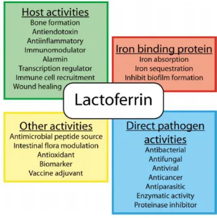
2009; Yen et al., 2011). Figure I.2 summarizes the multitude of activities that have been proposed for Lf.
Figure I.2: Multiple activities described for Lf (Vogel, 2012).
Concerning the anti-microbial properties of Lf, it is now well recognized that this protein plays a direct anti-microbial role in secretions and at the surface of epithelia. It has been documented that Lf exhibits strong anti-microbial activity against a broad spectrum of bacteria (Gram + and Gram -), fungi, yeasts, protozoa, viruses and parasites (reviewed by Legrand et al., 2008; González-Chávez et al., 2009; García-Montoya et al., 2012). The reasons why Lf exerts such a plethora of anti-microbial activities have been investigated and some mechanisms have been put forward, including iron sequestering (Zarember et al., 2007), direct interaction with the cell surfaces (Xu et al., 1999), impairment of adhesion to host cells (Diarra et al., 2003), inhibition of biofilm formation (Dashper et al., 2012), stimulation of the host immune system (Welsh et al., 2011),
among others.
Moreover, both immunomodulatory and anti-inflammatory properties have been attributed to Lf. In fact, this protein is now considered a keycomponent in the host
6
first line of defence having the ability to modulate the overall immune response (Puddu et al., 2009). This insight into the biological role of Lf arose when researchers found that this protein was abundant in neutrophils (Masson et al., 1969). In fact, Lf is a major component of the secondary granules of neutrophils, which is released through degranulation upon neutrophil activation, ultimately resulting in increased levels of Lf. Then, by interacting and promoting the recruitment of leukocytes and activation of dendritic cells, as well as by modulating the expression of soluble mediators like cytokines and chemokines, it controls the excessive inflammation and enhances the immune response (Cumberbatch et al., 2003; de la Rosa et al., 2008; Yamano et al., 2010).
Another fundamental feature of Lf is its ability to function as an enzyme. Besides the protease activity (Hendrixson et al., 2003), it was found that some subfractions of Lf obtained by chromatography possess five other enzymatic activities, namely DNase, RNase, ATPase, phosphatase, and amylase. This variety of activities can be attributed to variations in the protein characteristics like degree of glycosylation and oligomerization, and tertiary structure (Kanyshkova et al., 2003). The elucidation of the Lf enzymatic properties might help to understand its multi-activities as the nuclease activity is suggested to contribute to its anti-microbial properties.
The multiple functions of Lf are in part mediated by its specific receptors at the surface of target cells. Far more research is warranted in this area but some receptors have already been suggested in bacteria (Ling and Schryvers, 2006) and in mammals. Effectively, Lf receptors can be found in various mammalian tissues such as liver, monocytes, lymphocytes and bone (Suzuki et al., 2005), nonetheless the most studied is intelectin 1, the lactoferrin receptor in the human intestinal mucosa (Akiyama et al., 2013). Lf has been demonstrated to be internalized in some cell types (Lopez et al., 2008; Akiyama et al., 2013), however there are also some cells unable to internalize Lf or able to internalize only a small amount. This is the case of some cancer cells such as MCF-7 (Baumrucker et al., 2006), T-47D and MDA-MB-231 breast cancer cell lines (Zhang et al., 2014a).
7
I.1.1.1. Anti-Tumoral Activity of Lactoferrin
Cancer is a leading cause of death worldwide and accounted for 8.2 million deaths (22% of all deaths) in 2012 (WHO, 2014). It has been defined as a disease consisting of transformed cells acquiring cell autonomous hyper-proliferative, invasive and limitless survival capacities that then undergo a transition to expanding masses with metastatic propensity (van Kempen et al., 2003; Lorusso and Rüegg, 2008).
A growing number of reports suggest the Lf’s benefits against cancer and studies with various cancer cell lines and animal models have been reported, all showing the favorable effects of Lf (Gibbons et al., 2011). This topic has been thoroughly reviewed by Rodrigues et al., 2009; Tsuda et al., 2010; Vogel, 2012, among many others. One of the initial findings suggesting the anti-tumoral activity of Lf was published in 1995. In this study, it was established that the whey fraction of bovine milk could significantly inhibit the development of colon tumors in rats (Mclntosh et al., 1995). Then, new understanding on the biological role of Lf in cancer emerged when its expression was found to be downregulated in many types of cancer. In this context, Lf was suggested to negatively regulate the tumor progression, thus acting as a tumor suppressor (Zhou et al., 2008; Deng et al., 2013). Nowadays, there are numerous studies reporting the anti-tumoral activity of Lf in several types of cancer such as lung (Tung et al., 2013), colon (Fujita et al., 2004), breast (Duarte et al., 2011), stomach (Xu et al., 2010), cervix (Shi and Li, 2014), leukemia (Lee et al., 2009), head and neck (Wolf et al., 2007), bladder (Masuda et al., 2000) and melanoma (Roseanu et al., 2010).
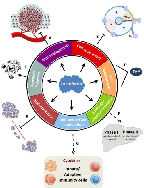
All those years of research allowed the identification of some mechanisms that might underlie the Lf anti-tumoral activity (Fig. I.3). One of them is dependent on Lf iron-binding ability (Fig. I.3 D). The carcinogenicity of iron compounds has been demonstrated in various experiments and free iron has been suggested to act as a mutagenic promoter by inducing oxidative damage to nucleic acids (reviewed by Toyokuni, 2009). Since Lf can bind iron locally in tissues, it may reduce the risk of oxidant-induced carcinogenesis (Rodrigues et al., 2009).
8
Figure I.3: Proposed mechanisms for the anti-tumoral activity of lactoferrin.
Legend: Induction Inhibition Modulation Sequestration (see text for references).
On the other hand, an immunomodulatory action for Lf in preventing carcinogenesis and impeding tumor progression has also been proposed (Fig. I.3 G). It has been reported that Lf has the ability to modulate the production of cytokines in cancer cells and to stimulate the production and/or activation of several immune cells (Ward et al., 2005; Wolf et al., 2007). For instance, Lf administration seems to increase the production of the pro-inflammatory cytokine interleukin-18 (IL-18) in the intestinal tract and to systemically activate the natural killer (NK) cells and CD8+ T lymphocytes in the circulation which exhibited marked cytotoxicity against murine colon carcinoma
9
26Lu (Co26Lu) cells and decreased the formation of lung metastatic colonies in vitro (Wang et al., 2000). Generally, Lf enhances the production of many cytokines including IFNα, β and γ, IL-18, IL-12 and IL-7, and it modulates the activation of cells involved either in innate or adaptive immunity, most particularly dendritic cells, macrophages, neutrophils, NK cells and various T cell subsets (reviewed by Legrand and Mazurier, 2010; Tsuda et al., 2010).
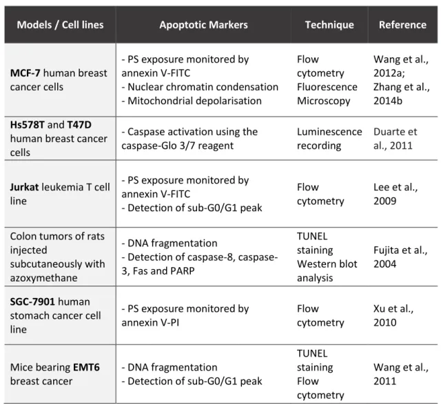
Lf anti-tumoral activity also relies on its ability to trigger apoptosis (Fig. I.3 C). Apoptosis is a cellular suicide process in which a programmed sequence of events culminates in the death of cells without the release of harmful substances into the surrounding area (Meier and Vousden, 2007). This process has been extensively studied in different models and various apoptotic markers induced by Lf have been described (Table I.1). Furthermore, the levels of some apoptosis-related proteins were found to be altered in Lf-treated tumor cells. In HeLa cervical carcinoma cells, Lf was shown to transactivate the tumor suppressor protein p53, known to induce apoptotic cell death, and its target genes mdm2 and p21 (Oh et al., 2004). In human stomach and breast cancer cell lines, Lf was found to downregulate the levels of the anti-apoptotic protein Bcl-2 (Xu et al., 2010; Zhang et al., 2014b). In mice bearing EMT6 breast cancer, a recombinant form of Lf induced apoptosis by decreasing the expression of Bcl-2 and increasing the expression of the pro-apoptotic Bax and the executioner caspase-3 at both mRNA and protein level (Wang et al., 2011).
Furthermore, accumulating evidence suggest that cell cycle arrest might play a critical role in Lf anti-tumoral activity (Fig. I.3 B). Several authors documented the Lf-induced cell cycle arrest, which occurs predominantly at the G1 phase (Wolf et al., 2007; Wang et al., 2011). Specifically, in head and neck cancer cell lines, Lf caused growth arrest at the G1 to S transition of the cell cycle by downregulating the G1 cyclin-dependent kinases (Cdk), and increasing the expression of p21 and p27, which are known inhibitors of the cell cycle (Xiao et al., 2004). A similar effect was found in Lf-treated breast cancer cells (Damiens et al., 1999). The retinoblastoma protein (Rb), a key tumor suppressor gene involved in the inhibition of cell cycle progression, was also affected by Lf in these and other cell lines, since its phosphorylated/inactive form was downregulated (Damiens et al., 1999; Xiao et al., 2004; Son et al., 2006). Moreover, it was also reported that the inhibitory effect of Lf in the cell cycle progression was cell
10
type-dependent, since in MDA-MB-231 breast cancer cells the arrest was observed at G2 phase, whereas in MCF-7 cells there was an arrest at G1 phase with low doses of Lf and at G2 phase with higher doses (Zhang et al., 2014a). In summary, the inhibition of cell proliferation, cell cycle arrest and modulation of the cell cycle regulatory proteins, together with the apoptosis induction are some of the main mechanisms described to explain the Lf anti-tumoral activity.
Table I.1: Apoptotic markers and respective techniques used to monitor the induction of apoptosis by lactoferrin. PS: phosphatidylserine; FITC: fluorescein isothiocyanate; TUNEL: terminal deoxynucleotidyl
transferase-mediated dUTP nick end labelling; PARP: poly (ADP-ribose) polymerase; PI: propidium iodide.
The anti-tumoral activity of Lf also stems from its anti-metastatic potential (Fig. I.3 E). In 1994, Bezault and co-workers demonstrated the inhibitory capacity of this protein in reducing the lung colonization derived from metastatic melanoma cells, in
Models / Cell lines Apoptotic Markers Technique Reference
MCF-7 human breast
cancer cells
- PS exposure monitored by annexin V-FITC
- Nuclear chromatin condensation - Mitochondrial depolarisation Flow cytometry Fluorescence Microscopy Wang et al., 2012a; Zhang et al., 2014b Hs578T and T47D
human breast cancer cells
- Caspase activation using the caspase-Glo 3/7 reagent
Luminescence recording
Duarte et al., 2011
Jurkat leukemia T cell
line
- PS exposure monitored by annexin V-FITC
- Detection of sub-G0/G1 peak
Flow cytometry
Lee et al., 2009 Colon tumors of rats
injected
subcutaneously with azoxymethane
- DNA fragmentation
- Detection of 8, caspase-3, Fas and PARP
TUNEL staining Western blot analysis Fujita et al., 2004 SGC-7901 human
stomach cancer cell line - PS exposure monitored by annexin V-PI Flow cytometry Xu et al., 2010
Mice bearing EMT6 breast cancer
- DNA fragmentation
- Detection of sub-G0/G1 peak
TUNEL staining Flow cytometry Wang et al., 2011
11
mice (Bezault et al., 1994). Lung metastatic colony formation was also inhibited by Lf in mice bearing a highly metastatic colon carcinoma 26Lu (Iigo et al., 1999). Additionally, Lf was shown to inhibit lung and liver metastases derived from the metastatic L5178Y-ML25 lymphoma cells (Yoo et al., 1997). The protective role of Lf against metastasis has generally been attributed to enhanced immunity by increased production of IL-18 and consequent activation of NK and T cells (Iigo et al., 2004).
In addition, it was found that bLf possesses anti-angiogenic properties as it was capable of inhibiting vascular endothelial growth factor (VEGF)-induced angiogenesis in a rat model (Fig. I.3 A). Since tumor growth is angiogenesis-dependent, the suppression of new blood vessel growth might be implicated in the Lf anti-tumoral effects (Norrby et al., 2001). Shimamura and co-workers showed that this inhibition of angiogenesis is dose-dependent (Shimamura et al., 2004). More recently, it was demonstrated that bLf decreases the expression of VEGF mRNA and protein in a lung cancer cell line and inhibits the in vivo formation of lung cancer dependent of VEGF overexpression (Tung et al., 2013). Curiously, in what concerns angiogenesis, human and bovine Lf exert opposite effects. bLf inhibits angiogenesis while hLf was reported to have a specific pro-angiogenic effect in VEGF-A-mediated angiogenesis, which might be explained by differences in their molecular features (Norrby, 2004).
Another mechanism by which Lf is capable of exerting anti-tumor activities was demonstrated in models of chemically induced carcinogenesis (Fig. I.3 F). This process encompasses two stages, namely initiation and post-initiation. The initiation stage requires the activity of enzymes belonging to the liver detoxication metabolism, namely phase I enzymes like cytochrome P450 species. These enzymes activate the carcinogens leading to DNA damage in the target organs. This activation is blocked by liver phase II enzymes responsible for detoxication and excretion. The compounds that supress the activation of phase I enzymes are called “blocking agents”, whilst those capable of inhibiting the post-initiation phase by supressing the proliferation of pre-malignant cells are defined as “supressing agents”. Authors using different rat and hamster models of chemical carcinogenesis, found that orally administered bLf significantly inhibited colon, esophagus, lung, bladder and buccal pouch carcinogenesis. The study of this inhibitory effect provided clear evidence that bLf acts as a blocking and supressing agent by
12
inhibiting phase I enzymes, enhancing the phase II enzymes, or by preventing the proliferation of pre-malignant cells (Tsuda et al., 2002; Mohan et al., 2006).
Regarding Lf iron saturation status, several studies have encompassed the anti-tumoral activities of apo- (iron-free) and holo-Lf (iron-saturated). The results are controversial depending on the context, but holo-Lf has been shown to be more effective against cancer. In fact, Kanwar et al., 2008 conducted a fascinating work in mice bearing EL-4 lymphomas, Lewis lung carcinoma or B16 melanoma tumours, in which it was proved that mice fed with holo-Lf prior to chemotherapy completely rejected their tumors, whereas feeding with other forms of Lf containing a lesser iron-saturation degree (apo-Lf, natural-Lf and 50% iron-saturated Lf) did not. Notably, holo-Lf treatment restored red and white blood cell numbers decreased by chemotherapy. Holo-Lf was also demonstrated to augment chemotherapeutic effects of tamoxifen in a mouse model representing metastatic 4T1 breast cancer (Sun et al., 2012).
Finally, high-throughput approaches concerning the anti-tumor role of Lf have already been carried out. Importantly, a proteomic profiling of human MDA-MB-231 breast cancer cell line exposed to hLf was performed, and the classification of the proteins up-regulated in the presence of hLf showed that the majority was involved in the maintenance of cellular homeostasis with proteins involved in cell signalling, cell cycle and apoptosis (Hoedt et al., 2014), which is in agreement with the proposed mechanisms for the Lf anti-tumoral activity. Despite the fact that of all these data gathered by several research groups point to a clear anti-tumoral role for Lf, the mechanisms by which it exerts these effects are not fully understood (Rodrigues et al., 2009). Therefore, further work on this subject is warranted, namely in what regards the Lf’s direct targets on cancer cells.
I.1.2. Clinical Applications of Lactoferrin
Being Lf a multifaceted protein with numerous interesting biological activities, its research advanced from basic research to clinical trials in which encouraging results were achieved. This, together with its established manufacturing process and consequent availability, expanded the Lf clinical potential namely regarding disease prevention, treatment and diagnosis (Tomita et al., 2009).
13
In the light of several in vitro and in vivo studies, many human clinical trials have been performed to attest the effectiveness of Lf administration against a large variety of human pathologies (reviewed by Rodrigues et al., 2009). Specifically on cancer therapeutics, some Lf forms have been investigated for the treatment of diverse types of cancer. In 2002, Morinaga Milk Industry Co Ltd. supported a human clinical trial, to determine whether oral intake of bLf would inhibit the growth of adenomatous colorectal polyps in human patients. It was found that a 1-year oral intake of 3 g of bLf per day induced statistically significant retardation of colorectal adenomatous polyp size in participants 63 years-old or younger (Kozu et al., 2009). Also, another company, Agennix, tested a recombinant form of human lactoferrin (talactoferrin), in phase II clinical trials for the treatment of non-small cell lung cancer. It was concluded that talactoferrin in combination with carboplatin and paclitaxel, revealed an apparent improvement in overall patient survival (Digumarti et al., 2011). Other clinical trials tested talolactoferrin in patients with various types of solid tumors concluding that it might be particularly effective against metastatic renal carcinoma and non-small cell lung cancer (Hayes et al., 2006, 2010; Jonasch et al., 2008).
The forms of Lf that have been mostly used in the field of health care and disease are Lf from bovine origin (bLf) and the recombinant talactoferrin referred above. bLf can easily be isolated from cow’s milk and it is nowadays produced by diverse manufacturing companies mainly by a cation-exchange chromatography system (Tomita et al., 2009). bLf has already been approved by the European Food Safety Authority as a safe ingredient for various applications, including for medical purposes, since no adverse effects were reported in several studies with humans (EFSA-NDA, 2012). Talactoferrin is produced in Aspergillus niger, a filamentous fungus, and it is structurally identical to the native hLf in all aspects differing only in its glycosylation (Jonasch et al., 2008).
As for the administration, it has been proved that the oral dosage of Lf is the most promising option as the dosing can be conducted easily and safely. Additionally, in
vitro and in vivo digestion studies have demonstrated the Lf gastric survival (Liao et al.,
2012). In fact, Troost and co-workers demonstrated that more than 60% of the administrated bLf, after the passage through adult human stomach and entrance in the small intestine, remains intact (Troost et al., 2001). Significant amounts of Lf were also found in fecal samples collected from exclusively breast-fed infants (Davidson and
14
Lönnerdal, 1987). Currently, Lf delivery systems are being developed in order to improve its clinical utility and also to reduce the dosing amount and the frequency (Onishi, 2011).
On the other hand, given that Lf levels are altered in certain pathologies, it has been considered a reliable biomarker for disease. In fact, the increased levels of fecal Lf were found to be specific for the detection of inflammation in patients with inflammatory bowel disease (Kane et al., 2003) and also to distinguish the aforementioned disease from irritable bowel disease (Zhou et al., 2014). In addition, Lf was suggested to be a potential biomarker for nasopharyngeal carcinoma since it was shown to be downregulated in these tumor cells and its increased expression has been associated with a good prognosis (Zhang et al., 2014c).
Further research on the clinical application of Lf is required, particularly on cancer therapeutics, in order to establish the usefulness of Lf as a pharmaceutical drug to be routinely applied. Therefore, it is very important that the commercial development of Lf goes hand-in-hand with basic research, particularly aiming at the elucidation of specific mechanisms of action (Brock, 2012).
I.2. TUMOR-MICROENVIRONMENT AND ITS ACIDITY
According to the current knowledge, carcinogenesis and tumor progression should be considered not as a cancer cell-centered condition, but rather as a disease involving complex multicellular interactions within a newly formed tissue, the cancer tissue (Lorusso and Rüegg, 2008). It was in this context that the term tumor microenvironment (TME) arose. This concept implies that cancer cells do not manifest the disease alone, but rather interact with normal cell types either physically or by the secretion of signalling molecules. van Kempen et al., 2003 postulated that the TME is the functional and structural collection of neoplastic and non-neoplastic cells, in addition to the dynamic microenvironment in which they live, with the emphasis on their functional interactions.
TME also contributes to the acquisition of cancer hallmark traits and its importance in the regulation of carcinogenesis is well-documented, like for example in the case of breast cancer progression (Kenny et al., 2010; Artacho-cordón et al., 2012; Hanahan and Coussens, 2012). Therefore, it acts as a “soil” in the formation, growth,
15
survival and metastasis of tumors (Wu et al., 2013). Notably, it seems that this microenvironment emerges during the course of tumorigenesis (Hanahan and Weinberg, 2011) and undergoes extensive changes during the multi-steps of this event (Spano and Zollo, 2012). TME is constituted of non-transformed host stromal cells, known to contribute in important ways to the biology of many tumors, such as endothelial cells, pericytes, cancer-associated fibroblasts, various immune cells, and a complex extracellular matrix (ECM) secreted by both the normal and neoplastic cells embedded in it (Hanahan and Weinberg, 2011; Ding et al., 2012).
The microenvironment of tumors comprises several features such as acidic pH, low nutrient levels, elevated interstitial fluid pressure (IFP), overexpressed proteases, and low levels of oxygenation (hypoxia) that is related to the abnormal vascular network that exists in tumors (Lunt et al., 2009; Wu et al., 2013). This hostile microenvironment provides the necessary signals that turn on several transcription factors resulting in the up-regulation of a great number of gene products known to promote malignant progression and metastatic dissemination (Rofstad et al., 2006). Moreover, there is a high level of heterogeneity in the pathophysiological TME both between different tumors and within an individual tumor (Lunt et al., 2009).
The feature of the TME that will be explored in the scope of this workis the acidic pH. Nowadays, it is known that cancer cells, regardless of their origin and genetic background, have an aberrant regulation of hydrogen ion dynamics leading to a reversal of the intracellular to extracellular pH gradient in tumors. Indeed, non-invasive measurements have shown that the extracellular pH (pHe) in tumors ranges from 6.5 to 6.9, whilst the intracellular pH (pHi) remains neutral to alkaline, creating an acid-outside pH gradient typically not observed in normal tissues (Wojtkowiak et al., 2011). This perturbation in pH dynamics rises very early in carcinogenesis and is one of the most common hallmarks of cancer (Reshkin et al., 2013). Consequently, the slightly acidic pH in the TME has become an important issue in the design of anti-tumor therapies, also because it contributes to the resistance to conventional therapies (Wu et al., 2013)
Tumor acidity is a complex and multifactorial process (Lunt et al., 2009). Although TME acidification can be related to an hypoxia-induced enhanced glycolytic activity that leads to production and secretion of H+ to the extracellular space (Wojtkowiak et al., 2011), it can also occur independently of hypoxia. Therefore,
16
acidification may be an intrinsic property of altered tumor cell metabolism, which likely evolved to provide tumor cells with a competitive advantage over stromal cells (Gillies et al., 2002). It appears that this pH dysregulation within TME remodels various physiological functions making solid tumors to become invasive and metastatic, and also contributing to tumor resistance to cytotoxic drugs (Barar and Omidi, 2013). In fact, there are several reports attesting that TME acidity promotes invasive growth and metastatic dissemination. In human melanoma cells it was shown that growth at acidic pHe in vitro enhances the potential to form experimental lung metastases in mice (Rofstad et al., 2006). This was also evidenced in the murine cell lines KHT-C2-LP1 fibrosarcoma and B16F1 melanoma where, following exposure to acidosis, cells showed an increase in experimental metastatic ability (Jang and Hill, 1997). Consequently, the concept of acid-mediated metastasis arose (Gatenby et al., 2006) as there are multiples steps of metastasis that seem to exhibit pH-sensitivity (Hashim et al., 2011). Importantly, methods to evaluate the metastatic potential based on intra-tumoral acidosis have been developed, reinforcing the importance of H+ dynamic in the metastatic process (Wang et al., 2014).
The establishment and maintenance of the acidic TME is a direct consequence of the cancer cells ability to secrete protons (Zhang et al., 2010). This H+ secretion is driven by a series of proton pumps that are up-regulated in cancer cells also protecting them from intracellular acidity and apoptosis (Bellone et al., 2013). These proton-exporting systems include vacuolar H+-ATPase, H+/Na+ exchangers, carbonic anhydrases (CAs), H+/Cl- symporter, monocarboxylate transporters (MCT, mainly MCT1) and Na+ -dependent Cl-/HCO
3- exchangers (Zhang et al., 2010; Reshkin et al., 2013). In the present work we are interested in the specific role of V-ATPase in the acidic TME of cancer cells.
I.2.1. V-ATPase and Cancer
Vacuolar H+-ATPases (V-ATPases) are a family of proton pumps that couple the energy of ATP hydrolysis to actively transport protons across both intracellular and plasma membranes of eukaryotic cells (Cipriano et al., 2008). These pumps are highly phylogenetically conserved among prokaryotes and eukaryotes and its importance is now well-recognized (Lee et al., 2010). Indeed, V-ATPases are crucial for numerous biological functions as they trigger a H+ transmembrane electrochemical potential that
17
is used to drive a variety of secondary active transport systems via H+-dependent symporters and antiporters, and channel-mediated transport systems (Beyenbach and Wieczorek, 2006). Thus, a diverse collection of physiological processes depend on V-ATPases, and a number of diseases have been associated with anomalies of these pumps (Bowman and Bowman, 2005).
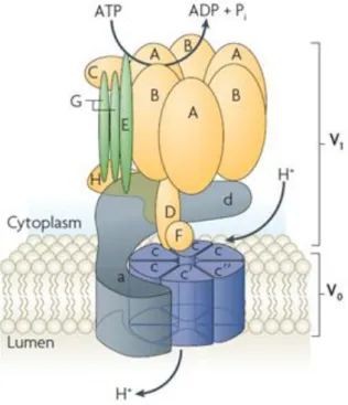
As for its structure, V-ATPase is a large multi-subunit complex organized into two major functional domains known as V1 and Vo. The V1 domain is soluble and comprises at least eight different subunits (A–H). This domain contains three catalytic sites for ATP hydrolysis formed by A and B subunits. The Vo is a membrane-bound domain that contains up to five subunits (a, c, c’, c’’ and d) and is responsible for proton translocation across the membranes (Fig. I.4) (Sun-Wada et al., 2004; Saroussi and Nelson, 2009). V-ATPase operates by a rotary mechanism that is driven by the hydrolysis of ATP within the V1 domain (Cipriano et al., 2008).
Figure I.4: Structure of V-ATPase (adapted from Casey et al., 2010).
V-ATPase was firstly discovered in lysosomes and in the central vacuoles of fungi and plants. Afterwards, it was identified in secretory vesicles, clathrin-coated vesicles, endosomes, Golgi-derived vesicles, among many others (Saroussi and Nelson, 2009). Besides its intracellular localization, V-ATPase is also present in plasma membrane of
18
cancer cells, as well as in a variety of specialized cells like osteoclasts, epididymal clear cells, renal alpha-intercalated cells, neutrophils and macrophages (Jefferies et al., 2008).
Focusing on cancer cell V-ATPase, it has been reported that this pump is overexpressed and localized at the plasma membrane in many types of metastatic cancers such as sarcomas (Perut et al., 2013), breast carcinomas (Sennoune et al., 2004) and melanomas (Baruthio et al., 2008). Moreover, highly metastatic cells were shown to preferentially use plasma membrane V-ATPase over Na+/H+ exchangers (Salyer et al., 2013). Authors have suggested that such an abnormal localization is much likely one of the initial steps of malignant cells transformation and its aberrant functioning, a continual enhancer of carcinogenesis and tumor progression (Lu and Qin, 2012). In this basis, the targeting of V-ATPase to the cell surface has been proposed to both contribute to the alkalinization of the tumor cell cytoplasm and to the acidification of the extracellular TME that aids invasion (Hinton et al., 2009; Pérez-Sayáns et al., 2009). The most important roles of V-ATPase in tumors are in cellular invasiveness, angiogenesis, proliferation, tumorigenesis and drug resistance (Boyd et al., 2001; Spugnini et al., 2010). However, it is in the metastatic process that V-ATPase has demonstrated major importance. Sennoune et al., 2004 demonstrated the plasma membrane localization of V-ATPase and its greater activity in highly than in lowly metastatic human breast cancer cells. The authors concluded that V-ATPase preferential expression at the cell surface is important for the acquisition of invasive and metastatic potential. a3 and a4 V-ATPase subunits isoforms were shown to be responsible for the translocation of this pump to the cell surface (Hinton et al., 2009; Capecci and Forgac, 2013). One fact that can explain the relevance of V-ATPase for tumor metastasis is that, by maintaining the aberrant acidic pH in the TME, it contributes to the activation, secretion, and cellular distribution of many proteases involved in the digestion of ECM including matrix metalloproteinases (MMP) (Fais et al., 2007; Pérez-Sayáns et al., 2009). In fact, in mouse malignant melanoma B16-F10 cells, it was found that the a3 subunit of V-ATPase promotes distant metastasis by stimulating invasiveness through increasing the expression and activity of MMP-2 and MMP-9 (Nishisho et al., 2011). V-ATPases may also play a role in tumor cell survival through pH regulation, prevention of acidosis-induced apoptosis and promotion of drug resistance (Casey et al., 2010).
19
More recently, a specific V-ATPase localization at the plasma membrane was proposed. In some cell types, plasma membrane V-ATPase was found to particularly localize at cholesterol-rich lipid rafts. Lipid rafts are microdomains of the cellular membranes that encompass high concentrations of lipids, especially cholesterol and sphingolipids, and also transmembrane or glycosylphosphatidylinisotol-anchored proteins. These structures represent authentic signalling platforms that are essential for signal transduction and protein trafficking, and are resistant to extraction with non-ionic detergents being generally isolated as detergent-resistant membranes (DRMs) (Staubach and Hanisch, 2011). Several lines of evidence indicate that deregulation of raft-dependent signalling favors tumor progression (reviewed by Murai, 2012). V-ATPase was already found to be a component of lipid rafts in highly metastatic melanoma cells (Baruthio et al., 2008), osteoclasts (Ryu et al., 2010), coronary arterial endothelial cells (Xu et al., 2012), synaptic plasma membrane (Yoshinaka et al., 2004), Jurkat T cells (Haller et al., 2001) and monocytic THP-1 cells (Li et al., 2003). In melanoma cells, it was demonstrated that V-ATPase is present in the rafts fraction of highly metastatic cells being otherwise inconspicuous in this fraction in non-metastatic cell lines. The authors suggested that the increased association of V-ATPase with rafts of the metastatic cells may reflect their increased activity (Baruthio et al., 2008). The reason for such a V-ATPase localization is still unknown, however, in coronary arterial endothelial cells, it was shown that V-ATPase is translocated and assembled into the lipid rafts where it provides an acidic microenvironment around these structures that promotes the formation of larger ceramide-enriched signalling platforms and amplifies raft-associated signals. When V-ATPase was inhibited by BafA1 or siRNA, the V-ATPase-mediated acidification was impaired thereby impeding lipid rafts clustering (Xu et al., 2012). In conclusion, when V-ATPase is localized at lipid rafts it is essential for their structural stability and functioning.
Given the V-ATPase importance for cancer cell biology and its substantial impact on pHi and pHe, it constitutes an especially attractive target for anticancer drugs like proton pump inhibitors (PPIs).
20
I.2.2. Proton Pump Inhibitors (PPIs): a promising anti-cancer therapy
Proton pump inhibitors (PPIs) are lipophilic and weak base pro-drugs that block/downregulate H+ transporters. They penetrate cell membranes and concentrate in acidic compartments, where they are very unstable being then converted into sulfonamide forms, which represent the active inhibitors (Bellone et al., 2013). Firstly, they were designed to act as potent inhibitors of the gastric acid pump H+/K+ ATPase, but later its application was extended to V-ATPase. PPIs have been used for decades as treatment for peptic diseases such as duodenal or gastric ulcers, with minimal side effects (Luciani et al., 2004; De Milito et al., 2010), and there are already various PPIs commercially available worldwide such as omeprazole, lansoprazole, esomeprazole and pantoprazole (De Milito and Fais, 2005).As previously mentioned, the low extracellular pH of the TME is a hallmark of cancer that is mainly dependent on the activity of proton pumps that are overexpressed in tumor cells. In view of these evidence, these pumps have attracted attention as candidates for anti-cancer therapeutic agents because, by inhibiting them, it is possible to reverse tumor specific H+ homeostasis, therefore acting on one of “cancer’s Achilles’ Heel” (Kroemer and Pouyssegur, 2008; Vishvakarma and Singh, 2011). Following this idea, after protonation, PPIs irreversibly bind the proton pump, dramatically disrupting H+ translocation and dynamics, and limiting TME acidosis (Fais et al., 2007). Thus, in the presence of PPIs, tumor cells are no longer able to control pH and undergo apoptosis which, consequently, results in inhibition of tumor proliferation and growth. The risk of minor adverse effects from PPIs is low, approximately 1-3%, with no significant differences noted between the PPIs (Thomson et al., 2010). PPIs are then perfect suited for a cancer-specific targeted strategy as their activation requires acidity such as that found in the TME (Bellone et al., 2013).
I.2.2.1. Targeting V-ATPase with PPIs
Given the V-ATPase significance and multifunctions in cancer, namely the maintenance of the acidic TME, many specific inhibitors have been developed (Bowman and Bowman, 2005). Actually, there are different classes of V-ATPase inhibitors that can be applied in cancer treatment (Pérez-Sayáns et al., 2009) and there are a plethora of
21
effects in the cell that have been demonstrated as a consequence of their application, including the variation in cytosolic pH homeostasis, cell cycle arrest, reactive oxygen species (ROS) production, modifications in cell signalling and increased chemosensitivity. These effects, if severe and sustained enough, can lead to cell death mostly apoptotic (Hernández et al., 2012).
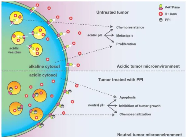
Although classic V-ATPase inhibitors (e.g., bafilomycins and concanamycins) or its molecular silencing can induce cell death in tumor cell lines, these compounds are highly toxic and not suitable for clinical use in humans (De Milito et al., 2007).Thus, PPIs have largely and successfully been applied due to their reduced cytotoxicity. The inhibition of V-ATPase with a PPI in cancer cells can have several consequences since the mechanisms that contribute to the malignant behavior are modified. These include neutralization of the TME and consequent apoptosis, reduction of the metastatic potential, chemosensitization and inhibition of tumor growth and survival (Fig. I.5) (Fais et al., 2007; Pérez-Sayáns et al., 2009).
Figure I.5: Effects of V-ATPase inhibition with PPIs on tumor microenvironment (adapted from Fais et
22
There are some PPIs targeting V-ATPase that have already been tested. De Milito et al., 2010 performed a pre-clinical study in human melanoma cells both in vitro and in
vivo, in which they demonstrated that esomeoprazol, a PPI that inhibits V-ATPase, is
capable of inhibiting tumor growth and significantly increase animal survival. On the other hand, pre-treatment with a PPI, through the inhibition of V-ATPase activity, was found to induce susceptibility of various human drug-resistant tumor cell lines to the cytotoxic effect of different anti-tumor drugs, with a marked reduction of drug efflux and decreased tumor acidity. Thus, these drugs can also be used to overcome and/or reverse the multi-resistance of the human tumors (Luciani et al., 2004).
23
I.3. AIMS
Although several studies have addressed the anti-tumoral activity of Lf, the underlying molecular mechanism is still elusive. Therefore, the general goal of the present study was to unveil the molecular target(s) of Lf in cancer cells. In particular, to explore the potential role of Lf as a PPI, three breast cell lines with distinct features were used, namely the highly metastatic cell line Hs 578T, the non-invasive cell line T-47D, and the non-tumorigenic cell line MCF-10-2A. In order to accomplish our goal, specific aims were design, to:
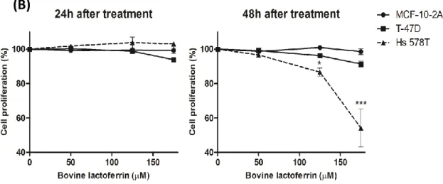
- investigate the susceptibilities of these three cell lines to bLf regarding cell proliferation and apoptosis, so that we could ascertain whether the susceptibilities were in line with our hypothesis. Also infer about the bLf specificity to cancer cells;
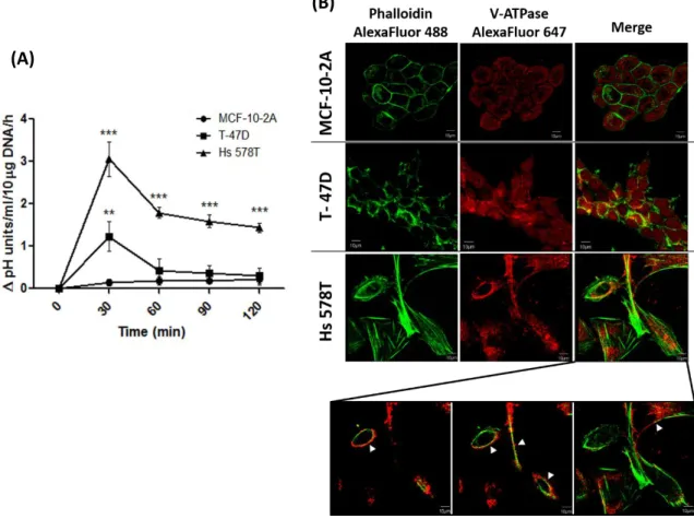
- study the distribution of V-ATPase in the breast cancer cell lines to evaluate if existing differences between the three cell lines can correlate with the observed susceptibilities to bLf;
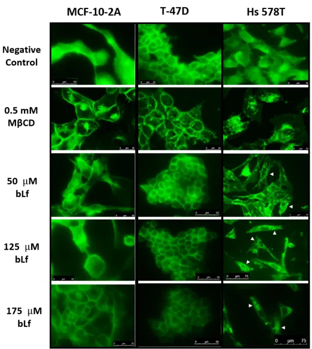
- assess key cellular events caused by bLf, such as extracellular and intracellular pH, that could be in agreement with the hypothetical V-ATPase inhibition; - determine the effect of bLf on cholesterol-enriched lipid rafts that were found
to contain V-ATPase in some types of metastatic tumor cells;
- provide in vitro evidences of the putative V-ATPase inhibition by bLf through the study of both proton pumping or hydrolytic activities.
The outcome of the present work may have great implications on cancer therapy, especially of highly metastatic tumors that are characterized by its acidic microenvironment.
24
I.4. REFERENCES
Adlerova, L., A. Bartoskova, and M. Faldyna. 2008. Lactoferrin : a review. Veterinarni
Medicina. 53(9):457–468.
Akiyama, Y., K. Oshima, K. Shin, H. Wakabayashi, F. Abe, D. Nadano, and T. Matsuda. 2013. Intracellular retention and subsequent release of bovine milk lactoferrin taken up by human enterocyte-like cell lines, Caco-2, C2BBe1 and HT-29. Biosci.
Biotechnol. Biochem. 77:1023–9. doi:10.1271/bbb.121011.
Alexander, D.B., M. Iigo, K. Yamauchi, M. Suzui, and H. Tsuda. 2012. Lactoferrin: an alternative view of its role in human biological fluids. Biochem. Cell Biol. 90:279– 306. doi:10.1139/o2012-013.
Andrés, M.T., and J.F. Fierro. 2010. Antimicrobial mechanism of action of transferrins: selective inhibition of H+-ATPase. Antimicrob. Agents Chemother. 54:4335–42. doi:10.1128/AAC.01620-09.
Andrés, M.T., M. Viejo-Díaz, and J.F. Fierro. 2008. Human lactoferrin induces apoptosis-like cell death in Candida albicans: critical role of K+-channel-mediated K+ efflux.
Antimicrob. Agents Chemother. 52:4081–8. doi:10.1128/AAC.01597-07.
Artacho-cordón, A., F. Artacho-cordón, S. Ríos-arrabal, I. Calvente, and M.I. Núñez. 2012. Tumor microenvironment and breast cancer progression: a complex scenario.
Cancer Biol. Ther. 13:14–24.
Baker, H.M., and E.N. Baker. 2004. Lactoferrin and Iron: structural and dynamic aspects
of binding and release. BioMetals. 17:209–216.
doi:10.1023/B:BIOM.0000027694.40260.70.
Baker, H.M., and E.N. Baker. 2012. A structural perspective on lactoferrin function.
Biochem. Cell Biol. 90:320–8. doi:10.1139/o11-071.
Barar, J., and Y. Omidi. 2013. Dysregulated pH in Tumor Microenvironment Checkmates Cancer Therapy. Bioimpacts. 3:149–62. doi:10.5681/bi.2013.036.
Baruthio, F., M. Quadroni, C. Rüegg, and A. Mariotti. 2008. Proteomic analysis of membrane rafts of melanoma cells identifies protein patterns characteristic of the tumor progression stage. Proteomics. 8:4733–47. doi:10.1002/pmic.200800169. Baumrucker, C.R., F. Schanbacher, Y. Shang, and M.H. Green. 2006. Lactoferrin
interaction with retinoid signaling: cell growth and apoptosis in mammary cells.
Domest. Anim. Endocrinol. 30:289–303. doi:10.1016/j.domaniend.2005.07.009.
Bellone, M., A. Calcinotto, P. Filipazzi, A. De Milito, S. Fais, and L. Rivoltini. 2013. The acidity of the tumor microenvironment is a mechanism of immune escape that can be overcome by proton pump inhibitors. Oncoimmunology. 2:e22058. doi:10.4161/onci.22058.