Central nervous system manifestations of COVID-19: A systematic review
Ali A. Asadi-Pooya, Leila Simani
PII: S0022-510X(20)30168-4
DOI: https://doi.org/10.1016/j.jns.2020.116832
Reference: JNS 116832
To appear in: Journal of the Neurological Sciences
Received date: 7 April 2020
Accepted date: 8 April 2020
Please cite this article as: A.A. Asadi-Pooya and L. Simani, Central nervous system manifestations of COVID-19: A systematic review, Journal of the Neurological Sciences (2019),https://doi.org/10.1016/j.jns.2020.116832
This is a PDF file of an article that has undergone enhancements after acceptance, such as the addition of a cover page and metadata, and formatting for readability, but it is not yet the definitive version of record. This version will undergo additional copyediting, typesetting and review before it is published in its final form, but we are providing this version to give early visibility of the article. Please note that, during the production process, errors may be discovered which could affect the content, and all legal disclaimers that apply to the journal pertain.
1
Central nervous system manifestations of COVID-19: a systematic review
Ali A. Asadi-Pooya, M.D. 1, 2, Leila Simani, Ph.D. 3
1 Epilepsy Research Center, Shiraz University of Medical Sciences, Shiraz, Iran.
2 Jefferson Comprehensive Epilepsy Center, Department of Neurology, Thomas Jefferson
University, Philadelphia, USA.
3 Skull Base Research Center, Loghman Hakim Hospital, Shahid Beheshti Univsersity of
Medical sciences, Tehran, Iran.
Address for Correspondence:
Ali A. Asadi-Pooya, M.D. Epilepsy Research Center,
Shiraz University of Medical Sciences, Shiraz, Iran.
E-mails: aliasadipooya@yahoo.com; l.simani62@gmail.com Phone: 98-9352274990
Running title: CNS & COVID-19
Number of characters in the title: 70; Number of characters in the running title: 14; Number of text pages: 5; Word count: 1213; Abstract word count: 228; Figures: 1; Tables: 3;
References: 20.
2 Page 2
Contributions
Name Location Contribution
Ali A. Asadi-Pooya, M.D.
Epilepsy Research Center, Shiraz University of Medical Sciences, Shiraz, Iran.
Jefferson Comprehensive Epilepsy Center, Department of Neurology, Thomas Jefferson University, Philadelphia, USA.
Design and conceptualized the study; analyzed the data; drafted and revised the manuscript
Leila Simani, Ph.D.
Skull Base Research Center, Loghman Hakim Hospital, Shahid Bhehsti University of Medical Sciences, Tehran,Iran
Major role in the acquisition of data; revised the manuscript
Disclosures
Ali A. Asadi-Pooya, M.D.: Honoraria from Cobel Daruo, RaymandRad and Tekaje; Royalty: Oxford University Press (Book publication). Leila Simani, PhD.: none.
3
Abstract
Objective: In this systematic review, we will discuss the evidence on the occurrence of
central nervous system (CNS) involvement and neurological manifestations in patients with COVID-19. Methods: MEDLINE (accessed from PubMed) and Scopus from December 01, 2019 to March 26, 2020 were systematically searched for related published articles. In both electronic databases, the following search strategy was implemented and these key words (in the title/abstract) were used: “COVID 19” OR “coronavirus” AND “brain” OR “CNS” OR “neurologic”. Results: Through the search strategy, we could identify two articles about neurological involvement by COVID-19. One of these publications was a narrative review and the other one was a viewpoint. However, the authors scanned the reference lists of the included studies and could identify multiple references. One study, specifically investigated the neurological manifestations of COVID-19 and could document CNS manifestations in 25% of the patients. Most of the studies investigated the manifestations of COVID-19 in general. Conclusion: While neurological manifestations of COVID-19 have not been studied appropriately, it is highly likely that some of these patients, particularly those who suffer from a severe illness, have CNS involvement and neurological manifestations. Precise and targeted documentation of neurological symptoms, detailed clinical, neurological, and electrophysiological investigations of the patients, attempts to isolate SARS-CoV-2 from cerebrospinal fluid, and autopsies of the COVID-19 victims may clarify the role played by this virus in causing neurological manifestations.
Key words: CNS; Coronavirus; COVID-19; Neurological; Seizure
4
1. Introduction
Coronavirus is one of the major viruses that primarily targets the human respiratory system, but it also has neuroinvasive capabilities and can spread from the respiratory tract to the central nervous system (CNS). Previous epidemics or pandemics of coronaviruses include the severe acute respiratory syndrome (SARS) in 2002 and the Middle East respiratory syndrome (MERS) in 2012. The most recent pandemic of coronavirus infection is coronavirus disease (COVID-19) that is caused by SARS-CoV21, 2. The symptoms of COVID-19 infection usually appear after an incubation period of about five days. The most common symptoms of COVID-19 illness are fever, cough, and fatigue; other symptoms include headache,
hemoptysis, and dyspnea, among others. In the most severe cases, patients may develop pneumonia, acute respiratory distress syndrome, acute cardiac problems, and multiorgan failure 1. The first cases of COVID-19 were reported in December 2019 1; however, when we searched the MEDLINE (accessed from PubMed), from December 01, 2019 to March 26, 2020, with the key word “COVID 19”, surprisingly 1655 articles were yielded. This shows that COVID-19 pandemic is of great global public health concern.
Coronavirus infections have been associated with neurological manifestations (e.g., febrile seizures, convulsions, change in mental status, and encephalitis)2, 3. Neurotropic and neuroinvasive capabilities of coronaviruses have been described in humans. Upon nasal infection, coronavirus enters the CNS through the olfactory bulb, causing inflammation and demyelination3. In this systematic review, we will discuss the evidence on the occurrence of CNS involvement and neurological manifestations in patients with COVID-19.
2. Methods
The report of this systematic review was made according to the recommendations of the Preferred Reporting Items for Systematic Reviews and Meta-Analyses (PRISMA) statement4,
5
5 (Figure 1). The review protocol was not previously registered. MEDLINE (accessed from
PubMed) and Scopus from December 01, 2019 to March 26, 2020 were systematically searched for related published articles. In both electronic databases, the following search strategy was implemented and these key words (in the title/abstract) were used: “COVID 19” OR “coronavirus” AND “brain” OR “CNS” OR “neurologic”. Articles written in English were all included in this search. To ensure literature saturation, the authors scanned the reference lists of the included studies or relevant reviews identified through the search. Both authors participated through each phase of the review independently (screening, eligibility, and inclusion). They independently screened the titles and abstracts yielded by the search against the inclusion criteria. They obtained full reports for all titles that appeared to meet the inclusion criteria or where there was any uncertainty. Authors screened the full text reports and decided whether these meet the inclusion criteria. They resolved any disagreement through discussions. Neither of the authors were blind to the journal titles or to the study authors or institutions.
The following data were extracted from the included studies: study authors, study designs, main results, and limitations. The methodological quality of the included studies was assessed by the authors. The class of evidence was defined following the American Academy of Neurology criteria for classification of evidence in studies of causation (Appendix 1)6.
Standard Protocol Approvals, Registrations, and Patient Consents
The Shiraz University of Medical Sciences Institutional Review Board approved this systematic review.
3. Results
Through the search strategy, we could identify two articles about neurological involvement by COVID-19 (Tables 1 and 2) 7, 8. One of these publications was a narrative review7 and the
6
other one was a viewpoint 8. However, to ensure literature saturation, the authors scanned the reference lists of the included studies and could identify multiple references 9-14. Table 3
shows the summary of these studies on the CNS manifestations of COVID-19. One study, specifically investigated the neurological manifestations of COVID-19 and could document CNS manifestations in 25% of the patients 9. However, the authors did not perform
electroencephalography (EEG) or cerebrospinal fluid (CSF) analysis. Another retrospective study investigated acute cerebrovascular disease occurrence following COVID-19 10. Other
studies investigated the manifestations of COVID-19 in general; they did not specifically pay attention to the neurological manifestations 11-14.
4. Discussion
In this study, we observed that the evidence on the CNS involvement and neurological manifestations of COVID-19 is scarce and of low quality. However, the only study that specifically investigated this issue documented that one-quarter of the hospitalized patients with a confirmed diagnosis of severe acute respiratory syndrome from coronavirus 2 infection had some manifestations of CNS involvement 9. Some patients with COVID-19 may show nonspecific neurological symptoms, such as confusion and headache. A few patients with COVID-19 showed more specific neurological manifestations, such as seizure or
cerebrovascular problems (Table 3). Furthermore, neuroinvasion of SARS‐CoV2 may partially explain why some patients develop respiratory failure, while others do not 7. Most coronaviruses share similar viral structures and infection pathways; hence, the pathomechanisms previously found for other coronaviruses may also be applicable for SARS‐CoV2. Human coronaviruses are not always confined to the respiratory tract; they can invade the CNS. A growing body of evidence shows that neuroinvasion and neurotropism is a common feature of human coronaviruses 2. Infection with SARS-CoV has been associated
7
with neurological manifestations. In the reported patients with SARS-CoV, CSF tested positive for the virus 15, 16. In one study of 183 hospitalized children with clinically suspected
acute encephalitis, 22 (12%) had coronavirus infection (type was not specified) by detection of anti-CoV IgM 17. In a study of 70 patients with MERS-CoV infection, altered mental status was reported in 26% of the patients and 9% of the people had seizure 18. Therefore, it is very likely to observe neurological manifestations in patients with COVID-19 if we carefully and specifically look for them.
Finally, patients with severe COVID-19 may have hypoxia, multiorgan failure, and metabolic and electrolyte derangements, and may require sophisticated medication regimens and
therapeutic interventions. Hence, it is plausible to expect clinical or subclinical acute symptomatic seizures and status epilepticus to happen in these patients. Impaired mental status has been reported in patients with severe COVID-19 9, 14 ; but, this manifestation has
never been studied appropriately in previous studies (Table 3). When visiting a patient who is in a critical medical condition and has a change in mental status, one should make sure that nonconvulsive status epilepticus (NCSE) is not a part of the clinical scenario. The diagnosis of NCSE is frequently overlooked, with patients in critical medical conditions having other serious problems. It is necessary to perform continuous EEG monitoring in any patient with a critical medical condition, who has a change in mental status, in order to make a timely diagnosis of NCSE 19. Salzburg Consensus Criteria for Non-Convulsive Status Epilepticus is a helpful guide to make a diagnosis of NCSE in critically ill patients 20.
5. Conclusion
While neurological manifestations of COVID-19 have not been studied appropriately yet, it is highly likely that some of these patients, particularly those who suffer from a severe illness, have CNS involvement and neurological manifestations. Precise and targeted documentation
8
of the neurological symptoms (e.g., headache, dizziness, etc.) and signs (e.g., change in mental status, meningeal signs, etc.), detailed clinical, neurological, and electrophysiological investigations (e.g., EEG) of the patients (particularly those with a change in mental status), attempts to isolate SARS-CoV-2 from CSF, and autopsies of the COVID-19 victims may clarify the roles played by this virus in causing neurological manifestations.
Acknowledgment
Shiraz University of Medical Sciences supported this study.
9
References
1. Rothan HA, Byrareddy SN. The epidemiology and pathogenesis of coronavirus disease (COVID-19) outbreak. Journal of Autoimmunity 2020:102433.
2. Desforges M, Le Coupanec A, Dubeau P, et al. Human Coronaviruses and Other Respiratory Viruses: Underestimated Opportunistic Pathogens of the Central Nervous System? Viruses 2020;12:14.
3. Bohmwald K, Galvez N, Ríos M, Kalergis AM. Neurologic alterations due to respiratory virus infections. Frontiers in cellular neuroscience 2018;12:386.
4. Hutton B, Salanti G, Caldwell DM, et al. The PRISMA extension statement for reporting of systematic reviews incorporating network meta-analyses of health care interventions: checklist and explanations. Annals of internal medicine 2015;162:777-784. 5. Moher D, Liberati A, Tetzlaff J, Altman DG. Preferred reporting items for systematic reviews and meta-analyses: the PRISMA statement. Annals of internal medicine
2009;151:264-269.
6. Gronseth GS CJ, Gloss D, Merillat S, Dittman J, Armstrong MJ, et al. . on behalf of the Guideline Development, Dissemination, and Implementation Subcommittee of the American Academy of Neurology. 2017. Clinical Practice Guideline Process Manual, 2017 ed. Minneapolis, MN: The American Academy of Neurology. 2017.
7. Li YC, Bai WZ, Hashikawa T. The neuroinvasive potential of SARS-CoV2 may be at least partially responsible for the respiratory failure of COVID-19 patients. Journal of
Medical Virology 2020.
8. Baig AM, Khaleeq A, Ali U, Syeda H. Evidence of the COVID-19 Virus Targeting the CNS: Tissue Distribution, Host–Virus Interaction, and Proposed Neurotropic
Mechanisms. ACS chemical neuroscience 2020.
9. Mao L, Wang M, Chen S, et al. Neurological Manifestations of Hospitalized Patients with COVID-19 in Wuhan, China: a retrospective case series study.
https://www.medrxiv.org/content/10.1101/2020.02.22.20026500v1/accessed on April 4, 2020. This article is a preprint and has not been peer-reviewed.
10. Li Y, Wang M, Zhou Y, et al. Acute Cerebrovascular Disease Following COVID-19: A Single Center, Retrospective, Observational Study. 2020.
https://papers.ssrn.com/sol3/papers.cfm?abstract_id=3550025/accessed on April 4, 2020. This article is a preprint and has not been peer-reviewed.
11. Huang C, Wang Y, Li X, et al. Clinical features of patients infected with 2019 novel coronavirus in Wuhan, China. The Lancet 2020;395:497-506.
12. Yang X, Yu Y, Xu J, et al. Clinical course and outcomes of critically ill patients with SARS-CoV-2 pneumonia in Wuhan, China: a single-centered, retrospective, observational study. The Lancet Respiratory Medicine 2020.
13. Wang D, Hu B, Hu C, et al. Clinical characteristics of 138 hospitalized patients with 2019 novel coronavirus–infected pneumonia in Wuhan, China. Jama 2020.
14. Chen N, Zhou M, Dong X, et al. Epidemiological and clinical characteristics of 99 cases of 2019 novel coronavirus pneumonia in Wuhan, China: a descriptive study. The Lancet 2020;395:507-513.
15. Hung EC, Chim SS, Chan PK, et al. Detection of SARS coronavirus RNA in the cerebrospinal fluid of a patient with severe acute respiratory syndrome. Clinical Chemistry 2003;49:2108-2109.
16. Lau K-K, Yu W-C, Chu C-M, Lau S-T, Sheng B, Yuen K-Y. Possible central nervous system infection by SARS coronavirus. Emerging infectious diseases 2004;10:342.
10
17. Li Y, Li H, Fan R, et al. Coronavirus infections in the central nervous system and respiratory tract show distinct features in hospitalized children. Intervirology 2016;59:163-169.
18. Saad M, Omrani AS, Baig K, et al. Clinical aspects and outcomes of 70 patients with Middle East respiratory syndrome coronavirus infection: a single-center experience in Saudi Arabia. International Journal of Infectious Diseases 2014;29:301-306.
19. Miró JM, de Terán FD, Singer PA, Prior MA-A. Emergency electroencephalogram: Usefulness in the diagnosis of nonconvulsive status epilepticus by the on-call neurologist. Neurología (English Edition) 2018;33:71-77.
20. Leitinger M, Beniczky S, Rohracher A, et al. Salzburg consensus criteria for non-convulsive status epilepticus–approach to clinical application. Epilepsy & Behavior 2015;49:158-163.
11
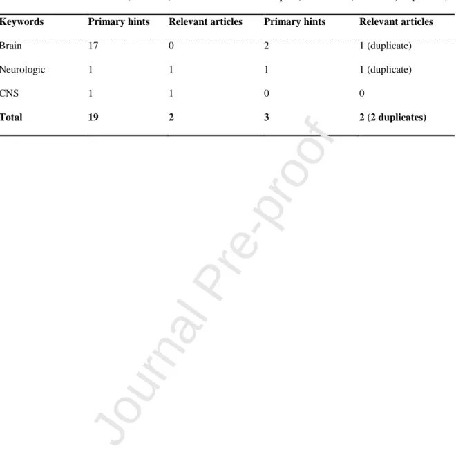
Table 1. The search keywords included “COVID 19” and those in the first column of the
table.
Medline (PubMed) Scopus (Article title, Abstract, Keywords)
Keywords Primary hints Relevant articles Primary hints Relevant articles
Brain 17 0 2 1 (duplicate)
Neurologic 1 1 1 1 (duplicate)
CNS 1 1 0 0
Total 19 2 3 2 (2 duplicates)
12
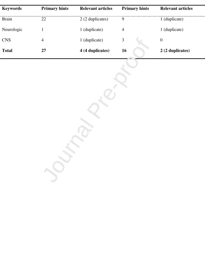
Table 2. The search keywords included “coronavirus” and those in the first column of the
table.
Medline (PubMed) Scopus (Article title, Abstract, Keywords)
Keywords Primary hints Relevant articles Primary hints Relevant articles
Brain 22 2 (2 duplicates) 9 1 (duplicate)
Neurologic 1 1 (duplicate) 4 1 (duplicate)
CNS 4 1 (duplicate) 3 0
Total 27 4 (4 duplicates) 16 2 (2 duplicates)
13
Table 3. Neurological manifestations of COVID-19.
Author/year Methods Neurological Manifestations Limitations Level of
evidence Mao/ 2020 (in press) 9 Retrospective case series of 214 admitted patients CNS manifestations: in 25%. Headache (13%), dizziness (17%), impaired consciousness (8%), acute cerebrovascular problems (3%), ataxia (0.5), and seizures (0.5%)
No CSF analysis; no EEG study; no clear definition of symptoms III Li/ 2020 10 Retrospective case series of 221 admitted patients
5% developed acute ischemic stroke, 0.5% had cerebral venous sinus thrombosis, and 0.5% had cerebral hemorrhage
Other related neurological manifestations were not studied. II Huang/ 202011 Prospective study of 41 admitted patients
Headache in 8% Not specifically studied neurological manifestations. No CSF or EEG studies I Yang/ 2020 12 Retrospective study of 52 critically ill adult patients
Headache in 6% Not specifically studied neurological manifestations. No CSF or EEG studies II Wang/ 2020 13 Retrospective case series of the 138 hospitalized patients
Dizziness in 9%; Headache in 7% Not specifically studied neurological manifestations. No CSF or EEG studies II Chen/ 2020 14 Retrospective case series of the 99 hospitalized patients
Confusion in 9%; Headache in 8% Not specifically studied neurological
manifestations. No CSF or EEG studies
II
CNS: central nervous system; CSF: cerebrospinal fluid; EEG: electroencephalography
Journal Pre-proof
14
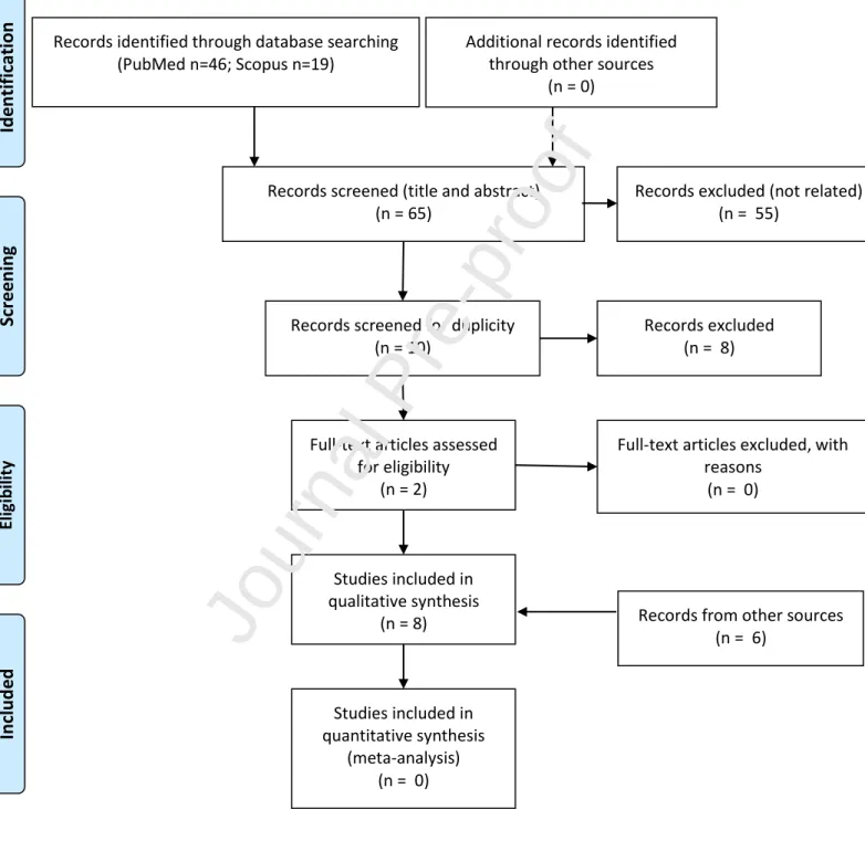
Figure 1 legend.
Preferred Reporting Items for Systematic Reviews and Meta-Analyses (PRISMA) Flow Diagram of the study.
15
Figure 1. Preferred Reporting Items for Systematic Reviews and Meta-Analyses (PRISMA)
Flow Diagram of the study.
Records identified through database searching (PubMed n=46; Scopus n=19) Scr e e n in g In cl u d e d El ig ib ili ty Id en ti fi cat
ion Additional records identified
through other sources (n = 0)
Records screened (title and abstract) (n = 65)
Records screened for duplicity (n = 10)
Records excluded (n = 8)
Full-text articles assessed for eligibility
(n = 2)
Full-text articles excluded, with reasons (n = 0) Studies included in qualitative synthesis (n = 8) Studies included in quantitative synthesis (meta-analysis) (n = 0)
Records excluded (not related) (n = 55)
Records from other sources (n = 6)
16
Highlights
Some patients with COVID-19 may show nonspecific neurological symptoms, such as confusion and headache.
A few patients with COVID-19 showed more specific neurological manifestations, such as seizure or cerebrovascular problems.
Neuroinvasion of SARS‐CoV2 may partially explain why some patients develop respiratory failure.