2015/2016
Vitor Agostinho Costa Pereira
Influence of wet AMD in the progression
of Geografic Atrophy in the fellow eye
Mestrado Integrado em Medicina
Área: Oftalmologia
Tipologia: Dissertação
Trabalho efetuado sob a Orientação de:
Doutor Manuel Alberto de Almeida e Sousa Falcão
Trabalho organizado de acordo com as normas da revista:
Acta Ophthalmologica
Vitor Agostinho Costa Pereira
Influence of wet AMD in the progression
of Geografic Atrophy in the fellow eye
DEDICATÓRIA
Influence of wet AMD in the progression of Geografic Atrophy in the
fellow eye
Vitor Costa Pereira
Faculty of Medicine, University of Porto [email protected]
+351 914983225
Estrada da Circunvalação, 7824 4200 – 162 Porto
Portugal
Manuel Falcão MD
Department of Sense Organs, Faculty of Medicine, University of Porto Department of Ophthalmology, Centro Hospitalar São João
Keywords
Geographic atrophy; Age-related macular degeneration; Fundus autofluorescence; Anti-VEGF.
Abstract
Purpose: To understand the influence of wet age-related macular degeneration (AMD), both
treated with anti-VEGF and not treated, in geographic atrophy (GA) progression in the fellow eye and to verify if the GA baseline area can also be a risk factor.
Methods: We accessed retrospectively data from Fundus Autofluorescence (FAF) of patients
with GA and we measured GA area through images collected in different visits. We defined 3 groups of patients: group 1 with GA and without wet AMD; group 2 with GA and concomitant wet AMD with a disciform scar; and group 3 with GA and wet AMD treated with anti-VEGF in the fellow eye. We compared differences of GA area progression between these three groups. Finally, we selected patients that had 3 or more images and we compared GA progression between the first and the last intervals of the measurements.
Results: The overall mean of GA area progression rate in the 3 groups was of 1.1±1.1; 0.7±0.6
and 1.2±1.1 mm2/year, respectively. Comparing the groups together there was no statistically
significant difference: group 1 and 2 (p=0.225); group 1 and 3 (p=0.918); group 2 and 3 (p=0.309). The mean of the GA area progression rate relative to the first and the last interval was of 1.17±1.3 and 0.79±0.9 mm2/year, respectively (p=0.095).
Conclusion: We found no association of GA progression rate with either the presence of
Introduction
Age-related macular degeneration (AMD) is a common chorioretinal degenerative condition and a main cause of blindness in elderly population, especially in developed countries (Nowak 2006, Fraser-Bell, Choudhury et al. 2010, Ambati and Fowler 2012, Sigler and Randolph 2013). There are some risk factors that are associated with the development of AMD, including soft drusen, genetic factors and smoking. On the other hand, nutritional supplements, on the basis of vitamin complexes, appear to slow the progression of late AMD (Age-Related Eye Disease Study Research 2001, Age-Related Eye Disease Study 2 Research, Chew et al. 2014). Therefore, its pathogenesis is likely due to the interaction of metabolic, functional, genetic and environmental factors (Nowak 2006, Sigler and Randolph 2013).
Two subgroups of AMD are classically distinguished: atrophic (dry form) and exudative (wet form). Dry AMD is a chronic disease that may cause some degree of visual impairment and sometimes progresses to severe blindness. On the other hand, wet AMD affects about 10-15% of AMD patients, emerges abruptly and rapidly progresses to blindness if left untreated (Wong, Chakravarthy et al. 2008, Ambati and Fowler 2012, Bhutto and Lutty 2012). A considerable number of patients develop wet AMD form on a background of dry AMD. Thus, some authors think that dry AMD can be a precursor state for wet AMD (Bhutto and Lutty 2012, Grunwald, Daniel et al. 2014, Schutze, Wedl et al. 2015).
Geographic atrophy (GA) represents the late stage manifestation of dry AMD, and it is characterized by a progressing course leading to degeneration of retinal pigment epithelium (RPE) and photoreceptors, which rely on the RPE for trophic support (Nowak 2006, Gobel, Fleckenstein et al. 2011, Ambati and Fowler 2012). Some etiologic mechanisms contribute to the development of GA and include senescence, ischemia, oxidative and photo-oxidative damage, inflammation, and improper RNA processing (Zarbin 2004, Kaneko, Dridi et al. 2011, Kumar, Mrejen et al. 2013). Atrophic areas enlarge continuously over time and are associated with a corresponding absolute scotoma. The extent of the foveal involvement determines visual acuity (VA) (Joussen and Bornfeld 2009, Gobel, Fleckenstein et al. 2011). Some risk factors promote the GA progression and they include hyperautofluorescence detected on the surrounding area of GA (Bearelly, Khanifar et al. 2011, Batioglu, Gedik Oguz et al. 2014), the pattern configurations and the number of GA regions (Sunness, Margalit et al. 2007). Furthermore, larger lesions appear to manifest higher growth rates, compared to smaller ones (Csaky, Richman et al. 2008, Biarnes, Arias et al. 2015). Smoking or body mass index do not appear to confer additional risk (Lindblad, Lloyd et al. 2009). There is to date no means of treatment to counteract or slow the progression of GA, contrary to wet AMD (Nowak 2006, Ambati and Fowler 2012, Kanagasingam, Bhuiyan et al. 2014).
the gold standard for evaluating progressive GA enlargement (Gobel, Fleckenstein et al. 2011). This method allows topographic mapping of lipofuscin in RPE and because in eyes with GA the atrophic areas have deficit of lipofuscin, they correspond to hypofluorescent dark areas (Fleckenstein, Charbel Issa et al. 2008).
On the other hand, the late stage of wet AMD is choroidal neovascularization (CNV). It is characterized by the growth of abnormal blood vessels from the choroid underneath the macula, haemorrhage and, in untreated patients, the final stage of this disorder is a disciform scar. This was thought to be a stable lesion. However, a study presented some cases of patients with disciform scars in which a ring of atrophy developed and extended around the scar (Sarks, Tang et al. 2006). In fact, the proposed etiologic stimuli for the development of CNV are similar as for GA and include ischemia, senescence, oxidative and photo-oxidative damage, and inflammation (Spaide, Armstrong et al. 2003, Zarbin 2004, Scholl, Fleckenstein et al. 2007). The overexpression of vascular endothelial growth factor (VEGF) is considered to be the cause of CNV and utility of anti-VEGF intravitreal therapy is well established in the treatment of CNV associated with AMD. Many studies have shown that ranibizumab, aflibercept and bevacizumab improve the visual acuity (Joussen and Bornfeld 2009, Amaro and Roller 2012, Jaffe, Martin et al. 2013, Grunwald, Daniel et al. 2014). However, macular morphological responses after anti-VEGF therapy are quite varied. One of the findings observed in some studies is the development of RPE and choriocapillary atrophy that suggests the appearance of de novo GA or, more frequently, faster progression of GA area in the eye submitted to treatment (Jaffe, Martin et al. 2013, Grunwald, Daniel et al. 2014, Schutze, Wedl et al. 2015).
Our study aims to understand whether the presence of wet AMD simultaneously with GA influences the progression rate of GA and to verify if the treatment with anti-VEGF in wet AMD has any effect on the rate of GA progression. This association has been proven within the treated eye (Grunwald, Daniel et al. 2014, Schutze, Wedl et al. 2015) and we will analyse that possible relation in the fellow eye. We will also try to comprehend the influence of the baseline GA area in GA area progression.
Methods
In this retrospective study we defined 3 groups of patients: group 1 with GA and without wet AMD; group 2 with GA and concomitant wet AMD that was not treated in the fellow eye due to a disciform scar; and group 3 composed of patients with GA and wet AMD treated with anti-VEGF in the fellow eye. The study was approved by the ethics committee of Centro Hospitalar São João.
injections per year and the drug used was also collected. All patients were imaged using the Spectralis® Heidelberg® SLO-OCT. Only FAF images with sufficient quality to be assessed using the Region Finder® software of the Spectralis® Heidelberg® were included in this study. Patients were selected from the Spectralis® Heidelberg® database of the SLO-OCT machine of the Centro Hospitalar São João by selecting the diagnosis “geographic atrophy”. To be included in the study the patients must have at least 2 funds autofluorescence images, and 1 year of follow up. In patients with several images each year, the images were graded with at least one year apart. We also defined the following exclusion criteria: area of GA larger than the window obtained from the Spectralis® AF image and the presence of other retinal disease such as diabetic retinopathy. For each patient included, baseline in this study was defined as the first autofluorescence image obtained, that could be either when GA (group 1) or wet AMD (group 2) was diagnosed in the fellow eye or after treatment started for this last disorder. The end of the follow up was considered to be the date of the last autofluorescence image.
We used data from patients that are being followed in the Retina Department of Hospital de São João. Here, patients with GA are followed through two non-invasive imaging modalities: FAF and Spectral-domain optical coherence tomography (SD-OCT), using the Spectralis® Heidelberg®. In routine clinical practice, clinicians recorded images with these two techniques. We used Heidelberg SPECTRALIS® Software to retrospectively access data from FAF modality.
To measure the GA area in each patient we accessed images recorded before, in medical appointments, and we outlined the area with the mouse-driven cursor using the Region FinderTM
(Heidelberg Engineering) software. Briefly, the Region Finder software identifies areas of similar fluorescent intensity in one retinal image that is a square of 20º. One operator manually selects the areas of interest to be measured. The software then automatically calculates the size of the areas of interest. When the same eye is re-measured on a latter image, the software calculates the differences in the size of the marked areas and automatically calculates the area change and respective growth rates.
For this study, in patients with bilateral Geographic Atrophy we arbitrarily selected the right eye to perform the analysis. In patients with more than two measurements, the sequential growth rates were calculated over time to understand if longstanding disease also affected progression of Geographic Atrophy or if continuous anti-VEGF treatment increased growth rates.
To analyse data obtained during the study we used IBM-SPSS software version 23.0 (IBM-SPSS, Inc, Chicago, IL) and we performed frequency and descriptive statistics. In all tests we considered statistical significance if P<0.05. We calculated statistically significant differences of
Results
Initially, 86 patients were collected from the data base and 58 of these were included. Of the 28 patients excluded, 18 (64%) did not have enough images to perform the analysis, lesions were larger than window size in 6 patients (21%), other ophthalmologic lesions were detected in 3 patients (11%) and 1 (4%) had no evidence of GA lesions.
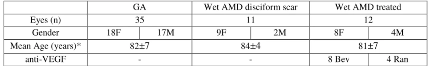
Of the 58 accepted eyes, 35 (60%) patients had GA without AMD; 11 (19%) had wet AMD not treated in the fellow eye and 12 (21%) had wet AMD treated with anti-VEGF in the fellow eye (Table 1). 23 (40%) were male patients and 35 (60%) were female.
The mean age of the patients was 82±6 years. Taking into account each group individually, the mean age from patients with GA without wet AMD was of 82±7 years, from patients with wet AMD not treated in the fellow eye was of 84±4 years and from patients with wet AMD treated with anti-VEGF in the fellow eye was of 81±7 years (Table 1). Comparing the groups, there was no statistically significant difference between group 1 and 2, group 1 and 3, and between group 2
and 3 (p=0,361; p=0,665; p=0,230, Student’s t-test).
Regarding to the measurements performed, 6 patients (10%) had two measurements included, 10 (17%) had 3 and 42 patients (73%) had four, resulting in a mean of 3.6 images per eye. The mean period of follow up was 35±14 months.
In relation to patients with wet AMD treated with anti-VEGF in the fellow eye, 8 of them (67%) were treated with bevacizumab, while 4 (33%) were treated with ranibizumab (Table 1). Patients initially treated with ranibizumab were eventually switched to bevacizumab due to hospital policy. No patients were treated with aflibercept. The mean number of injections performed was of 5 per patient per year.
The mean time of follow up was 35±14 months. Attending to the GA area at the first image, the mean was of 3.8±5.1 mm2 and at the last was of 6.6±7.0mm2. Once more, particularizing to
each group, the initial GA area of the groups 1, 2 and 3 was of 4.2±5.9 mm2, 4.4±3.9 mm2 and
2.2±2.8 mm2, respectively (Table 2). Comparing the GA area at the first image between groups 1 and 2, there was no statistically significant difference (p=0,918, Student’s t-test). The relation was
not statistically significant as well between group 1 vs group 3 and group 2 vs group 3 (p=0,264;
p=0,128, Student’s t-test).
Data concerning GA area progression rate between the images collected at different times during the study in all the 3 groups are shown at Table 2. In the first interval between measurements, the GA area progression rate for group 1 was of 1.3±1.5 mm2/year, for group 2
1.5±1.5mm2/year and for group 3 1.3±1.1 mm2/year. The same variable studied for the last
interval, in patients with more than 3 measurements performed, was of 0.9±0.9 mm2/year for
group 1, 0.5±0.9 mm2/year for group 2 and 0.8±0.8 mm2/year. In relation to the overall mean of
The relation between GA area progression rate between the patients with GA without wet AMD in the fellow eye and patients with wet AMD not treated in the fellow eye was not statistically significant, both in the first and last the interval and even in the overall period (p=0,701; p=0.195; p=0.225, respectively, Student’s t-test). Comparing patients with GA without wet AMD and
patients with wet AMD treated with anti-VEGF in the fellow eye, the relation was not statistically significant as well in the same 3 different intervals defined above (p=0.954, first interval; p=0.660, last interval; p=0.918, overall period; Student’s t-test). Furthermore, there was no statistically
significant difference between patients with wet AMD not treated and the ones treated with anti-VEGF (p=0.673, first interval; p=0,434, last interval; p=0.309, overall period; Student’s t-test).
Regarding to the comparison of the GA area progression rate between the first and the last interval of the measurements, there was no statistically significant difference (p=0.095, paired sample t-test). The mean of the GA area progression rate of the first interval was of 1.17±1.3
mm2/year while the mean of the last interval was of 0.79±0.9 mm2/year. The mean time between
the two measurements in the first interval was 12±6 months and in the last interval was 15±7 months.
Discussion
As mentioned above, several studies implicated the development of GA through various mechanisms and risk factors (Fraser-Bell, Choudhury et al. 2010, Ambati and Fowler 2012, Danis, Lavine et al. 2015).
In fact, in the past, the association between GA and wet AMD was not a focus of clinical interest. However, since the development of the most recent pharmacologic therapeutic for wet AMD with anti-VEGF agents that blocked neovascularization process, the affected eyes no longer progress to bleeding and fluid leakage. Instead, they start to develop gradual loss of RPE cells that lead to further vision loss. Therefore, this started to be an increasingly studied area (Danis, Lavine et al. 2015).
Of particular interest, a prospective study showed that the progression of GA area is significantly higher in patients that are being treated with anti-VEGF medication for the CNV developed in the same eye (Grunwald, Daniel et al. 2014).
A retrospective study analysed the same outcome and found that areas of GA have developed at the place of previous neovascular lesion in patients with CNV treated with anti-VEGF and these areas enlarged over time. However, patients submitted to anti-VEGF treatment do not appear to develop GA in eyes without AMD. (Tanaka, Chaikitmongkol et al. 2015).
pre-existing GA may continue to worsen (Grunwald, Daniel et al. 2014, Danis, Lavine et al. 2015).
We performed this study trying to understand if this processes explained above occur in the fellow eye. Patients with GA were compared with patients with wet AMD not treated with anti-VEGF. However no association has been found between GA progression and the presence of wet AMD in the fellow eye. We also analysed the possible influence of anti-VEGF in the development of GA but once more, no difference has been found between patients with wet AMD in the fellow eye treated with anti-VEGF and patients that had GA exclusively. There were no differences between the two groups of patients with wet AMD (treated and not treated with anti-VEGF).
Therefore treatment with anti-VEGF does not seem to influence the progression rate of geographic atrophy in the fellow eye, but physicians must have caution in doses applied of that medication because of the adverse effects that other studies have shown in the same eye (Kumar, Mrejen et al. 2013, Grunwald, Daniel et al. 2014, Schutze, Wedl et al. 2015, Tanaka, Chaikitmongkol et al. 2015). Even though there is data that supports the progression of geographic atrophy in the treated eye, in our study, no effect was found in the untreated eye, showing that the eventual systemic exposure in the doses used is irrelevant for the progression of this disease. In fact, taking into account the two anti-VEGF agents used in our study, bevacizumab seems to have increased systemic absorption, compared to Ranibizumab (Carneiro, Costa et al. 2012). Since most of the patients were treated with bevacizumab (even patients who were initially treated with ranibizumab, switched to bevacizumab in the last administrations) and there were no statistically significant differences regarding the GA progression comparing to the other groups of the study, ranibizumab would not theoretically show a different effect. Further studies with more patients are needed to understand the real effect of these two drugs.
Furthermore, a recent study suggested that one of the most prominent known drivers of GA growth is the baseline GA area and that enlargement of the area induces changes in FAF, with larger lesion areas leading to subsequent higher growth rates (Biarnes, Arias et al. 2015). This is against other proposed hypothesis, where elevated intracellular lipofuscin cause high levels of FAF, with consequent death of RPE cells and GA area progression (Biarnes, Arias et al. 2015).
analysing other important characteristics like FAF patterns and the influence of lipofuscin, namely its distribution.
Beyond its retrospective nature, our study had other important limitations. We did not have access to smoking history and anti-oxidant intake, what could be necessary to control biases. We also did not obtain images through SD-OCT modality. For that reason, it was not possible for us to evaluate anatomic characteristics and its relationship with the FAF images, what may provide us important information.
References
Age-Related Eye Disease Study 2 Research, G., et al. (2014). "Secondary analyses of the effects of lutein/zeaxanthin on age-related macular degeneration progression: AREDS2 report No. 3." JAMA Ophthalmol 132(2): 142-149.
Age-Related Eye Disease Study Research, G. (2001). "A randomized, placebo-controlled, clinical trial of high-dose supplementation with vitamins C and E and beta carotene for age-related cataract and vision loss: AREDS report no. 9." Arch Ophthalmol 119(10): 1439-1452.
Amaro, M. H. and A. B. Roller (2012). "Intravitreal ranibizumab and bevacizumab therapy for choroidal neovascularization in age-related macular degeneration with extensive pre-existing geographic atrophy." Arq Bras Oftalmol 75(4): 273-276.
Ambati, J. and B. J. Fowler (2012). "Mechanisms of age-related macular degeneration." Neuron
75(1): 26-39.
Batioglu, F., et al. (2014). "Geographic atrophy progression in eyes with age-related macular degeneration: role of fundus autofluorescence patterns, fellow eye and baseline atrophy area." Ophthalmic Res 52(2): 53-59.
Bearelly, S., et al. (2011). "Use of fundus autofluorescence images to predict geographic atrophy progression." Retina 31(1): 81-86.
Bhutto, I. and G. Lutty (2012). "Understanding age-related macular degeneration (AMD): relationships between the photoreceptor/retinal pigment epithelium/Bruch's membrane/choriocapillaris complex." Mol Aspects Med 33(4): 295-317.
Biarnes, M., et al. (2015). "Increased Fundus Autofluorescence and Progression of Geographic Atrophy Secondary to Age-Related Macular Degeneration: The GAIN Study." Am J Ophthalmol
160(2): 345-353 e345.
Carneiro, A. M., et al. (2012). "Vascular endothelial growth factor plasma levels before and after treatment of neovascular age-related macular degeneration with bevacizumab or ranibizumab." Acta Ophthalmol 90(1): e25-30.
Csaky, K. G., et al. (2008). "Report from the NEI/FDA Ophthalmic Clinical Trial Design and Endpoints Symposium." Invest Ophthalmol Vis Sci 49(2): 479-489.
Danis, R. P., et al. (2015). "Geographic atrophy in patients with advanced dry age-related macular degeneration: current challenges and future prospects." Clin Ophthalmol 9: 2159-2174.
Fleckenstein, M., et al. (2008). "High-resolution spectral domain-OCT imaging in geographic atrophy associated with age-related macular degeneration." Invest Ophthalmol Vis Sci 49(9):
4137-4144.
Fraser-Bell, S., et al. (2010). "Ocular risk factors for age-related macular degeneration: the Los Angeles Latino Eye Study." Am J Ophthalmol 149(5): 735-740.
Grunwald, J. E., et al. (2014). "Risk of geographic atrophy in the comparison of age-related macular degeneration treatments trials." Ophthalmology 121(1): 150-161.
Jaffe, G. J., et al. (2013). "Macular morphology and visual acuity in the comparison of age-related macular degeneration treatments trials." Ophthalmology 120(9): 1860-1870.
Joussen, A. M. and N. Bornfeld (2009). "The treatment of wet age-related macular degeneration." Dtsch Arztebl Int 106(18): 312-317.
Kanagasingam, Y., et al. (2014). "Progress on retinal image analysis for age related macular degeneration." Prog Retin Eye Res 38: 20-42.
Kaneko, H., et al. (2011). "DICER1 deficit induces Alu RNA toxicity in age-related macular degeneration." Nature 471(7338): 325-330.
Kumar, N., et al. (2013). "Retinal pigment epithelial cell loss assessed by fundus autofluorescence imaging in neovascular age-related macular degeneration." Ophthalmology 120(2): 334-341.
Lindblad, A. S., et al. (2009). "Change in area of geographic atrophy in the Age-Related Eye Disease Study: AREDS report number 26." Arch Ophthalmol 127(9): 1168-1174.
Nowak, J. Z. (2006). "Age-related macular degeneration (AMD): pathogenesis and therapy." Pharmacol Rep 58(3): 353-363.
Sarks, J., et al. (2006). "Development of atrophy of the retinal pigment epithelium around disciform scars." British Journal of Ophthalmology 90(4): 442-446.
Scholl, H. P., et al. (2007). "An update on the genetics of age-related macular degeneration." Mol Vis 13: 196-205.
Schutze, C., et al. (2015). "Progression of retinal pigment epithelial atrophy in antiangiogenic therapy of neovascular age-related macular degeneration." Am J Ophthalmol 159(6): 1100-1114
e1101.
Sigler, E. J. and J. C. Randolph (2013). "Comparison of macular choroidal thickness among patients older than age 65 with early atrophic age-related macular degeneration and normals." Invest Ophthalmol Vis Sci 54(9): 6307-6313.
Spaide, R. F., et al. (2003). "Continuing medical education review: choroidal neovascularization in age-related macular degeneration--what is the cause?" Retina 23(5): 595-614.
Sunness, J. S., et al. (2007). "The long-term natural history of geographic atrophy from age-related macular degeneration: enlargement of atrophy and implications for interventional clinical trials." Ophthalmology 114(2): 271-277.
Tanaka, E., et al. (2015). "Vision-threatening lesions developing with longer-term follow-up after treatment of neovascular age-related macular degeneration." Ophthalmology 122(1): 153-161.
Wong, T. Y., et al. (2008). "The natural history and prognosis of neovascular age-related macular degeneration: a systematic review of the literature and meta-analysis." Ophthalmology 115(1):
Tables
Table 1
GA Wet AMD disciform scar Wet AMD treated
Eyes (n) 35 11 12
Gender 18F 17M 9F 2M 8F 4M
Mean Age (years)* 82±7 84±4 81±7
anti-VEGF - - 8 Bev 4 Ran
Table 1: Baseline Characteristics: Number of eyes, gender, mean age and anti-VEGF used in patients with treated wet AMD, according to the groups of patients defined in our study. AMD: age-related macular degeneration; Bev: bevacizumab; F: female; GA: geographic atrophy; M: male; Ran: ranibizumab. *The differences in the mean age were not statistically significant between the three groups.
Table 2
GA Wet AMD disciform scar Wet AMD treated Baseline GAAa
(mm2) 4.2±5.9 4.4±3.9 2.2±2.8
Mean of overall GAA progression
rateb (mm2/year)
1.1±1.1 0.7±0.6 1.2±1.1
GAA progression rate in the first intervalc (mm2/year)
1.3±1.5 1.5±1.5 1.3±1.1
GAA progression rate in the last intervalc (mm2/year)
0.9±0.9 0.5±0.9 0.8±0.8
Table 2: Area of Geographic Atrophy at the first image and the GAA progression rate in different intervals among the images obtained, according to the groups of patients defined in our study. AMD: age-related macular degeneration; GA: geographic atrophy; GAA: geographic atrophy area. aThere was no statistically
significant differencebetween the three groups in relation to the baseline GAA. bThe differences in the
mean of overall GAA progression rate between the three groups were not statistically significant. cThere
AGRADECIMENTOS
Ao meu orientador, o Doutor Manuel Falcão, pela ajuda prestada e
acompanhamento constante ao longo da realização desta dissertação.
Anexos
1. Normas de publicação da revista Acta Ophthalmologica
ORIGINAL PAPERS
Arrangement of the manuscript: The manuscript should include the following: 1) title page; 2)
abstract, and key words; 3) main text (introduction, materials and methods, results, discussion); 4) acknowledgement; 5) references; 6) figure and figure legends; 7) tables; 8) illustrations and graphics. For more information on manuscript format, please refer to the following guidelines. 1) The Title Page should contain on separate lines author(s) name, institution, and the title of the
article. In addition it should contain the e-mail and postal addresses, plus telephone and fax numbers of the corresponding author. All author affiliations and corresponding authors addresses should be supplied in English.
2) The Abstract of original papers must be structured with the following headings: Purpose, Methods, Results, Conclusion, and should not exceed 250 words. Abstracts of review articles,
perspective of ophthalmology, historical articles, case reports and case series do not have to be structured in this same way. Diagnosis/Therapy in Ophthalmology contributions, editorials and Letters to the Editor do not have an abstract, please see separate instructions below. Key Words:
Four to nine key words for indexing purposes must be given.
3) Main text should be concise and as far as possible free of specialised language, and
unnecessary or not generally accepted abbreviations. Abbreviations must be spelled out on first mention.
The following order of presentation is recommended: Introduction stating the purpose of the
article and key aspects of present knowledge. Extensive literature reviews are not desirable. Primary sources are preferred. Material: Notice that the journal requests that all research has
followed the Tenets of the Declaration of Helsinki, and that the details are provided in the manuscript text. Methods of investigation with sufficient information to permit repetition of experiments. The journal requires that authors who report on eye cancer include in their manuscript the Union for International Cancer Control / American Joint Committee on Cancer (UICC / AJCC) Tumor, Node, Metastasis (TNM) categories and stages (7th Edition) in addition to any other cancer classification scheme the authors wish to use.
Statistics and mathematical analyses should be applied when appropriate and be described
under Methods. Authors are encouraged to take advice from an expert of statistics already when the study is designed. The following rules regarding reporting should be adhered to: - Report proportions if the number of subjects is smaller than 10 (e.g. 2 of 5), report percentages in integers if the number of subjects is less than 100 (e.g. 34%). If the number of subjects is larger, one decimal place can be given, but is seldom necessary (e.g. 34.5%). - Report summary statistics of normally distributed variables as mean with standard deviation, and other variable as medium with range. Use parametric and nonparametric statistical tests accordingly.
- Give exact p-values (e.g. p=0.15 and p=0.034); if p-value is smaller than 0.0001, report p - Give 95% confidence intervals for main findings.
- Mention the statistical test used with the p-value (e.g. p=0.015, paird t-test). If the same test is repeated, it does not need to be specified again.
here).
Results should be as clearly presented as possible. Scatterplots and similar graphical
presentations are often preferable to tables. Do not duplicate in the text data that is given in tables and figures. Discussion should be based directly on the author(s)' contributions and with reference to prior investigations, pointing out the significance and the limitations of the study.
4) Acknowledgements should indicate the name, society and date of the meeting if an abstract
of the article has been presented previously. Support for the study can also be published here. A statement regarding possible conflict of interest must be included here (e.g. disclose financial interest in the equipment or method described, research or travelling grant support, consulting services provided, or disclose absence of commercial or propriety interest).
5) References. The author(s) is responsible for accurate references that must conform to journal
style. If references are not in journal style, the manuscript will be returned for editing without review. References in the text should quote the last name(s) of the author(s) and the year of publication: (Brown & Smith 2003) or (Brown et al. 2003) when there are three or more authors. The reference list should include only those publications cited in the text and must be listed in alphabetic order with no numbering. Initials of forenames are placed after the surname with no commas, periods or spaces between initials. All articles should be cited in the original language of the reference, not as an English translation. References 'in press' must be filled in at latest in at the proof stage. Reference to unpublished material should state the author's name followed by 'unpublished' or 'personal communications'; such references should not appear in the reference list. Titles of journals are abbreviated according to the recommendations of the Index Medicus. We recommend the use of a tool such as Reference Manager for reference management and formatting.
Reference Manager reference styles can be searched for here: http://www.refman.com/support/rmstyles.asp
Examples of reference list:
Abrahamsson M, Ohlsson J & Abrahamsson H (2003): Clinical evaluation of an eccentric infrared photorefactor; the PowerRefractor. Acta Ophthalmol Scand 81: 605-610.
Sharaaway T (2003): Glaucoma surgery: Lest we forget. Acta Ophthalmol 81: 553-555.
Bailey IL (1998): Visual acuity. In: Benjamin WJ (ed.) Borish's Clinical Clinical Refraction. Philadelphia:W.B.Saunders179-202.
In papers with less than 9 authors, all author names should be listed in the reference list. In papers with 9 or more authors, please list the first three names followed by et al.
6) Figure Legends. Legends to figures should make the meaning of each illustration
understandable without reference to the text. Figure legends should be on separate pages. 7) Tables should be numbered consecutively in Arabic numerals and cited in the text. The
8) Illustrations and graphics. All photographs, drawings and graphs are referred to as figures,
abbreviated Fig., and should be numbered in sequence with Arabic numerals. Photographs and other images should be cropped so that only relevant parts of original figures are submitted. All figures should be planned to fit the printed column, 56, 117 or 178mm, and non-photographic illustrations should be professionally produced using modern software. Authors are encouraged to print their illustrations in the intended size before submission to make sure that sizes have been chosen correctly. The authors should also adapt graphics for the 3-column format and adjust the font size accordingly. Graphics are not re-drawn by the publisher. Histograms and similiar graphics should not be 3-dimensional. Complicated graphical illustrations can often be made more legible by the use of colour. Figures should be on separate pages. Please submit all line graphics as EPS or PDF files and photographs as TIF or PDF files. When submitting EPS files please embed fonts when you can. For more information on preparing and submitting figures please visit: http://www.blackwellpublishing.com/bauthor/illustration.asp
Consult the section on colour charges, colour policy and page charges below.
REVIEW ARTICLES
Review articles should be arranged in the following order: 1) title page; 2) abstract; 3) key words; 4) text, 5) acknowledgement; 6) references; 7) tables; 8) legends to figures; 9) illustrations and graphics.
Review articles have high priority. They are usually invited, but the journal may also accept unsolicited review articles. Articles with clinical relevance are preferred. Manuscripts of this type may be considerably longer than other contributions with numerous illustrations and more extensive reference lists. Review papers have an abstract that may be in a single segment.
CASE SERIES and CASE REPORTS
Case series and case reports should be arranged in the following order: 1) title page; 2) abstract; 3) key words; 4) text, 5) acknowledgement; 6) references; 7) tables; 8) legends to figures; 9) illustrations and graphics.
Acta Ophthalmologica receives many more case reports than it can publish. Published case
reports should provide unquestionably new knowledge, or be exceptionally well documented and educational. Authors should check before submission of case reports that their manuscript meets either of these requirements. Most manuscripts in this section are rejected without outside review for lack of new content and low priority. It is usually preferable to submit rare of unusual case observations in a much condensed format as a letter to the editor. Please consult also the Diagnosis/Therapy in Ophthalmology section below.
LETTERS TO THE EDITOR
Acta Ophthalmologica will consider letters for publication. Letters to the editor will be published
Letters should encourage scientific discussion on topics pertaining to ophthalmology, and may also consist of short reports or observations. A letter to the editor should start with 'Editor,' and be short and to the point. Long introductions and discussions are unacceptable. Letters should have title page, main text, no abstract, no key words, and no subheadings. Letters should not exceed 600 words and should have a maximum of 5 references and 1 table or 1 illustrations. It could be panel of figures. The editors reserve the right to edit letters for clarity and brevity.
DIAGNOSIS/THERAPY IN OPHTHALMOLOGY
The Diagnosis/Therapy in Ophthalmolgy section features photo essays, i.e. short contributions that focus on images rather than text. Submissions should be based on images which emphasise features relevant to contemporary diagnosis and treatment of eye disease. The aim of this section is to improve patient care by alerting readers to appropriate use of diagnostic techniques and important clinical signs. This section in not intended for reporting rare or unusual findings unless the technique described has wide applicability. The text should have no abstract, no subheadings and should start directly with the case report, followed by a short comment. The text should be about two doubly spaced manuscript pages. The legends are typed on a separate page. Illustrations can include any combination of clinical photographs, ultrasonographic and radiology images, histopathology slides, topographic maps, visual fields and other relevant pictures, provided that they have educational value to the general reader and cropped to show only essential details. The images must be accompanied by a concise comment and up to five references which put them in perspective. Acta will usually only print one article in this section in every issue. Acta has a section editor for this type of article.
THESES
Acta publishes full-text theses (academic dissertations) online. A one or two-page abstract is published in the printed version of the journal, while full text of the theses is available online only. Theses are freely available on the web both to subscribers and to non-subscribers. This means that theses can be found on Medline and are easily available over the world. The publication cost is low and the service is supported by Acta's owners, the Nordic Ophthalmological Societies. Clickhere for further information.
LANGUAGE
Papers should be written in English. The spelling should follow the Concise Oxford Dictionary, i.e. British English. For medical terms, Dorland's Medical Dictionary should be used. The minus sign should be shown as -. In decimal fractions use a full stop and not a comma. Footnotes should be avoided. In abbreviations, units, and symbols, standardised terms should be used.
COLOUR FIGURES AND POLICY
there will be no colour reproduction charge. For other manuscripts, a colourwork agreement formmust be completed and submitted, authorizing a charge of GBP 300. Please submit colour work forms to:
Customer Services (OPI) John Wiley & Sons Ltd, European Distribution Centre New Era Estate
Oldlands Way, Bognor Regis West Sussex PO22 9NQ
Invited articles are not charged for colour illustrations.
PAGE CHARGES
Any article which exceeds 4 pages will be charged. Excess pages must be paid for at a rate of
£80 (UK) per page unless specific written arrangements have been negotiated with the Editor-in-Chief. The page charges quoted in Sterling will be converted into the equivalent US Dollar or Euro rate depending on the billing area of the author if outside the UK. Invited papers are as a rule not charged for excess pages. Papers will be invoiced upon publication. One Acta page contains about 6,300 letters, space between words included. Please note that page charges apply to all authors, including subscribers and authors published in the online only section.
COPYRIGHT ASSIGNMENT
If your paper is accepted, the author identified as the formal corresponding author for the paper will receive an email prompting them to login into Author Services; where via the Wiley Author Licensing Service (WALS) they will be able to complete the license agreement on behalf of all authors on the paper.
For authors signing the Copyright Transfer Agreement
If the OnlineOpen option is not selected, the corresponding author will be presented with the Copyright Transfer Agreement (CTA) to sign. The terms and conditions of the CTA can be previewed in the samples associated with the Copyright FAQs below:
CTA Terms and Conditions http://authorservices.wiley.com/bauthor/faqs_copyright.asp
For authors choosing OnlineOpen
If the OnlineOpen option is selected the corresponding author will have a choice of the following Creative Commons License Open Access Agreements (OAA):
Creative Commons Attribution License OAA
Creative Commons Attribution Non-Commercial License OAA
Creative Commons Attribution Non-Commercial -NoDerivs License OAA
Services http://authorservices.wiley.com/bauthor/faqs_copyright.aspand
visit http://www.wileyopenaccess.com/details/content/12f25db4c87/Copyright--License.html. If you select the OnlineOpen option and your research is funded by The Wellcome Trust and members of the Research Councils UK (RCUK) you will be given the opportunity to publish your article under a CC-BY license supporting you in complying with Wellcome Trust and
Research Councils UK requirements. For more information on this policy and the Journal’s
compliant self-archiving policy please visit: http://www.wiley.com/go/funderstatement.
OPEN ACCESS
Online Open is available to authors of primary research articles who wish to make their article available to non-subscribers on publication, or whose funding agency requires grantees to archive the final version of their article. With OnlineOpen, the author, the author's funding agency, or the author's institution pays a fee to ensure that the article is made available to non-subscribers upon publication via Wiley Online Library, as well as deposited in the funding agency's preferred archive. For more information on Online Open including the full list of terms and conditions and the online order form please go to here.
Prior to acceptance there is no requirement to inform an Editorial Office that you intend to publish your paper OnlineOpen if you do not wish to. All OnlineOpen articles are treated in the same way as any other article. They go through the journal's standard peer-review process and will be accepted or rejected based on their own merit.
Please note: 50% discounted OnlineOpen fee for Members of the Danish Ophthalmological Society, the Finnish Ophthalmological Society, the Icelandic Ophthalmological Society, the Norwegian Ophthalmological Society, the Swedish Ophthalmological Society or the European Association for Vision and Eye Research (EVER).
PROOFS
Proofs will be sent to one author only. The corresponding author will receive an e-mail alert containing a link to a web site. A working e-mail address must therefore be provided for the corresponding author. The proof can be downloaded as a PDF (portable document format) file from this site. Acrobat Reader will be required in order to read this file. This software can be downloaded (free of charge) from the following web site:
http://www.adobe.com/products/acrobat/readstep2.html
AUTHOR SERVICES
Online production tracking is now available for your article through Wiley-Blackwell's
Author Services
Author Services enables authors to track their article - once it has been accepted - through the production process to publication online and in print. Authors can check the status of their articles online and choose to receive automated e-mails at key stages of production. The author will receive an e-mail with a unique link that enables them to register and have their article automatically added to the system. Please ensure that a complete e-mail address id provided when submitting the manuscript. Visit www.authorservices.wiley.com/bauthor/ for more details on online production tracking and a for a wealth of resources including FAQs and tips on article preparation, submission and more.
SUPPLEMENTS
Monographs of large original work, proceedings of symposia, etc. may be published as supplements, the full cost being paid by the author. Examples of material that may be accepted as supplements include theses with ophthalmic subjects or meeting proceedings from Nordic ophthalmic societies or ophthalmic meetings in the Nordic countries. Supplements are not subject to revision by the Editor. The quality of the language must meet the standards maintained by the journal, and all the above-mentioned instructions should be followed. All costs for production, postage to the subscribers, and translation or linguistic revision must be paid by the author. Supplements are sent free of charge to the subscribers. It is the policy to acknowledge in supplements any major sponsorship.
OFFPRINTS
There are no free offprints. Information on how to order offprints and reprints will accompany the proofs. To order offprints please
visit http://www.blackwellpublishing.com/bauthor/offprint.asp or email [email protected].
AUTHOR MATERIAL ARCHIVE POLICY
Please note that unless specifically requested, Wiley-Blackwell will dispose of all hardcopy or electronic material submitted two months after publication. If you require the return of any material submitted, please inform the editorial office or production editor as soon as possible if you have not already done so.
PAPER
DISCLAIMER
The Publisher, the Nordic Ophthalmological Societies or Editors cannot be held responsible for errors or any consequences arising from the use of information contained in this journal; the views and opinions expressed do not necessarily reflect those of the Publisher, the Nordic Ophthalmological Societies or Editors, neither does the publication of advertisements constitute any endorsement by the Publisher, the Nordic Ophthalmological Societies or Editors of the products advertised.
EDITORIAL OFFICE
Professor Einar Stefánsson University of Iceland
National University Hospital Department of Ophthalmology 101 Reykjavík, Iceland Bryndis Thordardottir Editorial Manager Tel: +354 897 9752 Fax: +354 543 4831
E-mail: [email protected]
PRODUCTION OFFICE