O
r i g i n a la
rt i c l e2 3 4 Arq Bras Oftalmol. 2017;80(4):234-7 http://dx.doi.org/10.5935/0004-2749.20170057
ABSTRACT
Purpose: We evaluated dynamic thiol/disulfide homeostasis (TDH), malondial-dehyde (MDA) levels, and catalase (CAT ) activity in patients with age-related macular degeneration (AMD). All analyzes were conducted on plasma samples. Methods: Thirty-two patients with AMD and 38 age-matched healthy con-trols were included. Native thiol, total thiol, and disulfide levels and TDH status were determined using a novel, automated assay. MDA levels and CAT activity were determined. Percentages were compared using the chi-squared test. The Student’s t-test and Mann-Whitney U-test were used to compare quantitative variables.
Results: Native thiol levels were significantly lower (p=0.004) in patients with AMD (272.02 ± 52.41 µmol/l) than in healthy individuals (307.82 ± 47.18 µmol/l), whereas disulfide levels were significantly higher (p<0.001) in patients with AMD than in controls (21.64 ± 5.59 vs. 14.48 ± 5.37 µmol/L). Dynamic TDH was also significantly lower (p<0.001) in patients with AMD than in controls (13.41 ± 4.3 vs. 25.41 ± 14.52 µmol/l). No significant differences were evident in total thiol or MDA levels. Mean CAT activity was significantly higher (p=0.043) in patients with AMD compared with controls (0.035 vs. 0.018 k/ml).
Conclusions: The antioxidant/oxidant balance demonstrated by dynamic TDH is shifted to the oxidative side in patients with AMD.
Keywords: Macular degeneration; Sulfhydryl compounds; Disulfides; Oxidative stress; Malondialdehyde
RESUMO
Objetivo: Avaliar a homeostase dinâmica de tiol/dissulfureto e os níveis de malon dialdeído (MDA) e catalase (CAT) em pacientes com degeneração macular relacionada à idade (DMRI). Todas as análises foram realizadas em amostras de plasma.
Métodos: Foram incluídos 32 pacientes com degeneração macular relacionada à
idade e 38 controles saudáveis de idade similar. Os níveis de tiol, tiol total, dissulfu reto e estado de homeostase de tiol/dissulfureto foram determinados utilizando um novo ensaio automatizado. Os níveis de atividade de MDA e CAT foram também determinados. As porcentagens foram comparadas pelo teste do quiquadrado. O teste t de Student e o teste U de Mann Whitney foram utilizados para comparar variáveis quantitativas.
Resultados: Os níveis de tiol nativo foram significativamente menores (p=0,004) nos pacientes com degeneração macular relacionada à idade (272,02 ± 52,41 μmol/l) do que nos indivíduos saudáveis (307,82 ± 47,18 μmol/l), enquan to os dissulfetos foram significativamente maiores em pacientes com degeneração macular relacionada à idade (21,64 ± 5,59 μmol/l versus 14,48 ± 5,37 μmol/l, res pectivamente, p<0,001). A homeostase dinâmica de tiol/dissulfureto também foi significativamente menor nos pacientes com degeneração macular re la cionada à idade (13,41 ± 4,3 μmol/l) versus os controles (versus 25,41 ± 14,52 μmol/l, p<0,001). Não foram observadas diferenças signiicativas nos níveis de tiol total ou MDA. A atividade média de CAT foi significativamente mais elevada (p=0,043) em doentes com degeneração macular relacionada à idade (0,035 k/ml vs. 0,018 k/ml).
Conclusões: O equilíbrio antioxidante/oxidante demonstrado pela homeostase
dinâmica de tiol/dissulfeto é deslocado para o lado oxidativo em pacientes com de generação macular relacionada à idade.
Descritores: Degeneração macular; Compostos de sulidrila; Dissulfetos; Estresse oxi dativo; Malondialdeído
Dynamic thiol/disulfide homeostasis in patients with age-related macular
degeneration
Homeostase dinâmica de tiol/dissulfureto em pacientes com degeneração macular relacionada à idade
Serdar aktaş1, Hacı Murat Sağdık1, MeHMet tetıkoğlu1, Hatıce aktaş2, F
atih Özcura1, Fatma uçar3, Murat alışık4, merve ergin4
Submitted for publication: November 24, 2016 Accepted for publication: February 12, 2017
1 Department of Ophthalmology, Dumlupinar University School of Medicine, Kutahya, Turkey. 2 Clinic of Ophthalmology, DPU Evliya Celebi Training and Research Hospital, Kutahya, Turkey. 3 Department of Clinical Biochemistry, Diskapi Yildirim Beyazit Training and Research Hospital,
Ankara, Turkey.
4 Department of Clinical Biochemistry, Ataturk Training and Research Hospital, Ankara, Turkey.
Funding: No specific financial support was available for this study.
Disclosure of potential conflicts of interest: None of the authors have any potential conflicts of interest to disclose.
Corresponding author: Serdar Aktaş. Department of Ophthalmology. Dumlupinar University School of Medicine, Kutahya - 43270 - Turkey - E-mail: [email protected]
Approved by the following research ethics committee: Diskapi Yildirim Beyazit Training and Research Hospital (#20/11).
INTRODUCTION
Age-related macular degeneration (AMD) is the most common cause of irreversible blindness among individuals aged over 65 in developed countries(1,2). The global prevalence of AMD is reportedly
8.69% in people aged 45-85 years, and the number of patients with AMD is projected to increase to approximately 196 million in 2020 and 288 million in 2040(3). Globally, the costs directly associated with
AMD are estimated to be $255 billion(4). Furthermore, the proportion
of people aged over 60 years is increasing rapidly in almost every country(5), resulting in a dramatic increase in the global societal and
economic burden of AMD.
The pathogenesis of AMD remains poorly understood. However, there is a general consensus that retinal damage occurs with aging as a result of factors such as oxidative damage, parainlammatory dysregulation, and vascular sclerosis(6,7). Moreover, the free-radical
theory of aging proposes that reactive oxygen intermediates (ROIs), such as free radicals, hydrogen peroxide, and singlet oxygen, are responsible for the development of age-related disorders(8). If the
free-radical theory applies, antioxidant/oxidant homeostasis may be disrupted in patients with AMD.
2 3 5 with the electrophilic groups of ROIs, forming disulfide bonds that
facilitate reversible thiol disulfide-exchange reactions(10). In this
study, we evaluated dynamic thiol/disulfide homeostasis (TDH) in plasma samples. We also measured malondialdehyde (MDA) levels and catalase (CAT ) activity.
METHODS
Thirty-two patients with exudative AMD and 38 age- and sex-matched healthy controls were enrolled at a university hospital. All patients provided written informed consent before enrollment. All procedures conformed to the tenets of the Declaration of Helsinki. The study was approved by the Ethics Committee of the Diskapi Yildirim Beyazit Training and Research Hospital, Ankara, Turkey.
Detailed ophthalmologic examinations were performed, in-cluding visual acuity testing, slit-lamp biomicroscopy, retinoscopy, intrao cular pressure measurement via Goldmann applanation tono-metry, optical coherence tomography, and luorescein angiography. Patients included in the study had either choroidal neovascularisa-tion or disciform scars. Participants with any systemic disease that could afect TDH, such as a chronic inlammatory disease, rheumato-logic disease, diabetes mellitus, hypertension, cardiovascular disease, or malignancy, as well as smokers, alcoholics, patients with a body mass index greater than 30 kg/m2, and anyone taking antioxidant
supplements, were excluded from the study.
All tests were conducted on plasma samples. Venous blood samples were collected in tubes containing ethylenediaminete-traacetic acid and centrifuged at 1500× g for 10 min. Plasma samples were frozen and stored at -80°C until analysis. Native thiol, total thiol, and disulide levels and TDH status were determined using a novel, automated assay developed by Erel and Neselioglu(11).
Brie-ly, the assay reduced dynamic disulide (-S-S-) bonds to form free functional thiol groups (-SH) using sodium borohydride. Then, the unreacted sodium borohydride was removed with formaldehyde. The thiol groups (reduced and native) were detected upon reaction with 5,5’-dithiobis-(2-nitrobenzoic) acid. The number of disulide bonds was calculated as half the diference between the total and native levels of thiol. Following the measurement of native thiol and disulide concentrations in the pretreated samples, the dynamic TDH was determined.
CAT activity was determined using the methods described by Aebi(12). The rate of decomposition of the H
2O2 substrate by the CAT
enzyme was measured spectrophotometrically at 240 nm. MDA levels were detected using the thiobarbituric acid method, and the formation of a pink-colored complex was measured spectrophoto-metrically at 532 nm(13).
S
TATISTICALANALYSISStatistical analysis was performed using SPSS software (Version 22.0; IBM Corp., Armonk, NY, USA). The Kolmogorov-Smirnov test
was used to determine the normality of data distribution. Because the distribution of everything except CAT activity was normal, the Student’s ttest for independent means was used to explore the signiicance of the diferences between measurements in the two study groups. The Mann-Whitney U-test was used to compare the diferences in CAT activity. p values <0.05 were considered to indicate statistical signiicance.
RESULTS
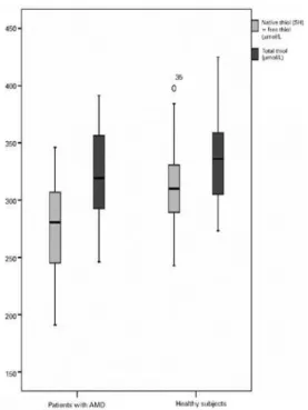
Table 1 presents the demographic details of the patient and control groups. There were no signiicant diferences between the patient and control groups in age (p=0.148) or sex (p=0.079). Native thiol, total thiol, and disulide levels, TDH status, MDA levels, and CAT activities are also given in table 1. Native thiol levels (272.02 ± 52.41 µmol/L) were signiicantly lower (p=0.004) in patients with AMD than in healthy individuals (307.82 ± 47.18 µmol/l; Figure 1). However, no significant differences were evident in total thiol levels (Figure 1). Disulfide levels were significantly higher (p<0.001) in patients with AMD (21.64 ± 5.59 µmol/l) than in controls (14.48 ± 5.37 µmol/L; Figure 2 A). Dynamic TDH was also significantly lower (p<0.001) in patients with AMD (13.41 ± 4.3 µmol/L) than in controls (25.41 ± 14.52 µmol/L; Figure 2 B). No significant differences were evident in MDA levels (Figure 2 C). Mean CAT activity was signifi-cantly higher (p=0.043) in patients with AMD (0.035 k/ml) than in controls (0.018 k/ml; Figure 2 D).
DISCUSSION
We used a novel and automated assay to determine TDH. Native thiol concentrations and thiol/disulfide ratios were significantly lower, and disulfide levels were significantly higher in patients with AMD than in controls. To the best of our knowledge, this is the second study to investigate dynamic TDH as a novel marker of oxidative stress in patients with AMD and compare the results with a control group.
Although the pathogenesis and etiology of AMD remain poorly understood, there is considerable evidence that oxidative stress plays a role in the onset and progression of the disease. The first di rect evidence of a causal relationship between oxidative stress and aging and age-related disorders was reported in a study invol-ving transgenic flies (Drosophila melanogaster): flies overexpressing genes for antioxidant enzymes (copperzinc superoxide dismutase and CAT) exhibited a longer lifespan and a delayed loss of physical performance(14).
Several studies that evaluated total thiol concentration in plas-ma samples reported significantly reduced thiol levels in patients with exudative AMD(18-20). To the best of our knowledge, the only
study to investigate TDH to date reported that patients with
advan-Table 1. Demographic and biochemical data of the AMD patients and control group
AMD patients (n=32) (mean ± SD) (minimum-maximum) Controls (n=38) (mean ± SD) (minimum-maximum) p*
Sex (female/male) 12/20 21/17 <0.079
Age 73.81 ± 6.3 (59-84) 71.66 ± 5.9 (57-85) <0.148
Native thiol, μmol/L 272.02 ± 52.41 (140.4-345.9) 307.82 ± 47.18 (167.6-418.8) <0.004
Native thiol/disulide, % 13.41 ± 4.3 (5.83-23) 25.41 ± 14.53 (11.34-79.54) <0.001
Disulide, μmol/L 21.64 ± 5.59 (12.35-35.1) 14.48 ± 5.38 (5-26.25) <0.001
Total thiol, μmol/L 315.3 ± 52.46 (188.6-391.5) 336.78 ± 49.31 (186.9-464.5) <0.082
MDA, nmol/ml 29.36 ± 8.34 (14.12-58.24) 33.29 ± 19.09 (23.24-144.12) <0.284
CAT, k/ml 0.035 ± 0.046 (0.002-0.194) 0.018 ± 0.049 (0.003-0.49) <0.043
*= comparisons with control eyes using the Student’s t-test. p<0.05 was considered statistically signiicant.
Dy n a m i ct h i o l/D i s u l f i D eh o m e o s ta s i si npat i e n t sw i t ha g e-r e l at e Dm a c u l a rD e g e n e r at i o n
2 3 6 Arq Bras Oftalmol. 2017;80(4):234-7
ced AMD exhibited significantly lower levels of TDH than healthy controls (20.3 ± 1.2 vs. 29.5 ± 3.1, p=0.005)(20). In this study, dynamic
TDH was also significantly lower (p<0.001) in patients with AMD (13.41 ± 4.3 µmol/L) than in controls (25.41 ± 14.52 µmol/L). There were no statistically significant differences in total thiol levels, but the native (free) thiol concentration (272.02 ± 52.41 µmol/L) was significantly lower (p=0.004) in patients with AMD than in healthy individuals (307.82 ± 47.18 µmol/L). This supports the conclusions of previous studies that rather than the total thiol concentration, the thiol/disulfide balance has a fundamental role in protection against oxidative stress(21,22).
CAT is an iron-dependent antioxidant enzyme that scaven-ges H2O2(23). Although this enzyme may play an important role in
antioxidant-defense in the retina, published reports regarding the relationship between systemic and retinal CAT activity and AMD are contradictory. Orr and Sohal(14) reported signiicantly reduced CAT
activity in the retinal pigment epithelium (RPE) of eyes with AMD. Tate et al.(24) observed increased CAT activity in the RPE in response
to a challenge with exogenous H2O2, suggesting that phagocytosis of the outer segments of the rods near the RPE is a response to oxi-dative stress that probably produces H2O2, which is believed to act as an intracellular signal that induces CAT activity. In this study, the mean CAT activity was signiicantly higher (p=0.043) in patients with AMD (0.035 k/ml) than in controls (0.018 k/ml). We believe that CAT activity, like other components of the antioxidant-defense system, is dependent on many factors, such as genetic variation, nutrition, and disorders, which may have inluenced the systemic or retinal CAT activities observed in previous studies. However, as discussed in a previous study(24), our indings suggest a relationship between
increased systemic CAT activity and AMD.
MDA is a common lipid peroxidation product and reliable marker of oxidative stress(25). Drusen, the hallmark of AMD, contains MDA,
which can damage the RPE(26). Studies that evaluated MDA
concen-trations in plasma samples reported signiicantly increased levels of MDA in patients with exudative AMD(27,28). In this study, no signiicant
diferences were observed in MDA levels. As addressed by our exclu-sion criteria, many factors can afect the antioxidant-defense system. However, other confounding factors that were not taken into consi-deration, such as genetic structure, nutrition, and disorders such as atherosclerosis, may explain the discrepancies between the results of diferent studies.
There were several limitations to this study. Because of the small sample size, our results are diicult to translate to all patients with AMD. Detection of TDH in vitreous samples can be useful for assessing oxidative stress in the retina, but this was not performed in this study. Aside from age-related diseases including AMD, many factors such as genes, environment, and diet may alter TDH and an tioxidant status. This situation may have resulted in a bias in par-ticipant enrollment.
In conclusion, this is the second study to examine TDH in patients with AMD. The antioxidant/oxidant balance of dynamic TDH shifts to the oxidative side in patients with AMD. These indings support pre-viously reported evidence of a causal relationship between oxidative stress and AMD. Oxidative stress markers may be disease speciic, and not all markers are disturbed in AMD. Further studies involving larger patient populations are warranted to determine the relationships between systemic markers of oxidative stress and AMD.
REFERENCES
1. Klein R, Klein BE, Linton KL. Prevalence of age-related maculopathy. The Beaver Dam Eye Study. Ophthalmology. 1992;99(6):933-43.
2. Mitchell P, Smith W, Attebo K, Wang JJ. Prevalence of age-related maculopathy in Australia. The blue mountains eye study. Ophthalmology. 1995;102(10):1450-60. 3. Wong WL, Su X, Li X, Cheung CM, Klein R, Cheng CY,et al. Global prevalence of
age-related macular degeneration and disease burden projection for 2020 and 2040: a systematic review and meta-analysis. Lancet Glob Health. 2014;2(2):106-16. Comment in: Lancet Glob Health. 2014;2(2):e65-6.
Figure 1. Comparison of native and total thiol levels in the plasma of patients with age-related macular degeneration versus healthy controls.
Figure 2. Comparative levels of oxidative stress markers in the blood of AMD patients and controls. A) Disulide levels in the plasma of patients with age-related macular de-generation (AMD) and healthy controls. B) Native thiol/disulide homeostasis in patients with AMD and healthy controls. C) Plasma levels of malondialdehyde in patients with AMD and healthy controls. D) Mean catalase activity in the plasma of patients with AMD and healthy controls.
B
D C
2 3 7 4. Gordois A, Pezzullo L, Cutler H. The global economic cost of visual impairment
[In-ternet]. AMD Alliance International; 2010. [cited 2016 Jan 21]. Available from: http:// www.icoph.org/dynamic/attachments/resources/globalcostofvi_inalreport.pdf 5. World Health Organization . World report on ageing and health [Internet]. Geneva:
WHO;2015. [cited 2015 Oct 16]. Available from: http://apps.who.int/iris/bitstream/ 10665/186463/1/9789240694811_eng.pdf?ua=1
6. Beatty S, Koh HH, Phil M, Henson D, Boulton M. The role of oxidative stress in the pathogenesis of age-related macular degeneration. Surv Ophthalmol. 2000;45(2): 115-34.
7. Danis RP, Lavine JA, Domalpally A. Geographic atrophy in patients with advanced dry age-related macular degeneration: current challenges and future prospects. Clin Ophthalmol. 2015;9:2159-74.
8. Harman D. The aging process. Proc Natl Acad Sci.1981;78(11):7124-8.
9. Wlodek L. Beneicial and harmful efects of thiols. Pol J Pharmacol. 2002;54(3):215-23. 10. Kemp M, Go YM, Jones DP. Non equilibrium thermodynamics of thiol/disulide redox
system: a prespective on redoxsystem biology. Free Radic Biol Med. 2008;44(6):921-37. 11. Erel O, Neselioglu S. A novel and automated assay for thiol/disulphide homeostasis.
Clin Biochem. 2014;47(18):326-32.
12. Aebi H. Catalase In: Bergmeyer U, editor. Methods of enzymatic analysis. New York: New York and London Academic Press; 1974. p. 673-7.
13. Esterbauer H, Cheeseman KH. Determination of aldehydic lipid peroxidation pro-ducts: malonaldehyde and 4-hydroxynonenal. Methods Enzymol. 1990;186:407-21. 14. Orr WC, Sohal RS. Extension of life-span by overexpression of superoxide dismutase
and catalase in Drosophilia melanogaster. Science. 1994;263(5150):1128-30. 15. Deneke SM. Thiol-based antioxidants. Curr Top Cell Regul. 2000;36:151-80. 16. Moriarty SE, Shah JH, Lynn M, Jiang S, Openo K, Jones DP, et al. Oxidation of
glutathio-ne and cysteiglutathio-ne in human plasma associated with smoking. Free Radic Biol Med. 2003; 35(12):1582-8.
17. Jiang S, Moriarty-Craige SE, Orr M, Cai J, Sternberg P, Jones DP. Oxidant-induced
apoptosis in human retinal pigment epithelial cells: Dependence on extracellular redox state. Invest Ophthalmol Vis Sci. 2005;46(3):1054-61.
18. Coral K, Raman R, Rathi S, Rajesh M, Sulochana KN, Angayarkanni N,et al. Plasma homocyctein and total thiol contend in patients with exudative age related macular degeneration. Eye (Lond). 2006;20(2):203-7.
19. Javadzadeh A, Ghorbanihaghjo A, Bahreini E, Rashtchizadeh N, Argani H, Alizadeh S. Plasma oxidized LDL and thiol-containing molecules in patients with exudative age-related macular degeneration. Mol Vis. 2010;16:2578-84.
20. Arikan Yorgun M, Toklu Y, Altınkaynak H, Tanrıverdi B, Ergin M, Biçer C. A novel tool for the assessment oxidative stress in age-related macular degeneration: thiol/disulide homeostasis revisited. Curr Eye Res. 2016;41(12):1584-9.
21. Sen CK. Cellular thiols and redox-regulated signal transduction. Curr Top Cell Regul. 2000;36:1-30.
22. Moran LK, Gutteridge JM, Quinlan GJ. Thiols in cellular redox signalling and control. Curr Med Chem. 2001;8(7):763-72.
23. Halliwell B, Gutteridge JM. The importance of free radicals and catalytic metal ions in human diseases. Mol Aspects Med. 1985;8(2):89-193.
24. Tate DJ Jr, Miceli MV, Newsome DA. Phagocytosis and H2O2 induce catalase and metallothionein gene expression in human retinal pigment epithelial cells. Invest Ophthalmol Vis Sci. 1995;36(7):1271-9.
25. Esterbauer H, Schaur RJ, Zollner H. Chemistry and biochemistry of 4-hydroxynonenal, malonaldehyde and related aldehydes. Free Radic Biol Med. 1991;11(1):81-128. 26. Schutt F, Bergmann M, Holz FG, Kopitz J. Proteins modiied by malondialdehyde,
4-hydroxynonenal, or advanced glycation end products in lipofuscin of human retinal pigment epithelium. Invest. Ophthalmol Vis Sci. 2003;44(8):3663-8. 27. Jia L, Dong Y, Yang H, Pan X, Fan R, Zhai L. Serum superoxide dismutase and
ma-lon dialdehyde levels in a group of Chinese patients with age-related macular de generation. Aging Clin Exp Res. 2011;23(4):264-7.
28. Ates O, Azizi S, Alp HH, Kiziltunc A, Beydemir S, Cinici E, et al. Decreased serum paraoxo-nase 1 activity and increased serum homocysteine and malondialdehyde levels in age-related macular degeneration. Tohoku J Exp Med. 2009;217(1):17-22.