Silk Hydrogels for Tissue Engineering
by KHOONS. LIM(∗) & TIMB. F. WOODFIELD
Christchurch Regenerative Medicine and Tissue Engineering (CReaTE) Group, Department of Orthopaedic Surgery and Musculoskeletal Medicine, University of Otago Christchurch, Christchurch 8011, New Zealand.
(*) Corresponding author Email: khoon.lim@otago.ac.nz
Abstract Silk hydrogels have been highlighted in the past decade as potential matrices for tissue engineering and regenerative medicine applications. In this mini review, the biological attributes of silk proteins, as well as methods reported in literature to fabricate silk hydrogels will be discussed.
1
Introduction
The field of tissue engineering (TE) which aims to re-pair, regenerate or replace damaged tissue has been ex-tensively researched in the past decade as an alterna-tive to organ transplantation. The general approach has been centred on combining cells, growth factors and tis-sue engineering matrices, with the goal of engineering functional tissues in vitro, which can then be implanted into the body. Hydrogels, which are highly hydrated poly-meric network have been highlighted as potential tissue engineering matrices, due to their structural similarity to the native extracellular matrix [1, 2]. Several materials ranging from synthetic to natural polymers, have been fabricated into hydrogels, and shown to support cellular growth and differentiation [2]. In particular, silk proteins, which have been traditionally used in biomedical applica-tions as sutures and drug delivery systems, have also been translated into tissue engineering matrices in the form of hydrogels. This review will focus on the main attributes of silk proteins as biomaterials, as well as methods to fab-ricate silk hydrogels for tissue engineering applications.
2
Hydrogels as tissue engineering
matrices
Hydrogels are defined as hydrophilic polymeric networks which are capable of absorbing water ranging from ten to a thousand times their dry weight [3]. They are
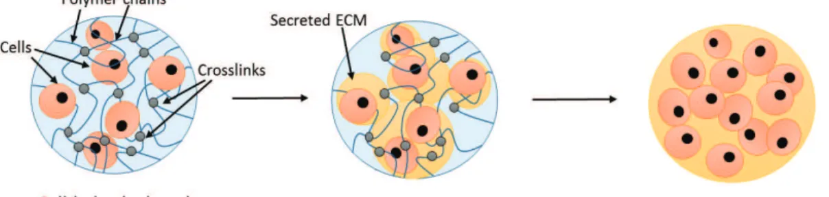
struc-turally similar to the native extracellular matrix (ECM) in their hydrated state, which enables facilitation of nu-trient diffusion and waste removal through the TE ma-trix. In an ideal scenario, when cells are encapsulated within a hydrogel, the hydrogel should be able to sup-port cell remodelling and ECM secretion, which results in a functional engineered tissue that can be transplanted (Figure 1). Hydrogels can be classified based on the forces between the crosslinked networks (physical or co-valent), or the nature of the polymer used for fabrica-tion (synthetic or natural) [2]. Physical hydrogels have either molecular entanglements, ionic bonding, or hy-drogen bonding holding the network together that can be reversible, while covalent gels are hydrogels with co-valently crosslinked networks that are permanent [3]. These gels can be fabricated from a range of synthetic or natural polymers.
In general, synthetic hydrogels have less batch-to-batch variability with good chemical and mechani-cal stability. Current synthetic polymeric hydrogel sys-tems reported in literature include poly(vinyl alcohol) (PVA), poly(ethylene oxide) (PEO), poly(ethylene gly-col) (PEG), poly(N-isoproplyacrylamide) (NIPAAm), and poly(propylene furmarate) (PPF) [1, 2, 4-8]. However, although the reported synthetic hydrogels have good physico-mechanical properties, their application as scaf-folds for TE remain limited due to the lack of biological sequences available to facilitate cellular functions. More-over, most of these polymers are hydrophilic and resist cell attachment [9, 10].
On the other hand, hydrogels fabricated from nat-urally derived polymers, such as chitosan, collagen, gelatin, hyaluronic acid, fibrinogen and fibrin, are known to have good bio-functionality to support cellular growth, proliferation and differentiation [11-16]. However, these natural hydrogels are generally weak with limited me-chanical stability as TE matrices. Therefore, there exists a need to engineer TE scaffolds that have the advantages of both synthetic and natural polymers, with good physico-mechanical properties and also with the ability to sup-port cellular function. In recent years, hydrogels fabri-cated from silk proteins have been identified as potential candidates to meet these criteria.
3
Silk proteins
Figure 2: Biological attributes of silk fibroin and sericin [18].
Silk is a combination of fibrous proteins synthesised in specialised epithelial cells that line glands in silkworms, and has been successfully used as a suture material for centuries with great potential in biomaterial applications [17]. Before converted to silk fibers, silk proteins are syn-thesized by silk gland cells and stored in the lumen of the silk glands [18]. The usage of silk fibers is advantageous in the biomaterial aspect as it has properties that can rival synthetic polymers but requires less harsh processing con-ditions [18]. Silks have impressive mechanical properties, environmental stability, biocompatibility, controlled pro-teolytic biodegradability, morphologic flexibility and the
ability for amino acid side change modification to immo-bilize growth factor [17].
The two major components in silk is fibroin and sericin, where fibroin is normally coated with sericin in the cocoons [18]. The biological attributes of these two silk proteins are listed in Figure 2. Sericin is secreted from the middle silk gland of a mature silkworm larva and acts as the glue that keeps fibroin together [19, 20]. Sericin produced by the most commonly researched do-mesticated type, Bombyx mori,(B. mori) consists of pep-tides with 3 major fractions of 150, 250, and 400 kDa [21]. This protein also exists in two kinds of conforma-tion, random coils or β-sheets. [18].
On the other hand, fibroin is the major structural pro-tein of silk which is secreted from the posterior silk gland [19]. Vepari et al reported that B. mori fibroin fibers are about 10-25 µm in diameter and contain a light protein chain (L-chain) with molecular weight of approximately 26 kDa, and heavy protein chain (H-chain) of approxi-mately 390 kDa, where both L- and H-chain are linked by a disulphide bond [17]. Similarly to sericin, fibroin also exists in random coils and β-sheets conformation. By heating, stretching or immersing fibroin in a polar sol-vent, the protein conformation undergoes transition from random coil to β-sheet (Figure 3). This β-sheet transfor-mation also corresponds to higher mechanical durability, where higher amount of β-sheet formation corresponds to higher mechanical strength [22].
Figure 3: Random coil to β-sheet transformation of silk fibroin.
5 CONCLUSION AND FUTURE OUTLOOKS
4
Silk hydrogels: Fabrication
tech-niques and applications
For the past decades, it has been thought that the sericin fraction of silk causes unwanted inflammatory responses and hence has not been the major focus of biomaterial research. Sericin, which is the glue component of the silk cocoons are normally isolated using heat in basic con-ditions. These isolation conditions have been shown to cause denaturation and degradation of the protein, which subsequently affects the mechanical properties of the re-sultant hydrogels. In order to prevent heat denaturation during sericin isolation, Teramoto et al. researched genet-ically modified silkworms (Sericin-Hope) whose cocoons contain 99% sericin. As these Sericin-Hope cocoons con-tain almost no fibroin, the heat treatment was not re-quired to separate the sericin from fibroin [23, 24]. How-ever, although these hydrogels were shown to have sig-nificantly higher elastic modulus than the conventionally used domesticated silkworms, their mechanical proper-ties were still not on par with fibroin hydrogels [23, 24]. Fibroin, the fibrous part of the silkworm cocoon, has been characterised to have abundance of hydrophobic amino acid groups such as glycine, sericin and alanine, which are capable to form physical crosslinks without ad-dition of any chemical crosslinkers [25]. This approach is beneficial as concerns associated with toxicity of chem-ical crosslinkers are eliminated. However, fibroin hydro-gels formed by this self-assembly approach require a long crosslinking time that can take up to days. Kim et al. showed that silk fibroin solution (2% w/v, pH 6.4 - 6.8, 37oC) required 30 days for complete gelation [20]. These
large hydrophobic domains of fibroin also allow its gela-tion time to be tailored by a number of factors such as temperature, ionic concentration, pH and salt concentra-tion [26]. The different factors used to control fibroin gelation are:
pH changes pH closer to fibroin isoelectric point
accel-erate gelation [27-29]
Temperature changes Fibroin solution crosslinks faster
at higher temperature. The resultant hydrogels are also mechanically stiffer [20, 27, 30]
Freeze-thawing Porosity of fabricated fibroin hydrogels
depend on the number of freeze-thaw cycles [31-33]
For example, Ayub et al. showed that decreasing the pH of the fibroin solution to 3-4 successfully facilitated gel formation within two days [34]. It was hypothe-sised that the lower pH initiated protonation of the car-boxyl groups, which subsequently reduce repulsion be-tween the fibroin polymer chains and led to formation of crosslinks [29, 35]. It has also been showed that the fi-broin protein structures are converted from random coils to β-sheets during the crosslinking process [34]. As the
physico-mechanical properties of the fibroin gels are di-rectly related to the amount of β-sheets formed, many researchers have focused on different methods to in-duce β-sheet transition [20, 27, 36, 37]. The compres-sive modulus of these fibroin gels fabricated using differ-ent methods can vary from 60 kPa to 7 GPa [26]. Chem-ical crosslinking of fibroin solution using different sol-vents and chemicals has also been studied to fabricate fibroin hydrogels. Although these gels are normally me-chanically stronger and require shorter gelation time, the chemicals or conditions used are normally quite harsh for cells in terms of TE applications. For example, typi-cal chemitypi-cal crosslinkers include glycerol, sodium dode-cyl sulfate and sodium N-lauroyl sarcosinate that are not cyto-compatible to cells at high concentrations [35, 38-40].
Fibroin hydrogels have gained huge popularity as TE matrices in the past decade for various applications such as bone engineering, cartilage engineering, nerve engi-neering, immunoisolation of cells, drug delivery and in-jectable void fillers. For example, mesenchymal stromal cells seeded into macroporous fibroin hydrogels were able to proliferate into osteoblasts and promote bone for-mation [41, 42]. Similarly. Fini et al. reported that the presence of fibroin gels in a cranial defect promoted bone healing by increasing the rate of osteoblasts proliferation and differentiation [43]. The rate of healing was signif-icantly better than the FDA approved synthetic polymer, poly(lactide-co-glycolic acid) (PLGA) [41]. Fibroin gels have also been used to repair peripheral nerve injuries, where conduits coated with fibroin gels and immobilised with nerve growth factors successfully promoted nerve regeneration of a 14 mm rat sciatic nerve injury [44]. These hydrogels have good chemical and mechanical sta-bility in vivo post implantation, which can be used as void fillers for surgical reconstruction and soft tissue augmen-tation [45].
All these examples highlighted the potential of fibroin hydrogels as tissue engineering matrices, mainly due to its inherent biocompatibility and biofunctionality proper-ties, as well as tailorable physico-mechanical properties.
5
Conclusion and future outlooks
In conclusion, this review has outlined the main at-tributes of the two silk proteins, sericin and fibroin, mainly focusing on methods to fabricate them into hy-drogels and their applications. Although these hyhy-drogels have shown great potential as TE matrices, there is still lack of data on the in vivo performance of these materials to demonstrate that they are safe for clinical use. As the TE field is trending towards bridging the gap between in vitro and in vivo studies, future experiments should focus on evaluating the in vivo degradation, functionality and mechanical properties of the silk hydrogels, to confirm that these gels meet the clinical requirements.
References
1. Lim, K.S., et al., Promoting Cell Survival and Prolifera-tion in Degradable Poly(vinyl alcohol)–Tyramine Hydro-gels. Macromolecular Bioscience, 2015. 10(15): p. 1423-1432.
2. Nicodemus, G. and S.J. Bryant, Cell encapsulation in biodegradable hydrogels for tissue engineering appli-cations. Tissue Engineering, Part B, 2008. 14(2): p. 149-65.
3. Rosiak, J.M. and F. Yoshii, Hydrogels and their med-ical applications. Nuclear Instruments and Methods in Physics Research Section B: Beam Interactions with Ma-terials and Atoms, 1999. 151(1-4): p. 56-64.
4. Lim, K.S., et al., Covalent incorporation of non-chemically modified gelatin into degradable PVA-tyramine hydrogels. Biomaterials, 2013. 34(29): p. 7097-7105.
5. Dadsetan, M., et al., Characterization of photo-cross-linked oli[poly(ethylene glycol) fumarate] hydro-gels for cartilage tissue engineering. Biomacromolecules, 2007. 8(5): p. 1702-1709.
6. Ali, S., et al., Immobilization of Cell-Adhesive Laminin Peptides in Degradable PEGDA Hydrogels Influ-ences Endothelial Cell Tubulogenesis. Biores Open Ac-cess, 2013. 2(4): p. 241-9.
7. Benoit, D.S.W. and K.S. Anseth, Heparin functional-ized PEG gels that modulate protein adsorption for hMSC adhesion and differentiation. Acta Biomaterialia, 2005. 1(4): p. 461-470.
8. Adrus, N. and M. Ulbricht, Rheological studies on PNIPAAm hydrogel synthesis via in situ polymerization and on resulting viscoelastic properties. Reactive and Functional Polymers, 2013. 73(1): p. 141-148.
9. Chen, Y.m., et al., Synthetic hydrogels as scaf-folds for manipulating endothelium cell behaviors. Chi-nese Journal of Polymer Science (English Edition), 2010: p. 1-19.
10. Chen, M.W., M. Gupta, and K. Cheung, Alginate-based microfluidic system for tumor spheroid forma-tion and anticancer agent screening. Biomedical Microde-vices, 2010. 12(4): p. 647-654.
11. Bhattarai, N., J. Gunn, and M. Zhang, Chitosan-based hydrogels for controlled, localized drug delivery. Advanced Drug Delivery Reviews, 2010. 62(1): p. 83-99. 12. Benton, J.A., et al., Photocrosslinking of gelatin macromers to synthesize porous hydrogels that promote valvular interstitial cell function. Tissue Eng Part A, 2009. 15(11): p. 3221-30.
13. Masters, K.S., et al., Crosslinked hyaluronan scaf-folds as a biologically active carrier for valvular intersti-tial cells. Biomaterials, 2005. 26(15): p. 2517-2525.
14. Park, S.H., et al., Tissue-engineered cartilage us-ing fibrin/hyaluronan composite gel and its in vivo im-plantation. Artificial organs, 2005. 29(10): p. 838-845.
15. Elvin, C.M., et al., Evaluation of photo-crosslinked fibrinogen as a rapid and strong tissue adhesive. J Biomed
Mater Res A, 2010. 93(2): p. 687-695.
16. Janmey, P.A., J.P. Winer, and J.W. Weisel, Fibrin gels and their clinical and bioengineering applications. Journal of The Royal Society Interface, 2009. 6(30): p. 1-10.
17. Vepari, C. and D.L. Kaplan, Silk as a biomaterial. Progress in Polymer Science, 2007. 32(8-9): p. 991-1007. 18. Mondal, M., K. Trivedy, and S. Nirmal Kumar, The silk proteins, sericin and fibroin in silkworm, Bombyx mori Linn., - a review. Caspian Journal of Environmen-tal Sciences, 2007. 5(2): p. 63-76.
19. Dash, R., et al., Purification and biochemical char-acterization of a 70 kDa sericin from tropical tasar silk-worm, Antheraea mylitta. Comparative Biochemistry and Physiology Part B: Biochemistry and Molecular Biology, 2007. 147(1): p. 129-134.
20. Kim, U.J., et al., Structure and Properties of Silk Hydrogels. Biomacromolecules, 2004. 5(3): p. 786-792.
21. Dash, R., S. Mukherjee, and S.C. Kundu, Iso-lation, purification and characterization of silk protein sericin from cocoon peduncles of tropical tasar silkworm, Antheraea mylitta. International Journal of Biological Macromolecules, 2006. 38(3): p. 255-258.
22. Rujiravanit, R., et al., Preparation of Crosslinked Chitosan/Silk Fibroin Blend Films for Drug Delivery Sys-tem. Macromolecular Bioscience, 2003. 3(10): p. 604-611.
23. Teramoto, H., et al., Role of Hydroxyl Side Chains in Bombyx mori Silk Sericin in Stabilizing Its Solid Struc-ture. Macromolecules, 2007. 40(5): p. 1562-1569.
24. Teramoto, H., K. Nakajima, and C. Takabayashi, Preparation of elastic silk sericin hydrogel. Bioscience, Biotechnology, and Biochemistry, 2005. 69(4): p. 845 -847.
25. Fournier, A., Quantitative data on the Bombyx mori L. silkworm: a review. Biochimie, 1979. 61(2): p. 283-320.
26. Kapoor, S. and S.C. Kundu, Silk protein-based hy-drogels: Promising advanced materials for biomedical ap-plications. Acta biomaterialia, 2015.
27. Motta, A., et al., Fibroin hydrogels for biomedical applications: preparation, characterization and in vitro cell culture studies. Journal of Biomaterials Science, Poly-mer Edition, 2004. 15: p. 851-864.
28. Fang, J.-Y., et al., Characterization and evaluation of silk protein hydrogels for drug delivery. Chemical and pharmaceutical bulletin, 2006. 54(2): p. 156-162.
29. Zhou, P., et al., Effects of pH and calcium ions on the conformational transitions in silk fibroin using 2D Ra-man correlation spectroscopy and 13C solid-state NMR. Biochemistry, 2004. 43(35): p. 11302-11311.
30. Ribeiro, M., et al., The role of dialysis and freezing on structural conformation, thermal properties and mor-phology of silk fibroin hydrogels. Biomatter, 2014. 4(1): p. e28536.
31. Min, S., et al., Preparation and characterization of crosslinked porous silk fibroin gel. Sekiyu Gakkaishi,
5 CONCLUSION AND FUTURE OUTLOOKS
1998. 54(2): p. 85-92.
32. Morita, Y., et al., Visco-elastic properties of carti-lage tissue regenerated with fibroin sponge. Bio-medical materials and engineering, 2002. 12(3): p. 291-298.
33. Guziewicz, N., et al., Lyophilized silk fibroin hy-drogels for the sustained local delivery of therapeutic monoclonal antibodies. Biomaterials, 2011. 32(10): p. 2642-2650.
34. Ayub, Z.H., M. Arai, and K. Hirabayashi, Mech-anism of the gelation of fibroin solution. Bioscience, biotechnology, and biochemistry, 1993. 57(11): p. 1910-1912.
35. Matsumoto, A., et al., Mechanisms of silk fibroin sol-gel transitions. The Journal of Physical Chemistry B, 2006. 110(43): p. 21630-21638.
36. Motta, A., et al., Stabilization of Bombyx mori silk fibroin/sericin films by crosslinking with PEG-DE 600 and genipin. Journal of Bioactive and Compatible Polymers, 2011. 26(2): p. 130-143.
37. Minoura, N., et al., Attachment and growth of cul-tured fibroblast cells on silk protein matrices. Journal of Biomedical Materials Research, 1995. 29: p. 1215-1221.
38. Wu, X., et al., Sodium dodecyl sulfate-induced rapid gelation of silk fibroin. Acta Biomaterialia, 2012.
8(6): p. 2185-2192.
39. Zhang, H., et al., Preparation and characterization of a novel spongy hydrogel from aqueous Bombyx mori sericin. e-Polymers, 2008(066).
40. Zhang, F., et al., Potential of biocompatible regen-erated silk fibroin/sodium N-lauroyl sarcosinate hydro-gels. Journal of Biomaterials Science, Polymer Edition, 2015. 26(12): p. 780-795.
41. Meinel, L., et al., Silk implants for the healing of critical size bone defects. Bone, 2005. 37(5): p. 688-698. 42. Kim, H.J., et al., Influence of macroporous protein scaffolds on bone tissue engineering from bone marrow stem cells. Biomaterials, 2005. 26(21): p. 4442-4452.
43. Fini, M., et al., The healing of confined critical size cancellous defects in the presence of silk fibroin hydrogel. Biomaterials, 2005. 26(17): p. 3527-3536.
44. Tang, S., et al., The effects of gradients of nerve growth factor immobilized PCLA scaffolds on neurite out-growth in vitro and peripheral nerve regeneration in rats. Biomaterials, 2013. 34(29): p. 7086-7096.
45. Etienne, O., et al., Soft tissue augmentation using silk gels: an in vitro and in vivo study. Journal of peri-odontology, 2009. 80(11): p. 1852-1858.
![Figure 2: Biological attributes of silk fibroin and sericin [18].](https://thumb-eu.123doks.com/thumbv2/123dok_br/18632470.911155/2.892.117.379.404.849/figure-biological-attributes-silk-fibroin-sericin.webp)
