R
eviewA
RticleAbstract
Objectives: To present recent scientiic evidence on the effects of stem cell transplantation in animal models of neonatal hypoxic-ischemic brain injury and address the translational relevance of cell therapy for clinical application in this context.
Sources: The PubMed and Scopus databases were used to select articles. The selection criterion was the speciicity of articles regarding the subject studied, preferably articles published from 2000 onward. We also reviewed classic articles from previous years that were applicable to this review.
Summary of the indings: Stem cells from different exogenous sources may exhibit neuroprotective properties in experimental models of neonatal hypoxia-ischemia. In most animal experiments, the morphological and functional beneits observed were independent of neural differentiation, suggesting associated mechanisms of action, such as the release of trophic factors and inlammatory modulation.
Conclusions: Based on the experimental studies analyzed, cell therapy may become a promising therapeutic approach in the treatment of children with hypoxic-ischemic encephalopathy. However, further studies are warranted to elucidate potential mechanisms of action of these cells and to deine safe and effective clinical strategies.
J Pediatr (Rio J). 2010;86(6):451-464: Hypoxic-ischemic encephalopathy, stem cells, asphyxia, cell therapy.
Copyright © 2010 by Sociedade Brasileira de Pediatria
451 Introduction
Neonatal hypoxic-ischemic (HI) brain injury is a major cause of neurological morbidity and mortality in infants. Statistical data suggest an incidence of asphyxia of 2-4 per 1,000 full-term births. In Brazil, neonatal asphyxia is estimated to occur in approximately 2% of live births.1 Furthermore, 20-50% of asphyxiated newborns die within the neonatal period, and up to 25% of survivors may
exhibit permanent neuropsychological impairments, such as mental retardation, cerebral palsy, epilepsy, and learning disability.2
The most frequent cause of HI encephalopathy is severe intrauterine asphyxia, and the main pathogenic mechanism attributed to its neuropathology is impaired cerebral blood low.3 Associated neurotoxic events, such as
Use of stem cells in perinatal asphyxia:
from bench to bedside
Simone de Paula,1 Samuel Greggio,1 Jaderson Costa DaCosta2
1. Mestre. Programa de Pós-Graduação em Saúde da Criança, Pontifícia Universidade Católica do Rio Grande do Sul (PUCRS), Porto Alegre, RS, Brazil. 2. Doutor. Professor titular, Neurologia, Faculdade de Medicina, PUCRS, Porto Alegre, RS, Brazil. Diretor, Instituto do Cérebro do Rio Grande do Sul (InsCer),
PUCRS, Porto Alegre, RS, Brazil.
No conflicts of interest declared concerning the publication of this article.
Suggested citation: de Paula S, Greggio S, DaCosta JC. Use of stem cells in perinatal asphyxia: from bench to bedside. J Pediatr (Rio J). 2010;86(6):451-464.
energy failure, membrane depolarization, excitatory amino acid release, accumulation of free radicals, and apoptosis, occur simultaneously and contribute to cellular dysfunction and neuronal death after HI insults.4
Despite the technological and scientiic advances in perinatal care of at-risk newborns, the clinical management of asphyxiated infants has been limited to maintenance of oxygenation, control of blood pressure and homeostasis, treatment of seizures, and control of intracranial hypertension.5
New neuroprotective strategies have been investigated in experimental studies and clinical trials due to the clinical signiicance and socioeconomic impact generated by neonatal brain damage. Calcium blockers, inhibitors of excitatory amino acids and free radicals, use of nitric oxide, growth factors, neuropeptides and hypothermia are some of the current therapeutic approaches that aim to interrupt the cascade of neurochemical events triggered by hypoxia-ischemia.4Except for hypothermia, which shows satisfactory outcomes only in infants with moderate HI injury, these therapies have limited results.6
In this context, cell therapy has been explored because it is an up-to-date and promising approach for treatment of severe neurological diseases. Stem cells represent a natural unit of embryonic development and tissue repair, characterized as a subset of immature, undifferentiated and unspecialized cells that have the capacity for self-renewal and differentiation into speciic cell lineages.7 Such cells have been found in all postnatal organs and tissues, including the central nervous system (CNS), previously known by the lack of progenitor cells and regenerative potential.8 Recent discoveries have revolutionized the ield of stem cell biology by demonstrating the clinical potential of these cells in a variety of human diseases. Initially used in the treatment of hematologic malignancies and autoimmune disorders, transplantation of immature and undifferentiated cells is being currently proposed as a potential source of new cells and trophic factors to minimize cell damage and regenerate necrotic tissues resulting from CNS injury.9 Experimental studies have shown that stem cell transplantation can improve functional recovery in experimental models of cerebral ischemia, Parkinson’s disease, Huntington’s disease, epilepsy, and spinal cord injury.10-14
Basically, stem cells that have been used in experimental studies of neonatal HI injury fall into two categories: neural stem cells from neuronal embryonic or adult tissue; and somatic stem cells of non-neural origin, particularly those isolated from bone marrow and umbilical cord blood.
The objective of this review was to present the state of the art of experimental studies of cell therapy in animal models of neonatal HI brain injury (Table 1), addressing potential mechanisms of action of this therapeutic resource and the translational relevance of stem cell transplantation for clinical application in HI encephalopathy.
Transplantation of fetal neocortical tissue
In 1996, Elsayed et al.15 conducted the irst study on the use of cellular resources in the treatment of experimental HI brain injury. In that investigation, the authors assessed the effects of intracerebral transplantation of fetal neocortical tissue blocks performed 7 days after induction of HI brain injury in neonatal rats. Although successful transplants were observed in 63% of cases, the study failed to demonstrate signiicant therapeutic effects on brain atrophy. Additionally, that study did not conduct a functional assessment of transplanted animals. Another research group also performed intracerebral transplantation of fetal neocortical tissue 3 days after induction of HI brain injury.16 Instead of using tissue blocks, the authors used cell suspensions to facilitate the transplantation procedure. The results showed improvement in motor function and asymmetry in treated animals. However, although transplants were identiiable in 72% of the animals 10-12 weeks after implantation, the authors observed absence of cortical cytoarchitecture recovery.
Neural stem cell transplantation
Neural stem cells (NSC) have the capacity for self-renewal and limited capability to generate cells of neuronal and glial lineages. Such cells can be isolated from different regions of the embryonic nervous system or harvested from two speciic regions of the adult brain: the subventricular zone of the lateral ventricles and the subgranular zone of the hippocampal dentate gyrus.7,9 Studies have shown that NSC can migrate and survive in injured brain areas and can be induced to differentiate in vivo and in vitro into neurons, oligodendrocytes and astrocytes, indicating a potential alternative to replacement of cell types affected in HI brain injury.9,17
Once isolated, NSC can proliferate in vitro in response to the presence of speciic growth factors, generating cell clusters called neurospheres. Basically, these structures are composed of multipotent NSC and neural progenitors with more impaired development.9,18,19 Some studies have shown that subventricular zone astrocytes that express a glial protein may also be considered as NSC.20
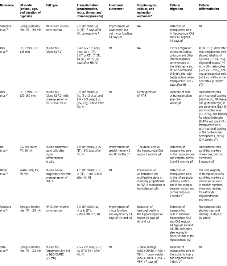
Table 1 - Review of publications on stem cell therapy in experimental hypoxia-ischemia in the neonatal period
BDNF = brain-derived neurotrophic factor; BHK-GDNF = baby hamster kidney cells (BHK) transfected with glial cell line-derived neurotrophic factor (GDNF) complementary DNA; MAPC = multipotent adult progenitor cells; ChABC = chondroitinase ABC; HUCBSC = human umbilical cord blood stem cells; MSC = mesenchymal stem cells; NSC = neural stem cells; CXCR4 = CXC chemokine receptor 4; FGF-2 = fibroblast growth factor 2; GDNF = glial cell line-derived neurotrophic factor; HI = hypoxia-ischemia; ic = intracerebral route; ica = intracardiac route; icv = intracerebroventricular route; ip = intraperitoneal route; iv = intravenous route; NA = not analyzed; NGF = nerve growth factor; NT3 = neurotrophin 3; P = postnatal day; PGA = polyglycolic acid; pT = post-transplantation; SDF-1 = stromal cell-derived factor 1; T-PX = transplantation on postnatal day X; NI = no immunosuppression; CT = contralateral transplant; IT = ipsilateral transplant; VEH = vehicle.
* Treated animals vs. control animals. ↑= increase.
↓= reduction. Reference
Elsayed et al.15
Jansen et al.16
Park et al.27
Imitola et al.23
Katsuragi et al.32
Katsuragi et al.33
Zheng et al.21
Meier et al.36
HI model (animal, age, and duration of hypoxia)
Long-Evans black-hooded rats; P7-8; 60, 120 or 120-150 min
Wistar rats; P7; 150 min
CD-1 mice; P7; 120 min
C57Bl/6 mice; P7; 120 min
Wistar rats; P14; 120 min
Wistar rats; P9; 120 min
Sprague-Dawley rats; P7; 120 min
Wistar rats; P7; 80 min
Cell type
Murine fetal neocortical tissue (embryonic day 13)
Murine fetal neocortical tissue (embryonic day 16)
Murine NSC (clone C17.2) in PGA scaffold (NSC-PGA)
Five strains of human and murine NSC
Encapsulated BHK-GDNF cells
Encapsulated BHK-GDNF cells
Multipotent astrocytic stem cells from murine subventricular zone
HUCBSC
Transplantation (concentration, route, timing, and imunosuppression)
1-2 mm3 block transplant; ic (IT); 7 days after HI; NI
5 x 10⁴ cells/µL; ic (IT); 3 days after HI; NI
1 or 2 NSC-PGA complexes (1 x 10⁷ cells/mL; 100-200 µL); ic (IT); 7 days after HI; NI
5 x 10⁵ cells/mL; ic (CT); 3 days after HI; cyclosporine
1 capsule (1 x 10⁸ cells/mL); ic (IT); 2 days pre-HI; NI
1 capsule (1 x 10⁸ cells/mL); ic (IT); 2 days pre-HI; NI
5 x 10⁴ cells/µL; ic (IT); 24 h after HI; NI
1 x 10⁷ cells/500 µL; ip; 24 h after HI; NI
Functional outcomes*
NA
Improvement of motor function (3-8 weeks pT) and asymmetry (9 weeks pT)
↓ unilateral rotation NA NA Improvement of cognitive performance after 6 weeks pT
NA
Improvement of locomotor function 21 days pT
Morphological, cellular, and molecular outcomes*
No effect on brain atrophy. Presence of axonal connectivity between graft and adjacent cortical areas of host tissue
Absence of cortical cytoarchitecture recovery;
transplants stained for neurochemical markers and absence of glial labeling; presence of astrocytes surrounding the transplant (9-11 weeks pT)
Reduction of brain damage; formation of interconnections between NSC/PGA and host tissue; neovascularization; ↓ monocyte iniltration and astroglial scar; restoration of long-distance neuronal projections
NSC express CXCR4; ↑ expression of SDF-1α
in the infarcted brain region (astrocytic and endothelial cells); interaction of SDF-1α/CXCR4 pathway
↑ serum GDNF; ↓ incidence and severity of neuronal damage (7 days pT)
Reduction of brain injury 17 weeks pT
NA
No effect on brain atrophy within 21 days pT
Cellular Migration
Transplants with satisfactory (63%) and poor (19%) development, located in the cortex or adjacent areas 6 weeks pT
Identiication of 72% of transplants located in areas adjacent to the corpus callosum of the sensorimotor cortex 9-11 weeks pT
Transplant showed satisfactory uptake in the infarcted brain cavity 2-6 weeks pT
Positive correlation between the presence of transplanted cells in the ischemic region and SDF-1α expression
BHK-GDNF cells remained viable 7 days pT
NA
Detection of transplanted cells in the cortical and periventricular ischemic region within 21 days pT
Presence of transplanted cells surrounding brain injury 21 days pT
Cellular Differentiation
NA
NA
Transplanted cells exhibited markers of neurons (5%) and oligodendrocytes, in the cortical penumbra of injury, 2 weeks pT
Transplanted cells with neuronal labeling in the cortical penumbra of injury NA NA Transplanted cells exhibited markers of astrocytes and neurons 3-21 days pT
Table 1 - Review of publications on stem cell therapy in experimental hypoxia-ischemia in the neonatal period (continuation)
Reference
Yasuhara et al.58
Park et al.22
Park et al.29
Ma et al.24
Dayer et al.30
Yasuhara et al.57
Sato et al.28
HI model (animal, age, and duration of hypoxia)
Sprague-Dawley rats; P7; 150 min
CD-1 mice; P7; 138 min
CD-1 mice; P7; 120-180 min
C57Bl/6 mice; P7; 90 min
Wistar rats; P7; 30 min
Sprague-Dawley rats; P7; 150 min
Sprague-Dawley rats; P7; 120 min
Cell type
MAPC from murine bone marrow
Murine NSC (clone C17.2)
Murine NSC (clone C17.2) with overexpression of NT-3 (NSC-NT3)
Murine embryonic stem cells after neural differentiation in vitro
Murine neural progenitor cells with overexpression of FGF-2
MAPC from murine bone marrow
Murine NSC (embryonic day 14) or NSC-ChABC (25 mU)
Transplantation (concentration, route, timing, and imunosuppression)
2 x 10⁵ cells/3 µL; ic (IT); 7 days after HI; cyclosporine A
0.4-1.6 x 10⁵ cells/ 4 µL; ic; 1 (IT), 3 (IT or CT), 7 (IT), 14 (IT), or 35 (IT) days after HI; NI
3 x 10⁵ cells/8 µL (ic, IT at 2 sites) and 1.0 x 10⁵ cells/2 µL (icv, CT); 3 days after HI; NI
1 x 10⁴ cells/µL; icv (IT); 2-3 days after HI; NI
4 x 10⁴ cells/0.5 µL; ic (IT); 1 and 3 days after HI; NI
2 x 10⁵ cells/3 µL; iv or ic (IT); 7 days after HI; NI
2.5 x 10⁵ cells/5 µL; icv (IT); 24 h after HI; NI
Functional outcomes*
Improvement of asymmetry, but not motor function, 14 days pT
NA
NA
Improvement of spatial memory 2 and 8 months pT
NA
Improvement of motor function and asymmetry 14 days pT (iv and ic)
NA Morphological, cellular, and molecular outcomes* NA NA Overexpression of NT-3
↑ neuronal cells in the hippocampal CA1 region 8 months pT
Preservation of an immature and proliferative state is inversely proportional to FGF-2 expression in transplanted cells
Reduction of neuronal death in the hippocampal CA3 region 14 days pT (iv and ic)
↓ brain damage (NSC+ChABC < NSC = VEH); ↑ brain weight (NSC+ChABC > NSC = VEH) (7 days pT)
Cellular Migration
Detection of transplanted cells in hippocampal CA2 and CA3 regions 14 days pT
CT: cell migration across the corpus callosum and other interhemispheric commissures to the infarcted area; IT: cells remained at injury site, with better uptake when transplanted 3 to 7 days after HI
Presence of cells at transplantation sites 2-4 weeks pT
Detection of transplanted cells in the hippocampus and cerebral cortex 2 and 8 months pT
Detection of transplanted cells in the infragranular ischemic cortex and in the margin between cortex and corpus callosum 2 weeks pT
Detection of transplanted cells in ischemic hippocampal CA2 and CA3 regions 14 days pT (iv and ic). The cells were also located in blood vessels in the hippocampus (iv)
Presence of transplanted cells in the ischemic injury and adjacent areas 7 days pT
Cellular Differentiation
NA
IT vs. CT (3 days after HI): transplanted cells showed labeling of neurons (~5 vs. 0%), oligodendrocytes (~4 vs. ~1%), astrocytes (~23 vs. ~15%), and neural progenitor cells (~18 vs. ~6%) in the neocortex (1 month pT)
Transplanted cells with neuronal labeling (cholinergic, GABAergic and glutamatergic) in the penumbra (81.4%) and infarcted area (10-20%), and labeling for oligodendrocyte (0.4%) and glia (1%); transplanted cells with neuronal labeling in the contralateral hemisphere (~90%) (2-4 weeks pT)
Transplanted cells exhibited markers of neurons, but not astrocytes, 8 months pT
The vast majority of transplanted cells exhibited markers of immature neurons; in smaller numbers, there was labeling for astrocyte, oligodendrocyte and neuron
Transplanted cells showed neuronal labeling 14 days pT (iv and ic)
NA
BDNF = brain-derived neurotrophic factor; BHK-GDNF = baby hamster kidney cells (BHK) transfected with glial cell line-derived neurotrophic factor (GDNF) complementary DNA; MAPC = multipotent adult progenitor cells; ChABC = chondroitinase ABC; HUCBSC = human umbilical cord blood stem cells; MSC = mesenchymal stem cells; NSC = neural stem cells; CXCR4 = CXC chemokine receptor 4; FGF-2 = fibroblast growth factor 2; GDNF = glial cell line-derived neurotrophic factor; HI = hypoxia-ischemia; ic = intracerebral route; ica = intracardiac route; icv = intracerebroventricular route; ip = intraperitoneal route; iv = intravenous route; NA = not analyzed; NGF = nerve growth factor; NT3 = neurotrophin 3; P = postnatal day; PGA = polyglycolic acid; pT = post-transplantation; SDF-1 = stromal cell-derived factor 1; T-PX = transplantation on postnatal day X; NI = no immunosuppression; CT = contralateral transplant; IT = ipsilateral transplant; VEH = vehicle.
* Treated animals vs. control animals. ↑= increase.
Table 1 - Review of publications on stem cell therapy in experimental hypoxia-ischemia in the neonatal period (continuation)
Reference
de Paula et al.38
Yasuhara et al.40
Pimentel-Coelho et al.37
Jenny et al.31
van Velthoven et al.56
Lee et al.55
Xia et al.41
HI model (animal, age, and duration of hypoxia)
Wistar rats; P7; 120 min
Sprague-Dawley rats; P7; 150 min
Lister-Hooded rats; P7; 90 min
Wistar rats; P7; 30 min
C57Bl/6 mice; P9; 45 min
Sprague-Dawley rats; P7; 210 min
Sprague-Dawley rats; P7; 150 min
Cell type
HUCBSC
HUCBSC or HUCBSC+M (mannitol 1.1 mol/L)
HUCBSC
Murine neural progenitor cells with overexpression of FGF-2
MSC from murine bone marrow
MSC from human bone marrow MSC from human umbilical cord blood Transplantation (concentration, route, timing, and imunosuppression)
1 x 10⁷ cells/100 µL; iv; 24 h after HI; NI
1.5 x 10⁴ cells/ 200 µL; iv; 7 days after HI; NI
2 x 10⁶ cells/200 µL; ip; 3 h after HI; NI
2-5 x 10⁴ cells/1 µL; ic (IT); 4 days after HI; NI
1 x 10⁵ cells/2 µL; ic (IT); 3 days (T-P12) or 10 days (T-P19) after HI; NI
1 x 10⁶ cells/µL; ica; 3 days after HI; NI
5 x 10⁴ cells/µL; ic (IT); 3 days after HI; cyclosporine
Functional outcomes*
No effect on spatial memory deicit 3 weeks pT
Improvement of motor function and asymmetry 7 and 14 days pT (HUCBSC < HUCBSC+M) Improvement of sensorimotor relexes 4 days pT
NA
Improvement of motor asymmetry 7 and 18 days pT (T-P12) or 11 and 18 days pT (T-P19)
Despite no effect on motor function deicit, there was ↓ motor asymmetry 20 days pT
Improvement of neurological function 14, 21 and 28 days pT
Morphological, cellular, and molecular outcomes*
No effect on brain atrophy 3 weeks pT
↑ brain levels of GDNF, BDNF and NGF 3 days pT (HUCBSC < HUCBSC+M); ↑ dendritic density in the hippocampal CA1 region 14 days pT
↓ neuronal death and caspase 3 expression in the ischemic striatum 2 days pT; ↓ microglial activation in the cortex 7 days pT
Formation of cell clusters with overexpression of FGF-2 in perivascular areas
T-P12: ↓ brain injury (18 days pT); ↑ neuro- and oligodendrogenesis, and ↓ microglial proliferation in the hippocampus and ischemic cortex (7 and 18 days pT); ↑ astrocyte proliferation in the hippocampus, and ↓ in the ischemic cortex (7 days pT). T-P19: ↓ brain injury (18 days pT)
No effect on brain atrophy 6 weeks pT
↓ brain injury 28 days pT
Cellular Migration
Detection of few transplanted cells 24 h, 1 and 3 weeks pT
Presence of few transplanted cells in the ischemic hippocampus 14 days pT
Presence of few transplanted cells in the ischemic cortex and striatum 2 days pT
Presence of transplanted cells in areas of cortical injury close to blood vessels 7 days pT
Less than 1% of proliferating cells in the ischemic hemisphere derived from transplanted cells Detection of transplanted cells equally distributed in both hemispheres 6 weeks pT
Presence of transplanted cells in the cortex and dispersion into the hippocampus 7 days pT
Cellular Differentiation NA NA NA Transplanted cells showed labeling of immature neurons 7 days pT
NA
Transplanted cells showed labeling of astrocyte and microglia (> number), neuron and oligodendrocyte (< number), equally distributed in both hemispheres 6 weeks pT
Transplanted cells exhibited markers of astrocytes, but not neurons, 7 days pT
BDNF = brain-derived neurotrophic factor; BHK-GDNF = baby hamster kidney cells (BHK) transfected with glial cell line-derived neurotrophic factor (GDNF) complementary DNA; MAPC = multipotent adult progenitor cells; ChABC = chondroitinase ABC; HUCBSC = human umbilical cord blood stem cells; MSC = mesenchymal stem cells; NSC = neural stem cells; CXCR4 = CXC chemokine receptor 4; FGF-2 = fibroblast growth factor 2; GDNF = glial cell line-derived neurotrophic factor; HI = hypoxia-ischemia; ic = intracerebral route; ica = intracardiac route; icv = intracerebroventricular route; ip = intraperitoneal route; iv = intravenous route; NA = not analyzed; NGF = nerve growth factor; NT3 = neurotrophin 3; P = postnatal day; PGA = polyglycolic acid; pT = post-transplantation; SDF-1 = stromal cell-derived factor 1; T-PX = transplantation on postnatal day X; NI = no immunosuppression; CT = contralateral transplant; IT = ipsilateral transplant; VEH = vehicle.
* Treated animals vs. control animals. ↑= increase.
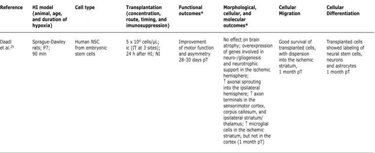
Table 1 - Review of publications on stem cell therapy in experimental hypoxia-ischemia in the neonatal period (continuation)
Reference
Daadi et al.25
HI model (animal, age, and duration of hypoxia)
Sprague-Dawley rats; P7; 90 min
Cell type
Human NSC from embryonic stem cells
Transplantation (concentration, route, timing, and imunosuppression)
5 x 10⁴ cells/µL; ic (IT at 3 sites); 24 h after HI; NI
Functional outcomes*
Improvement of motor function and asymmetry 28-30 days pT
Morphological, cellular, and molecular outcomes*
No effect on brain atrophy; overexpression of genes involved in neuro-/gliogenesis and neurotrophic support in the ischemic hemisphere; ↑ axonal sprouting into the ipsilateral hemisphere; ↑ axon terminals in the sensorimotor cortex, corpus callosum, and ipsilateral striatum/ thalamus; ↑ microglial cells in the ischemic striatum, but not in the cortex (1 month pT)
Cellular Migration
Good survival of transplanted cells, with dispersion into the ischemic striatum, 1 month pT
Cellular Differentiation
Transplanted cells showed labeling of neural stem cells, neurons and astrocytes 1 month pT
In an elegant study, Park et al.22 presented interesting observations on the behavior of NSC when transplanted into the brain of neonatal mice subjected to HI injury. When transplanted into the contralateral hemisphere of the HI brain injury, NSC migrated across the corpus callosum and other interhemispheric commissures to the infarcted area. However, when transplanted into the ipsilateral hemisphere, the cells remained at the site of injury. In both cases, NSC differentiated in the ischemic site, exhibiting labeling for neurons, oligodendrocytes, astrocytes, and neural progenitor cells. In addition, that same research group elucidated the inlammatory mechanisms involved in the migration of NSC in response to HI injury.23
Embryonic stem cells from the inner cell mass of blastocyst-stage embryos also have the ability to differentiate into neurons by speciic culture protocols and may be considered as alternative cell sources for treatment of brain injury. Two studies used embryonic stem cells differentiated into neurons in HI injury models. In the irst study, transplantation of embryonic stem cells differentiated into neurons in vitro resulted in improved spatial memory in HI rats.24 However, although the number of neuronal cells increased in the ischemic
hippocampus, there is no clear correlation between the outcomes and neuronal repopulation, differentiation or release of trophic factors by the delivered cells. In another recently published study, restoration of motor function and symmetry was observed in HI rats after transplantation of human embryonic stem cells differentiated into neurons. However, the authors found no restorative effects on the structural brain damage.25
The remaining ischemic environment can inluence tissue repair due to the severity of the HI process. HI injuries may associate with massive tissue necrosis, resulting in a porencephalic cyst. Such situation provides an impaired microenvironment for homing of transplanted cells due to lack of blood supply and adequate extracellular matrix. Through studies in the ield of tissue engineering, a new treatment approach has been proposed based on the use of biodegradable scaffolds within the infarcted cavity in order to foster the formation of connections between graft and host tissue.9,26 Using this therapeutic approach, Park et al.27 demonstrated successful implantation of scaffolds associated with reduction in infarction cavities and motor deicit in rats subjected to HI brain injury. Surprisingly, the authors observed the formation of interconnections BDNF = brain-derived neurotrophic factor; BHK-GDNF = baby hamster kidney cells (BHK) transfected with glial cell line-derived neurotrophic factor (GDNF) complementary DNA; MAPC = multipotent adult progenitor cells; ChABC = chondroitinase ABC; HUCBSC = human umbilical cord blood stem cells; MSC = mesenchymal stem cells; NSC = neural stem cells; CXCR4 = CXC chemokine receptor 4; FGF-2 = fibroblast growth factor 2; GDNF = glial cell line-derived neurotrophic factor; HI = hypoxia-ischemia; ic = intracerebral route; ica = intracardiac route; icv = intracerebroventricular route; ip = intraperitoneal route; iv = intravenous route; NA = not analyzed; NGF = nerve growth factor; NT3 = neurotrophin 3; P = postnatal day; PGA = polyglycolic acid; pT = post-transplantation; SDF-1 = stromal cell-derived factor 1; T-PX = transplantation on postnatal day X; NI = no immunosuppression; CT = contralateral transplant; IT = ipsilateral transplant; VEH = vehicle.
* Treated animals vs. control animals. ↑= increase.
between graft and host tissue, in addition to restoration of long-distance neuronal projections previously damaged by the HI process.
Other compounds may also be associated with cell therapy in order to provide support to NSC development in HI injury models. An example is the use of chondroitinase ABC (ChABC) combined with intracerebroventricular NSC transplantation in a neonatal HI rat model, which resulted in a signiicant reduction of brain damage.28 Due to the ability to promote brain plasticity, the authors suggest that ChABC would have facilitated NSC adhesion and migration to ischemic areas.
Considering NSC as possible cellular vehicles of trophic factors, some studies have used genetically engineered stem cells to express increased levels of these substances. Using NSC overexpressing neurotrophin-3 (NSC/NT-3) in the HI rat brain, the researchers observed an increase in neuronal differentiation of 5 to 10-20% in the infarcted area and > 80% in the penumbra, when compared to non-genetically engineered NSC lines. The authors suggest that the production of NT3 by NSC may have functioned in an autocrine/paracrine fashion by binding to adjacent cells of the host tissue and stimulating endogenous neurogenesis.22,29 Other studies have also shown that NSC overexpressing speciic trophic factors migrate to the injured brain site and generate niches of immature and proliferative cells available for brain repair.30 Additionally, the perivascular environment has been shown to be critical to maintaining these cells in an active immature and proliferative state.31 Studies have also demonstrated that the use of cellular vehicles to release certain trophic factors can reduce the incidence and extent of brain damage and cognitive impairment in HI experimental studies.32,33
Umbilical cord stem cell transplantation
Human umbilical cord blood stem cells (HUCBSC) have been used in experimental studies of HI brain injury due to their ability to differentiate into neurons and glial cells in vitro and in vivo. However, there are only few studies using non-human umbilical cord blood cells in animal models of neurological diseases, with conlicting results concerning the concomitant use of immunosuppressants.34,35
The irst study to test the effect of HUCBSC transplantation in HI rats was performed by Meier et al.36 The authors demonstrated that intraperitoneal injection of HUCBSC 24 h after HI injury in neonatal rats resulted in improved walking pattern in the animals. The authors also found that the stem cells migrated to the injured brain region, but no reduction in brain atrophy or transdifferentiation of transplanted cells was observed. Using the same route of delivery, a group of researchers demonstrated that intraperitoneal transplantation of HUCBSC 3 h after HI injury can improve primitive relexes in these animals.37 In addition, the
authors demonstrated reduced cell death in the striatum and an anti-inlammatory effect in the cortex. However, the presence of few transplanted cells could be observed in the ischemic striatum and cortex of treated animals.
That same year, our research group published a study on the effects of intravenous delivery of HUCBSC in an experimental model of HI brain injury.38 However, our results demonstrated only a trend toward reduction of behavioral and morphological deicits in treated rats 30 days after transplantation. Moreover, few transplanted cells were identiied in the brain of treated animals. Aspects such as dosage, length of follow-up for evaluation and route of delivery may have inluenced the outcomes of the study.
In parallel, we also developed an experimental model of hypoxia-ischemia in newborn piglets for the study of cell therapy in medium-sized animals.39 Newborn pigs that received HUCBSC via the common carotid artery showed better neurological scores compared to animals subjected only to the HI injury model or the group transplanted via the umbilical artery. Additionally, the presence of few transplanted cells could be observed only in some animals in the group transplanted via the carotid artery.
An interesting study showed that intravenous injection of HUCBSC 7 days after HI brain injury, improved motor coordination and asymmetry in treated rats.40 Furthermore, increased levels of growth factors were detected in the ipsilateral hemisphere 3 days after transplantation. In that study, the authors also included a group of HI animals receiving the same dose of HUCBSC combined with mannitol, a substance that increases blood-brain barrier permeabilization. The results of this experimental group showed that mannitol enhanced further the functional effects and expression of trophic factors, compared to the group that received HUCBSC alone.
The potential therapeutic feasibility of mesenchymal stem cells derived from human umbilical cord blood has been recently analyzed in an experimental model of HI brain injury.41 Transplantation was performed 3 days after HI injury by intracerebral route. Treated animals showed a progressive improvement of neurological function, associated with reduction of brain tissue injury. In addition, the authors veriied the presence of transplanted cells in the cortex, with dispersion into the hippocampus, 7 days after transplantation, showing astrocytic, but not neuronal differentiation.
Mechanism of action Description
Cell transdifferentiation Direct transformation of stem cells (various sources) into mature neurons
Release of trophic factors Stem cells may act as cellular vehicles for the production and/or release of neurotrophic factors
Stimulation of endogenous neurogenesis Transplanted stem cells are believed to potentiate these intrinsic mechanisms of repair
Modulation of the inlammatory process Stem cells may inhibit the activation of various immune cells and thereby increase neurogenesis and production of trophic factors
Stimulation of angiogenesis Transdifferentiation of stem cells into new blood vessels (angiogenesis),
increased vascularization in the penumbra and indirect formation of new blood vessels by the release of growth factors
Induction of neuroplasticity Increase in afferent and efferent connections between the site of injury and brain regions, restoring local synaptic activity by synaptogenesis
Table 2 - Main mechanisms of action of stem cells in perinatal asphyxia Bone marrow stem cell transplantation
Studies of bone marrow stem cells have also been widely used in animal models of cerebral ischemia, such as adult rats,42-45 and, more recently in clinical trials, showing promising results.46,47
Bone marrow-derived blood contains, at least, two cell populations with great potential for clinical use: hematopoietic stem cells and mesenchymal stem cells (MSC).34 The inding that hematopoietic stem cells also have the ability to transdifferentiate into neuronal lineage has expanded the scope of use of this source of immature cells.48,49 Speciically, MSC derive from bone marrow stromal cells and are deined according to the presence of speciic cell-surface markers, in vitro behavior, and differentiation potential.50 This cell type has an increased capacity to express neuronal phenotypes in vitro51,52 and in vivo.53,54
Recently, two research groups have presented their results using bone marrow-derived MSC in an experimental model of HI brain injury. Lee et al.55 used the intracardiac route for transplantation 72 h after injury. The authors observed the presence of transplanted cells equally distributed in both hemispheres 6 weeks after transplantation. Such cells expressed more frequently markers of astrocyte and microglia, in addition to markers of neurons and oligodendrocytes, in smaller numbers. Furthermore, motor asymmetry improved in HI rats 40 days after transplantation, although repair of damaged ischemic tissue was not observed.
In another study, intracerebral injection of MSC 72 h after injury, increased proliferation and neuronal differentiation in the ischemic hemisphere.56 Histological and motor outcomes were also favorable 10 and 21 days after treatment. However, less than 1% of new neurons derived from the transplanted cells. The study suggests that MSC have favored endogenous formation of new neuronal cells via suppression of microglial inlammatory activity.
Using a puriied fraction of MSC, known as multipotent adult progenitor cells (MAPC), Yasuhara et al.57 demonstrated that intracerebral or intravenous delivery of MAPC derived from the mouse bone marrow, 7 days after HI injury, resulted in improved motor function in transplanted animals. Interestingly, recovery of motor deicit was similar between animals that received allogeneic transplants (genetically different animals, but of the same species) and those that received syngeneic transplants (genetically identical animals). Associated with decreased neuronal loss, the authors also detected the presence of MAPC in ischemic hippocampal regions and neuronal labeling 14 days after transplantation.57,58
Mechanisms of action involved in cell therapy in experimental hypoxia-ischemia
Understanding the mechanisms of action involved in the process of stem cell-induced neuroregeneration is essential to optimize the clinical beneits of cell therapy. Several factors (summarized in Table 2) may be responsible for the positive results shown in recent studies of experimental HI brain injury. However, despite the signiicant beneits demonstrated in these studies, the variables responsible for the successful transplantation of stem cells in HI brain injury have yet to be established.
Cell transdifferentiation
into cells derived from the ectodermal layer is still under debate. In in vitro studies, transdifferentiation is detected by the positivity of speciic markers of neurons and glial cells. However, the characterization of electrophysiological properties of these transdifferentiated cells is as yet poorly documented.59-61 Due to these indings, other mechanisms have been proposed.62
Release of trophic factors
Trophic factors are a family of polypeptides essential for the survival and differentiation of normal developing neurons and also play an important role in neuroprotection of mature neurons under pathological conditions.63 Stem cells can serve as vehicles for speciic molecules, acting as vectors for the production and/or release of neurotrophic factors.29,64 By interacting with their receptors, stem cells may release growth factors and cytokines, thus inhibiting apoptosis, increasing angiogenesis, and/or stimulating the differentiation of endogenous precursor cells.12,40Data from the study by Borlongan et al.65 reported a 15% increase in the production of trophic factors in the circulating blood of adult rats subjected to cerebral ischemia after HUCBSC transplantation. Recently, the same group has reported similar results after stem cell transplantation in a neonatal HI injury model in rats.40 Although endogenous release of trophic factors by neuronal tissue occurs in response to ischemic injury, this compensatory mechanism is insuficient to promote tissue regeneration and/or functional recovery. Therefore, the authors of these studies suggest that the increase of speciic trophic factors occurs probably due to the transplantation of stem cells.
Stimulation of endogenous neurogenesis
Several studies have demonstrated increased endogenous neurogenesis in response to neonatal HI injury.66,67 However, transplanted stem cells are believed to potentiate these intrinsic mechanisms of repair. In the study by van Velthoven et al.,56 transplantation of stem cells into HI animals was shown to reduce microglial proliferation and increase neurogenesis. The authors suggest that the transplanted cells had the ability to modulate post-HI injury inlammatory response, thus facilitating the process of endogenous neurogenesis.
Modulation of the inlammatory process
While beneicial to the recruitment and migration of stem cells to the site of injury, inlammatory process determined by brain damage appears to be restrictive for cell differentiation.23 In this context, recent studies have shown that different sources of stem cells can inhibit the activation of various immune cells. Pluchino et al.68 showed that NSC promoted neuroprotection through anti-inlammatory cytokines and immunomodulatory molecules.
In another study, intravenous injection of HUCBSC increased the survival of neuronal tissue and reduced leukocyte iniltration and expression of proinlammatory proteins in the ischemic brain.69
In the CNS, microglial cells are the irst line of immune defense and comprise a major inlammatory process that contributes to brain damage. This cell population releases a variety of cytokines and growth factors that react rapidly in the injured brain tissue to promote phagocytosis and present antigens to T cells. However, microglial activation is commonly associated with decreased production of trophic factors and reduced neurogenesis.9,63,70Two studies of cell therapy in experimental HI injury reported that transplantation of stem cells reduced microglial expression in the ischemic cortex and hippocampus and, consequently, improved functional outcomes in treated animals.37,56 In contrast, Daadi et al.25 found increased proliferation of microglial cells in the striatum of HI animals that received NSC transplants. These data support the idea that microglial cells play a pro- and anti-inlammatory role, depending on their activation state and functional phenotype.71
Stimulation of angiogenesis
Some studies of ischemic injury in adult rats have shown increased vascularization in the penumbra within a few days after transplantation of stem cells.72,73 Furthermore, transdifferentiation of transplanted stem cells into new blood vessels (angiogenesis) has been observed.34,74 It is also likely that indirect cellular events occur in blood vessel formation. Chen et al.75 showed that stem cells from the bone marrow stroma promoted angiogenesis in the ischemic region by increasing the endogenous levels of vascular endothelial growth factor. To date, there is only one study indicating neovascularization of brain tissue in HI animals that received NSC transplants.27 The authors suggest that post-transplantation angiogenic signals were generated, thus allowing the formation of a new and vascularized parenchyma at the site of the porencephalic cyst.
Induction of neuroplasticity
Aspect Advantages Disadvantages
Route of delivery
Intravenous Less invasive and safer Need for chemotactic signals
Cells may migrate to other organs
Need to assess adverse effects
Intra-arterial Increased targeting of stem cells Risk of microembolism and ischemia
to injured sites Routine technique
Intracranial Facilitates migration of stem cells More invasive, risk of injury
at the site of ischemic injury
Intraperitoneal Practical, simple and less Cells need to travel long distances
invasive technique Few studies
Cell source and type
Embryo or fetus Increased capability to differentiate Ethical and religious barriers
and proliferate Dificulty in obtaining samples
Impractical for use in the acute phase of injury
Umbilical cord blood Ease of obtaining samples Little characterization of transplanted cells
(mononuclear cells) Large amounts of cells
Possibility of autologous transplantation
Bone marrow (mononuclear cells) Large amounts of cells Little characterization of transplanted cells
Possibility of autologous transplantation Painful technique and impractical for newborns
Neural stem cells Cells of the same embryonic origin Restricted migration
Widely studied Cell death
Gold standard for treatment of Dificulty in obtaining samples and
brain injury immediate use
Post-transplantation timing
Acute Plays a neuroprotective role Limited to the use of little processed
Presence of chemotactic signals and cell types (culture)
vascular permeability Impractical for established cases
Consistent preclinical results
Chronic Possibility of use in patients with Absence of appropriate chemotactic signals
established sequelae Absence of vascular permeability
Few studies
Table 3 - Relevant aspects to the clinical use of cell therapy in perinatal asphyxia Translational aspects of cell therapy in perinatal
asphyxia
Translational research can be deined as a process which leads from evidence-based medicine to sustainable solutions for public health problems.78Preclinical studies are the irst step in developing new therapies. However, the transfer of knowledge acquired through studies in vitro and in animal models of HI brain injury to the clinical use of cell therapy requires attention to differences between species and critical analysis of experimental results. Moreover, aspects still poorly understood (Table 3), such as the route of delivery, the number of transplanted cells, the cell type used, and the timing of intervention after injury, are important variables that might signiicantly inluence clinical response. Therefore,
after clarifying key issues concerning safety of treatment, it is possible that phase I clinical studies may be initiated to evaluate this novel therapeutic approach.
By intracerebral transplantation, it is possible to reach directly a speciic brain area. Despite the precision of the technique, this is an invasive procedure and several applications are needed to cover an ischemic injury, resulting in further brain impairment.79 Intracerebroventricular injections represent another intracranial approach to deliver stem cells. However, despite allowing a diffuse cellular distribution, this route is not capable of reaching lesions distant from the ventricles and faces the same issues that are common to the intracerebral transplantation. In addition, transplanted cells may adhere to the ventricular wall and cause obstructive hydrocephalus.79,80 Although most experimental studies of cell therapy in neonatal HI brain injury use intracerebral transplantation, other less invasive delivery routes are more likely to be established for clinical practice. In the experimental context, intravenous and intracerebral transplantation of stem cells showed similar extent of motor and morphological recovery in an animal model of neonatal HI brain injury.57
A recent meta-analysis demonstrated that intravenous cell delivery can improve outcomes in animal models of neurological disorders, and apoptosis inhibition is the main molecular change in the brain after transplantation.81 Intravenous cell delivery is a less invasive, simple and safe procedure, allowing widespread distribution of transplanted cells to HI brain areas.38,40,57 In addition, this procedure enables exposure of cells to chemotactic signals originating from the brain injury, which can selectively accumulate cells within the target tissue.79,82 However, only a small amount of stem cells may be able to reach the site of brain injury, since they can be trapped in the lungs, kidneys, liver, and spleen. Therefore, ectopic growth and toxicity in other organs need to be assessed before clinical use.80
An alternative approach to avoid the systemic circulation system has been to deliver stem cells intra-arterially. Recent data show equally distributed migration and favorable neurological outcomes using this therapeutic approach in a rat model of HI brain injury.55 This route bypasses the uptake by the systemic organs, allowing a large number of stem cells to reach the ischemic site, once vessels are reperfused. In the case of a permanent vascular occlusion, the transplanted cells will be distributed only in the penumbra region.79 However, recent studies have reported high mortality rates with intra-arterial delivery in HI transplanted animals, suggesting the formation of microvascular occlusion and ischemia.79,80,83 In medical practice, this delivery method is commonly used in interventional vascular catheterization techniques. In a Brazilian clinical trial of cell therapy in patients with chronic ischemic stroke (NCT00473057), promising results were observed after intra-arterial transplantation of autologous bone marrow stem cells.46
The intraperitoneal route has also been used for cell transplantation in neonatal HI injury, demonstrating cell migration from the peritoneal cavity into the injured brain
regions.36,37 The study indings suggest the presence of precise chemotactic signals and the need to break the blood-brain barrier so that cell migration can occur over such long distances. Despite being a practical procedure, further studies are needed to support this alternative route of transplantation.
The choice of time interval between the establishment of HI injury and intervention is also important, since the brain environment changes dramatically over time after HI injury. As a result, most studies using systemic administration of stem cells in HI injury are conducted in the acute stages of injury. Early injection of stem cells may exert an effect on the preservation and survival of neuronal tissue. Additionally, van Velthoven et al.56 suggest that cell transplantation be performed between 2 and 3 days after HI injury, at which time the brain has greater capacity for cell proliferation and activation of endogenous repair mechanisms. In the study by Park et al.,22 the therapeutic window for effective cell transplantation covered 3-7 days following HI injury, due to high metabolic, biochemical and molecular activity in this period which could facilitate the migration of transplanted cells. In the case of transplantation at later time intervals, stem cells might stimulate the release of trophic factors to restore lost function.80 However, despite the functional improvement observed in adult rats that received stem cells one month after cerebral ischemia,84 there are no studies demonstrating therapeutic beneits in animal models of chronic HI brain injury, thus limiting clinical applicability to HI injury established due to the absence of appropriate chemotactic signals and vascular permeability.
Blood-brain barrier permeabilization may also facilitate CNS entry of stem cells or neurotrophic factors, especially when peripheral routes are used for transplantation. However, it has been recently shown that the number of intravenously delivered stem cells in the ischemic hippocampus of HI transplanted animals did not differ signiicantly when comparing animals that received stem cells alone to those that received a combination of mannitol and stem cells.40 In contrast, transplanted animals that were administered mannitol showed increased brain levels of some trophic factors. There was also a marked improvement in behavioral and histological outcomes with the combined use of stem cells and mannitol, possibly through a paracrine effect of these cells. This evidence points out the importance of blood-brain barrier permeabilization in cell therapy, but also questions whether neuroprotection is really dependent on cell migration and differentiation.
References
1. Souza FM. Fatores associados à asixia perinatal no Brasil: estudo populacional com base no Sistema de Informações de Nascidos Vivos. [Tese de Doutorado em saúde da criança e da mulher]. Rio de Janeiro: Fundação Oswaldo Cruz; 2003.
2. Vannucci SJ, Hagberg H. Hypoxia-ischemia in the immature brain.
J Exp Biol. 2004;207:3149-54.
3. Perlman JM. Summary proceedings from the neurology group on hypoxic-ischemic encephalopathy. Pediatrics. 2006;117: S28-33.
4. Procianoy RS, Silveira RC. Síndrome hipóxico-isquêmica. J Pediatr (Rio J). 2001;77 Suppl 1:S63-70.
5. Vannucci RC. Hypoxic-ischemic encephalopathy. Am J Perinatol. 2000;17:113-20.
6. Sahni R, Sanocka UM. Hypothermia for hypoxic-ischemic encephalopathy. Clin Perinatol. 2008;35:717-34, vi.
7. Li L, Xie T. Stem cell niche: structure and function.Annu Rev Cell Dev Biol. 2005;21:605-31.
8. da Silva Meirelles L, Chagastelles PC, Nardi NB. Mesenchymal stem cells reside in virtually all post-natal organs and tissues. J Cell Sci. 2006;119:2204-13.
9. Burns TC, Verfaillie CM, Low WC. Stem cells for ischemic brain injury: a critical review. J Comp Neurol. 2009;515:125-44. 10. Costa-Ferro ZS, Vitola AS, Pedroso MF, Cunha FB, Xavier LL,
Machado DC, et al. Prevention of seizures and reorganization of hippocampal functions by transplantation of bone marrow cells in the acute phase of experimental epilepsy. Seizure. 2010;19:84-92.
11. Dunnett SB, Rosser AE. Cell therapy in Huntington’s disease. NeuroRx. 2004;1:394-405.
12. Haas S, Weidner N, Winkler J. Adult stem cell therapy in stroke.
Curr Opin Neurol. 2005;18:59-64.
13. Kim JH, Auerbach JM, Rodriguez-Gomez JA, Velasco I, Gavin D, Lumelsky N, et al. Dopamine neurons derived from embryonic stem cells function in an animal model of Parkinson’s disease.
Nature. 2002;418:50-6.
14. Koda M, Okada S, Nakayama T, Koshizuka S, Kamada T, Nishio Y, et al. Hematopoietic stem cell and marrow stromal cell for spinal cord injury in mice.Neuroreport. 2005;16:1763-7.
15. Elsayed MH, Hogan TP, Shaw PL, Castro AJ. Use of fetal cortical grafts in hypoxic-ischemic brain injury in neonatal rats. Exp Neurol. 1996;137:127-41.
16. Jansen EM, Solberg L, Underhill S, Wilson S, Cozzari C, Hartman BK, et al. Transplantation of fetal neocortex ameliorates sensorimotor and locomotor deicits following neonatal ischemic-hypoxic brain injury in rats. Exp Neurol. 1997;147:487-97.
use of this cell type in neonatal brain injury is the dificulty in obtaining samples. Biopsy tissue or postmortem tissue specimens may be insuficient to produce signiicant amounts of these cells.85 Ethical and religious issues involved in the harvesting of embryonic and fetal tissue and concerns about the risks of immune response or formation of malignant tumors also inhibit the clinical applicability of these cells. In vitro neuronal differentiation for future use of these cells in newborns with HI injury raises further concerns about potential risks of contamination, immune reaction to the factors added to the cell culture medium, and dificulty of use in acute transplantation. Similar disadvantages related to safety and dificulties of immediate use are also presented by MSC from bone marrow and umbilical cord blood, widely used in experimental HI brain injury.
In contrast, the processing of mononuclear stem cells derived from umbilical cord blood and bone marrow is relatively simple and rapid. In the neonatal context, mononuclear stem cells derived from umbilical cord blood offer some advantages over bone marrow aspiration. Obtaining stem cells from umbilical cord blood offers no risk or discomfort to the newborn, and cells can be transplanted after autologous collection. In addition, umbilical cord blood can be used therapeutically in the perinatal period or cryopreserved for later use.62,86 However, further studies are needed to characterize cell populations and evaluate effectiveness and safety.80,87
Dose-response analysis of stem cells in animal models also needs to be performed to provide a basis for their clinical use. According to Janowski et al.,81 there is a dose-response association between the number of cells injected and the effects of the treatment of neurological diseases in experimental studies. However, the optimal dose of stem cells is partly dependent on the delivery route as well as on the cell type used, the timing of intervention, and the amount of transplanted cells that reach the brain injury. From a clinical standpoint, outcomes related to safety of cell transplantation, such as ectopic tissue formation and behavioral abnormalities, should be incorporated in the methodology of investigations and patient follow-up. It is also important to consider safe and effective methods for the noninvasive monitoring of migration of transplanted cells, thus establishing a proper understanding of patients’ responses to the proposed treatment.
Conclusions
Basic research has shown promising results in the ield of neonatal brain injury. Despite the variety of methods employed, most studies cited in this review indicate that stem cells may have neuroprotective properties, resulting in improved functional outcomes in treated animals. Certainly, the success of cell therapy in rodents can be regarded as the irst step in developing a therapeutic approach for
future clinical use. However, it is noteworthy that human brain diseases have more complex mechanisms of damage and regeneration than in experimental animals. Therefore, issues related to dose, cell type, route of delivery and proper timing of intervention need to be deined. Although not crucial to clinical application, further investigations on the mechanisms of action of stem cells in HI injury are also an important tool in establishing the safety and effectiveness of transplantation.
Acknowledgements
17. Hess DC, Borlongan CV. Stem cells and neurological diseases.
Cell Prolif. 2008;41 Suppl 1:94-114.
18. Rice CM, Scolding NJ. Adult stem cells-reprogramming neurological repair? Lancet. 2004;364:193-9.
19. Kornblum HI. Introduction to neural stem cells. Stroke. 2007;38:810-6.
20. Doetsch F, Caille I, Lim DA, Garcia-Verdugo JM, Alvarez-Buylla A.
Subventricular zone astrocytes are neural stem cells in the adult mammalian brain. Cell. 1999;97:703-16.
21. Zheng T, Rossignol C, Leibovici A, Anderson KJ, Steindler DA, Weiss MD. Transplantation of multipotent astrocytic stem cells into a rat model of neonatal hypoxic-ischemic encephalopathy.
Brain Res. 2006;1112:99-105.
22. Park KI, Hack MA, Ourednik J, Yandava B, Flax JD, Stieg PE, et al.
Acute injury directs the migration, proliferation, and differentiation of solid organ stem cells: evidence from the effect of hypoxia-ischemia in the CNS on clonal ”reporter” neural stem cells.Exp Neurol. 2006;199:156-78.
23. Imitola J, Raddassi K, Park KI, Mueller FJ, Nieto M, Teng YD, et al.
Directed migration of neural stem cells to sites of CNS injury by the stromal cell-derived factor 1alpha/CXC chemokine receptor 4 pathway. Proc Natl Acad Sci U S A. 2004;101:18117-22. 24. Ma J, Wang Y, Yang J, Yang M, Chang KA, Zhang L, et al. Treatment
of hypoxic-ischemic encephalopathy in mouse by transplantation of embryonic stem cell-derived cells. Neurochem Int. 2007; 51:57-65.
25. Daadi MM, Davis AS, Arac A, Li Z, Maag AL, Bhatnagar R, et al.
Human neural stem cell grafts modify microglial response and enhance axonal sprouting in neonatal hypoxic-ischemic brain injury.Stroke. 2010;41:516-23.
26. Orive G, Anitua E, Pedraz JL, Emerich DF. Biomaterials for promoting brain protection, repair and regeneration. Nat Rev Neurosci. 2009;10:682-92.
27. Park KI, Teng YD, Snyder EY. The injured brain interacts reciprocally with neural stem cells supported by scaffolds to reconstitute lost tissue. Nat Biotechnol. 2002;20:1111-7.
28. Sato Y, Nakanishi K, Hayakawa M, Kakizawa H, Saito A, Kuroda Y, et al. Reduction of brain injury in neonatal hypoxic-ischemic rats by intracerebroventricular injection of neural stem/ progenitor cells together with chondroitinase ABC. Reprod Sci. 2008;15:613-20.
29. Park KI, Himes BT, Stieg PE, Tessler A, Fischer I, Snyder EY. Neural stem cells may be uniquely suited for combined gene therapy and cell replacement: Evidence from engraftment of Neurotrophin-3-expressing stem cells in hypoxic-ischemic brain injury.Exp Neurol. 2006;199:179-90.
30. Dayer AG, Jenny B, Sauvain MO, Potter G, Salmon P, Zgraggen E, et al. Expression of FGF-2 in neural progenitor cells enhances their potential for cellular brain repair in the rodent cortex. Brain. 2007;130:2962-76.
31. Jenny B, Kanemitsu M, Tsupykov O, Potter G, Salmon P, Zgraggen E, et al. Fibroblast growth factor-2 overexpression in transplanted neural progenitors promotes perivascular cluster formation with a neurogenic potential. Stem Cells. 2009;27:1309-17.
32. Katsuragi S, Ikeda T, Date I, Shingo T, Yasuhara T, Ikenoue T.
Grafting of glial cell line-derived neurotrophic factor secreting cells for hypoxic-ischemic encephalopathy in neonatal rats. Am J Obstet Gynecol. 2005;192:1137-45.
33. Katsuragi S, Ikeda T, Date I, Shingo T, Yasuhara T, Mishima K, et al. Implantation of encapsulated glial cell line-derived neurotrophic factor-secreting cells prevents long-lasting learning impairment following neonatal hypoxic-ischemic brain insult in rats. Am J Obstet Gynecol. 2005;192:1028-37.
34. Bliss T, Guzman R, Daadi M, Steinberg GK. Cell transplantation therapy for stroke. Stroke. 2007;38:817-26.
35. Sanberg PR, Willing AE, Garbuzova-Davis S, Saporta S, Liu G, Sanberg CD, et al. Umbilical cord blood-derived stem cells and brain repair. Ann N Y Acad Sci. 2005;1049:67-83.
36. Meier C, Middelanis J, Wasielewski B, Neuhoff S, Roth-Haerer A, Gantert M, et al. Spastic paresis after perinatal brain damage in rats is reduced by human cord blood mononuclear cells. Pediatr Res. 2006;59:244-9.
37. Pimentel-Coelho PM, Magalhães ES, Lopes LM, deAzevedo LC, Santiago MF, Mendez-Otero R. Human cord blood transplantation in a neonatal rat model of hypoxic-ischemic brain damage: functional outcome related to neuroprotection in the striatum. Stem Cells Dev. 2010;19:351-8.
38. de Paula S, Vitola AS, Greggio S, de Paula D, Mello PB, Lubianca JM, et al. Hemispheric brain injury and behavioral deicits induced by severe neonatal hypoxia-ischemia in rats are not attenuated by intravenous administration of human umbilical cord blood cells.
Pediatr Res. 2009;65:631-5.
39. de Paula D. Células-tronco de cordão umbilical em modelo experimental de asixia neonatal em suínos [Tese]. Porto Alegre: Pontifícia Universidade Católica do Rio Grande do Sul; 2010. 40. Yasuhara T, Hara K, Maki M, Xu L, Yu G, Ali MM, et al. Mannitol
facilitates neurotrophic factor upregulation and behavioral recovery in neonatal hypoxic-ischemic rats with human umbilical cord blood grafts. J Cell Mol Med. 2009 Feb 4.
41. Xia G, Hong X, Chen X, Lan F, Zhang G, Liao L. Intracerebral transplantation of mesenchymal stem cells derived from human umbilical cord blood alleviates hypoxic ischemic brain injury in rat neonates. J Perinat Med. 2010;38:215-21.
42. Borlongan CV, Evans A, Yu G, Hess DC. Limitations of intravenous human bone marrow CD133+ cell grafts in stroke rats. Brain Res. 2005;1048:116-22.
43. Brenneman M, Sharma S, Harting M, Strong R, Cox CS Jr, Aronowski J, et al. Autologous bone marrow mononuclear cells enhance recovery after acute ischemic stroke in young and middle-aged rats. J Cereb Blood Flow Metab. 2010;30:140-9.
44. Liu Z, Li Y, Zhang ZG, Cui X, Cui Y, Lu M, et al. Bone marrow stromal cells enhance inter- and intracortical axonal connections after ischemic stroke in adult rats. J Cereb Blood Flow Metab.
2010;30:1288-95.
45. Chen JR, Cheng GY, Sheu CC, Tseng GF, Wang TJ, Huang YS.
Transplanted bone marrow stromal cells migrate, differentiate and improve motor function in rats with experimentally induced cerebral stroke. J Anat. 2008;213:249-58.
46. Barbosa da Fonseca LM, Gutilen B, Rosado de Castro PH, Battistella V, Goldenberg RC, Kasai-Brunswick T, et al. Migration and homing of bone-marrow mononuclear cells in chronic ischemic stroke after intra-arterial injection. Exp Neurol. 2010;221:122-8.
47. Correa PL, Mesquita CT, Felix RM, Azevedo JC, Barbirato GB, Falcao CH, et al. Assessment of intra-arterial injected autologous bone marrow mononuclear cell distribution by radioactive labeling in acute ischemic stroke. Clin Nucl Med. 2007;32:839-41.
48. Eglitis MA, Mezey E. Hematopoietic cells differentiate into both microglia and macroglia in the brains of adult mice. Proc Natl Acad Sci U S A. 1997;94:4080-5.
49. Mezey E, Chandross KJ, Harta G, Maki RA, McKercher SR. Turning blood into brain: cells bearing neuronal antigens generated in vivo from bone marrow. Science. 2000;290:1779-82.
50. Dominici M, Le Blanc K, Mueller I, Slaper-Cortenbach I, Marini F, Krause D, et al. Minimal criteria for deining multipotent mesenchymal stromal cells. The International Society for Cellular Therapy position statement. Cytotherapy. 2006;8:315-7. 51. Alexanian AR, Maiman DJ, Kurpad SN, Gennarelli TA. In vitro
and in vivo characterization of neurally modiied mesenchymal stem cells induced by epigenetic modiiers and neural stem cell environment. Stem Cells Dev. 2008;17:1123-30.
52. Barnabe GF, Schwindt TT, Calcagnotto ME, Motta FL, Martinez G Jr, de Oliveira AC, et al. Chemically-induced RAT mesenchymal stem cells adopt molecular properties of neuronal-like cells but do not have basic neuronal functional properties. PLoS One. 2009;4: e5222.
53. Mezey E, Key S, Vogelsang G, Szalayova I, Lange GD, Crain B.
54. Brazelton TR, Rossi FM, Keshet GI, Blau HM. From marrow to brain: expression of neuronal phenotypes in adult mice. Science. 2000;290:1775-9.
55. Lee JA, Kim BI, Jo HC, Choi CW, Kim EK, Kim HS, et al. Mesenchymal stem-cell transplantation for hypoxic-ischemic brain injury in neonatal rat model. Pediatr Res. 2010;67:42-6.
56. van Velthoven CT, Kavelaars A, van Bel F, Heijnen CJ. Mesenchymal stem cell treatment after neonatal hypoxic-ischemic brain injury improves behavioral outcome and induces neuronal and oligodendrocyte regeneration. Brain Behav Immun. 2010;24:387-93.
57. Yasuhara T, Hara K, Maki M, Mays RW, Deans RJ, Hess DC, et al. Intravenous grafts recapitulate the neurorestoration afforded by intracerebrally delivered multipotent adult progenitor cells in neonatal hypoxic-ischemic rats. J Cereb Blood Flow Metab. 2008;28:1804-10.
58. Yasuhara T, Matsukawa N, Yu G, Xu L, Mays RW, Kovach J, et al.
Behavioral and histological characterization of intrahippocampal grafts of human bone marrow-derived multipotent progenitor cells in neonatal rats with hypoxic-ischemic injury. Cell Transplant. 2006;15:231-8.
59. Fernandes KJ, Kobayashi NR, Gallagher CJ, Barnabe-Heider F, Aumont A, Kaplan DR, et al. Analysis of the neurogenic potential of multipotent skin-derived precursors. Exp Neurol. 2006;201:32-48.
60. Phinney DG, Isakova I. Plasticity and therapeutic potential of mesenchymal stem cells in the nervous system. Curr Pharm Des. 2005;11:1255-65.
61. Wislet-Gendebien S, Wautier F, Leprince P, Rogister B. Astrocytic and neuronal fate of mesenchymal stem cells expressing nestin.
Brain Res Bull. 2005;68:95-102.
62. Santner-Nanan B, Peek MJ, McCullagh P, Nanan R. Therapeutic potential of stem cells in perinatal medicine. Aust N Z J Obstet Gynaecol. 2005;45:102-7.
63. Boucherie C, Hermans E. Adult stem cell therapies for neurological disorders: beneits beyond neuronal replacement? J Neurosci Res. 2009;87:1509-21.
64. Muller FJ, Snyder EY, Loring JF. Gene therapy: can neural stem cells deliver? Nat Rev Neurosci. 2006;7:75-84.
65. Borlongan CV, Hadman M, Sanberg CD, Sanberg PR. Central nervous system entry of peripherally injected umbilical cord blood cells is not required for neuroprotection in stroke. Stroke. 2004;35:2385-9.
66. Ong J, Plane JM, Parent JM, Silverstein FS. Hypoxic-ischemic injury stimulates subventricular zone proliferation and neurogenesis in the neonatal rat.Pediatr Res. 2005;58:600-6.
67. Plane JM, Liu R, Wang TW, Silverstein FS, Parent JM. Neonatal hypoxic-ischemic injury increases forebrain subventricular zone neurogenesis in the mouse.Neurobiol Dis. 2004;16:585-95. 68. Pluchino S, Zanotti L, Rossi B, Brambilla E, Ottoboni L, Salani
G, et al. Neurosphere-derived multipotent precursors promote neuroprotection by an immunomodulatory mechanism. Nature. 2005;436:266-71.
69. Vendrame M, Gemma C, de Mesquita D, Collier L, Bickford PC, Sanberg CD, et al. Anti-inlammatory effects of human cord blood cells in a rat model of stroke. Stem Cells Dev.
2005;14:595-604.
70. Ekdahl CT, Claasen JH, Bonde S, Kokaia Z, Lindvall O. Inlammation is detrimental for neurogenesis in adult brain. Proc Natl Acad Sci U S A. 2003;100:13632-7.
71. Schwartz M. Macrophages and microglia in central nervous system injury: are they helpful or harmful? J Cereb Blood Flow Metab.
2003;23:385-94.
72. Senior K. Angiogenesis and functional recovery demonstrated after minor stroke. Lancet. 2001;358:817.
73. Zhang ZG, Zhang L, Jiang Q, Chopp M. Bone marrow-derived endothelial progenitor cells participate in cerebral neovascularization after focal cerebral ischemia in the adult mouse.
Circ Res. 2002;90:284-8.
74. Carroll JE, Borlongan CV. Adult stem cell therapy for acute brain injury in children. CNS Neurol Disord Drug Targets. 2008;7:361-9.
75. Chen J, Zhang ZG, Li Y, Wang L, Xu YX, Gautam SC, et al.
Intravenous administration of human bone marrow stromal cells induces angiogenesis in the ischemic boundary zone after stroke in rats.Circ Res. 2003;92:692-9.
76. Zhang C, Saatman KE, Royo NC, Soltesz KM, Millard M, Schouten JW, et al. Delayed transplantation of human neurons following brain injury in rats: a long-term graft survival and behavior study.
J Neurotrauma. 2005;22:1456-74.
77. Shen LH, Li Y, Chen J, Zhang J, Vanguri P, Borneman J, et al. Intracarotid transplantation of bone marrow stromal cells increases axon-myelin remodeling after stroke. Neuroscience. 2006;137:393-9.
78. Lean ME, Mann JI, Hoek JA, Elliot RM, Schoield G. Translational research. BMJ. 2008;337:a863.
79. Walczak P, Zhang J, Gilad AA, Kedziorek DA, Ruiz-Cabello J, Young RG, et al. Dual-modality monitoring of targeted intraarterial delivery of mesenchymal stem cells after transient ischemia.
Stroke. 2008;39:1569-74.
80. Stem Cell Therapies as an Emerging Paradigm in Stroke Participants.
Stem Cell Therapies as an Emerging Paradigm in Stroke (STEPS): bridging basic and clinical science for cellular and neurogenic factor therapy in treating stroke. Stroke. 2009;40:510-5. 81. Janowski M, Walczak P, Date I. Intravenous route of cell delivery for
treatment of neurological disorders: a meta-analysis of preclinical results. Stem Cells Dev. 2010;19:5-16.
82. Guzman R, Choi R, Gera A, De Los Angeles A, Andres RH, Steinberg GK. Intravascular cell replacement therapy for stroke. Neurosurg Focus. 2008;24:E15.
83. Li L, Jiang Q, Ding G, Zhang L, Zhang ZG, Li Q, et al. Effects of administration route on migration and distribution of neural progenitor cells transplanted into rats with focal cerebral ischemia, an MRI study. J Cereb Blood Flow Metab. 2010;30:653-62. 84. Shen LH, Li Y, Chen J, Zacharek A, Gao Q, Kapke A, et al. Therapeutic
beneit of bone marrow stromal cells administered 1 month after stroke. J Cereb Blood Flow Metab. 2007;27:6-13.
85. Crook JM, Kobayashi NR. Human stem cells for modeling neurological disorders: accelerating the drug discovery pipeline.
J Cell Biochem. 2008;105:1361-6.
86. Harris DT. Cord blood stem cells: a review of potential neurological applications. Stem Cell Rev. 2008;4:269-74.
87. Borlongan CV. Cell therapy for stroke: remaining issues to address before embarking on clinical trials.Stroke. 2009;40:S146-8.
Correspondence: Jaderson Costa DaCosta
Laboratório de Neurociências, InsCer, PUCRS Avenida Ipiranga, 6690, Prédio 60, 2° andar, sala 07 Jardim Botânico
CEP 90619-900 – Porto Alegre, RS – Brazil Tel.: +55 (51) 3320.3250