KARINE ROSÁRIO DA SILVA TEIXEIRA
Expression of matrix gla protein (MGP) in breast cancer:
a molecular and cellular approach
Dissertação para obtenção do Grau de
Mestre em Oncobiologia
mES
UNIVERSIDADE DO ALGARVE
Departamento de Ciências Biomédicas e Medicina
Trabalho efetuado sob a orientação científica da Doutora Natércia
Conceição e Doutora Sofia Braga
ii
UNIVERSIDADE DO ALGARVE
Departamento de Ciências Biomédicas e Medicina
KARINE ROSÁRIO DA SILVA TEIXEIRA
Expression of matrix gla protein (MGP) in breast cancer:
a molecular and cellular approach
Dissertação para obtenção do Grau de
Mestre em Oncobiologia
mES
Trabalho efetuado sob a orientação científica da Doutora Natércia
Conceição e Doutora Sofia Braga
iii Declaro ser o(a) autor(a) deste trabalho, que é original e inédito. Autores e trabalhos consultados estão devidamente citados no texto e constam da listagem de referências incluída.
Declaração Copyright
A Universidade do Algarve tem o direito, perpétuo e sem limites geográficos, de arquivar e publicitar este trabalho através de exemplares impressos reproduzidos em papel ou de forma digital, ou por qualquer outro meio conhecido ou que venha a ser inventado, de o divulgar através de repositórios científicos e de admitir a sua cópia e distribuição com objetivos educacionais ou de investigação, não comerciais, desde que seja dado crédito ao autor e editor.
Karine Rosário da Silva Teixeira
Expression of matrix gla protein (MGP) in breast cancer: a
molecular and cellular approach
iv
Abstract
Every year around 8.2 million people die due to cancer, in Portugal, breast cancer is the leading cause of women death (in cancer patients). Several risk factors could be pointed being age (over 50 years old), gender (females), reproductive and hormonal factors and genetic predisposition (presence of mutations in BRCA 1 and BRCA 2) the most commons. Staging is done regarding several aspects, e.g.: histology, histopathologic localization, hormone receptors, mutations or expression of cluster genes.
Research from last decades is focused into targeted therapies. MGP, matrix Gla protein, was pointed as a possible target. MGP primary structure consists in a signal peptide, a phosphorylation domain and a γ-carboxylase recognition site. MGP has also five Gla residues. For a long time, only an isoform with four exons was known, but recently was discovered a second isoform with five exons, with three additional putative binding sites, conferring, possible, a higher binding capability as well as a presumably higher calcification inhibitory power.
Although MGP role in cancer is not fully understood, being upregulated in some types of tumors and downregulated others, in breast cancer, was established, as a prognostic factor, being overexpressed in poor diagnose cases.
Being MGP role in tumorigenesis to be defined, the aims of this work were: clarify MGP expression in normal vs tumoral tissue (using in silica method – TCGA database); as well as clarify regulatory mechanisms of MGP (TCGA database); analyze expression of each isoform in breast cancer cell lines (results were obtained performing qRT-PCR followed by electrophoresis and sequencing of the extracted bands) and assess the result of overexpression of each isoform (using XTT and wound healing assay).
We could conclude that both variants could be expressed in simultaneous. Every cell line presented E4 isoform but just a few presented E5 isoform. Hound healing assay revealed that an overexpression of E4 and E5 variants slow down the rate of the area invaded by migrating cells, specially an overexpression of E4 isoform. After in silica analyze we could conclude that MGP is more expressed in tumoral tissue vs normal tissue, existing a negative correlation between MGP expression and mir155 expression and methylation, that means that: in tumoral tissue mir155 is less expressed than in normal tissue and occurs an hypomethylation in tumoral tissue. Knowing that MGP
v inhibits calcification and is correlated with angiogenesis, our hypothesis is that overexpression of MGP in tumoral tissue is a mechanism of cancer to facilitate angiogenesis – a hallmark of cancer.
Keywords: Breast Cancer; MGP, mir155; prognostic factor, angiogenesis,
vi
Resumo
Introdução: o cancro é caracterizado por um crescimento anormal e incontrolado de células que podem adquirir características malignas por acumulação de mutações. Hanahan and Weinberg (2000) descreveram que o desenvolvimento tumoral baseado em vários passos está assente na aquisição, por parte do tumor, de seis princípios biológicos: i) manter sinalização proliferativa; ii) desenvolver mecanismos de defesa contra supressores de crescimento; iii) resistir á morte celular; iv) ativar imortalidade replicativa, v) induzir angiogénese e vi) ativar invasão e metástase. Se uma visão mais tradicional atribuía estas alterações ao resultado de mutações genéticas, uma visão mais atual do cancro engloba também a epigenética como fator crucial na repressão e ativação de genes capazes de conferir as características malignas aos tumores; a metilação genómica é um exemplo de um mecanismo já descrito com um papel importante na atividade tumoral, sendo que está associado a hipometilação do genoma com o cancro.
Determinar os mecanismos moleculares desta doença aproximam a população científica de terapias alvo que permitam ajudar a controlar a disseminação desta doença que é considerada pela organização mundial de saúde uma doença epidémica em ascensão. Todos os anos morrem no mundo 8.2 milhões de pessoas devido a cancro (cerca de 13% de todas as mortes do mundo), sendo a segunda causa de morte em Portugal, EUA e Europa. Em Portugal, o cancro do pulmão e brônquios é a primeira causa de morte por cancro nos homens, enquanto que a primeira causa de morte por cancro nas mulheres é o Cancro da Mama (CM). Existem vários fatores de risco para o CM, sendo os principais: i) idade acima dos 50 anos; ii) ser do sexo feminino (apenas 1% dos casos corresponde a homens); iii) história pessoas ou familiar de CM; iv) fatores reprodutivos e hormonais; v) exposição endógena a estrogénios (como p.e. uso da pílula) e vi) predisposição genética (comumente mutação nos genes BRCA1 e BRCA2). O estadiamento da doença é feito usando TNM (avalia tamanho do Tumor, Nódulos e Metástases). Contudo, outras características são usadas em cancro para definir o tratamento, nomeadamente: localização histopatológica (divide os carcinomas in situ dos carcinomas invasivos); expressão de determinados recetores hormonais e expressão de genes em clusters/aglomerados (conferindo a divisão em Luminal A, Luminal B, HER2 e tipo basal), sendo que nas últimas décadas tem se procurado
vii encontrar terapias direcionas, sendo que cada vez mais se procuram novos alvos terapêuticos.
Um possível alvo terapêutico é a MGP (proteína Gla da matriz). Está localizada no braço pequeno do cromossoma 12 (12p12.3) e é uma proteína dependente da vitamina K. A sua estrutura primária consiste num péptido de sinal, um domínio de fosforilação e um local de reconhecimento da γ-carboxilase; tem também cinco resíduos Gla que após conversão pela enzima γ-glutamil carboxilase passam a ter uma maior afinidade para minerais e iões minerais (como o cálcio, fosfato e cristais de hidroxiapatite – componentes minerais do esqueleto). Durante muito tempo apenas uma variante era conhecida E4 (quatro exões) contudo foi identificada uma nova isoforma: E5 (cinco exões), que apresenta três locais putativos adicionais de ligação a γ-carboxilação; esta característica pode provocar um aumento da sua capacidade de ligação, levando a um poder inibitório mais forte. MGP está associada à inibição de calcificação (devido à sua afinidade de ligação mineral e iónica), e mutações neste gene estão associadas a aterosclerose (calcificação vascular), síndrome de Keutel e cancro. No caso do cancro estudos revelam um papel na diferenciação, proliferação e migração celular, no início da angiogénese e na tumorigénese. Contudo o seu papel na oncogénese e a correlação entre a sua expressão e o tipo de tumores está ainda por determinar; sendo que para alguns tipos de tumores, apresenta maior expressão no tecido tumoral e em outros mais em tecido normal. No cancro da mama contudo, tem sido apontado como fator de prognóstico, estando a sua sobreexpressão associada a um pior prognóstico.
Objectivos: Estando o papel da MGP no cancro ainda por definir, o principal
objetivo deste trabalho passa por tentar correlacionar a sua expressão em tecido normal e tumoral assim como em subgrupos de pacientes; clarificar a expressão das variantes em linhas celulares do cancro da mama, assim como qual o resultado a sobreexpressão das variantes na proliferação e migração celular e, identificar mecanismos regulatórios da MGP e o sua expressão em tecido normal vs tumoral.
Materiais e métodos: Para cada linha celular foi averiguado qual a(s)
variante(s) expressa(s), recorrendo para isso a qRT-PCR e posteriormente a uma confirmação por sequenciação das bandas obtidas por electroforese, das amostras do qRT-PCR. O efeito da sobreexpressão das isoformas na proliferação e migração celular foi avaliado em duas linhas celulares da mama (linhas não comerciais) previamente transfetadas com o plasmídeo de interesse (E4, E5 e um vector vazio usado como
viii controlo) que foram sujeitadas a ensaios de XTT e do risco. A relação entre a expressão da MGP em tecidos tumorais, assim como a análise da expressão do mir155 foi averiguada recorrendo a um estudo in silico a partir da base de dados The Cancer Genome Atlas (TCGA). Usou-se esta ferramenta também para avaliação da metilação de quatro CpGs da MGP. Contudo, fez-se também uma avaliação de uma CpGs na linha celular de cancro da mama T47D, fazendo inicialmente um tratamento com 5-azacitidina para desmetilar o genoma. A análise da metilação foi feita recorrendo a um tratamento com bissulfito e posterior análise por PCR.
Conclusão: As duas variantes podem ser expressas em simultâneo, sendo que
em todas as linhas celulares em estudo foi comprovada a expressão da variante E4, já a variante E5 não foi detetada em várias linhas celulares. Ensaio do risco mostrou que uma maior expressão da MGP (tanto variante E4 como E5, sendo que na variante E4 os resultados foram mais significativos) parece promover uma diminuição da taxa de migração celular, sendo que após as 72h de ensaio as células não transfectadas (ou transfetadas com vetor vazio) tinham já ocupado 100% da área, enquanto que as células transfetadas com E5 só foi possível verificar os 100% às 96h, e nas células transfetadas com E4 ao fim das 96h ainda não tinha ocorrido 100% de área invadida.
Os ensaios in silico demostraram que a MGP está mais expressa no tecido tumoral, e que há uma correlação negativa entre expressão da MGP e metilação (ocorrendo uma hipometilação no tecido tumoral) assim como com a expressão de mir155. Sabendo que MGP inibe calcificação e, está relacionada com a angiogénese, parece que a maior expressão da MGP em tecido tumoral seja um mecanismo do tumor para promover a angiogénese – uma marca do cancro (Hallmark of Cancer).
Palavras-chave: Cancro da mama; MPG; mir155; factor de prognóstico,
ix
Abbreviations and acronyms
5-Aza – 5-Azacytidine A - adenine
Aa – amino acid
AG – astrocytic gliomas
AJCC – American joint committee on cancer ALND – axillary lymph node
BC – breast cancer BGP – bone gla protein bp – base pairs
BRCA – Breast invasive Carcinoma (as well as a gene name) C – cytosine
cDNA – complementar DNA CH3 – methyl group
CO2 – carbon dioxide CTL – control
DMEM – Dulbecco’s Modified Eagle Medium DNA – deoxyribonucleic acid
DNM – DNA methyl transferse ECM – Extracellular matrix ER – estrogen receptor ESR1 – estrogen receptor 1 F – forward
FBS – fetal bovine serum FS – first strand
G - guanine
GAPDH – glycealdehyde-3-phosphate Dehydrogenese Gla – γ carboxylated glutamic acid
HER2 – epidermal growth factor receptor 2 HRT – hormonal replacement therapy IHM – immunohistochemistry
kD – kilo Dalton LN – lymph node
x MGP – Matrix Gla Protein
miRNA – micro RNA
M-MLV – moloney-murine leukemia virus mRNA – messenger RNA
NHGRI – National Human Genome Research Institute NIH – The National Cancer Institute
NT – normal tissue ºC – Celsius degrees
P/S – penicillin/ streptomycin PBS - phosphate buffered saline PCR - polymerase chain reaction PR – progesterone receptor pre-mRNA – pre-messenger RNA pT – pathologic stage
qRT-PCR – quantitative reverse transcriptase polymerase chain reaction R – reverse
RNA - ribonucleic acid RNases – RNA nucleases
RPMI – Roswell Park Memorial Institute
RT-PCR – reverse transcriptase polymerase chain reaction SLND – sentinel lymph node
T – timine
TAE – tris acetate edta
TCGA – The Cancer Genome Atlas
TNBC – triple negative breast cancer (all tumors with negative receptors and a cell line)
TNM – tumor, node and metastasis TP53 – tumor protein 53
TT – tumoral tissue U – units
UK – United Kingdom
USA – United States of America V – volts
xi WT – wild typ
xii
Index
Abstract ... iv
Resumo ... vi
Abbreviations and acronyms ... ix
Chapter I – Introduction ... 1
The genetic material of life ... 2
Constitution and Structure of DNA ... 2
Cancer Genetics ... 3 DNA Methylation ... 5 Epidemiology ... 6 Breast Cancer ... 8 Epidemiology ... 8 Risk factors ... 9 Staging ... 11 Histopathologic Type ... 19 Types of tumor ... 20
MGP – Matrix Gla Protein ... 23
MGP variants ... 23
Characterization of the protein and its functions ... 24
MGP and cancer ... 25
Cell culture ... 27
Aims ... 28
Chapter II - Materials and Methods ... 29
Cell lines and cultures ... 30
RNA extraction and purification... 30
Synthesis of cDNA by Reverse Transcriptase reaction ... 32
xiii
Confirming results of qRT-PCR by Electrophoresis and Sequencing ... 33
Transient transfection ... 34
Cell Viability Assays ... 34
Wound healing assay – motility assay ... 35
Demethylating treatment with 5 – Azacytidine ... 35
Genomic DNA extraction from culture cells ... 36
Methylation analysis by bisulfite conversion ... 36
Bioinformatic database – The Cancer Genome Atlas (TCGA) ... 39
Patients selection ... 39
Chapter III – Results and Discussion ... 40
MGP expression in different cell lines ... 41
Proliferation and Migration Assays ... 43
XTT – CFI-TERT and TNBC ... 43
Wound healing assay ... 45
Methylation analysis ... 46
Analysis breast cancer genomics data using the cBioPortal ... 46
MGP mRNA expression is higher in tumor tissue ... 47
Methylation pattern and mir155 expression are correlated with MGP expression ... 48
MGP in tumoral tissue is hypomethylated in comparison with matched normal tissue ... 48
mir155 is downregulated in tumoral tissue, comparing to matched normal tissue ... 51
Final Remarks and Future Perspectives ... 52
Final Remarks and Future Perspectives ... 53
Bibliography ... 55 Annexes ... a
xiv
Index of Figures
Figure 1 | Scheme of semiconservative replication ... 3
Figure 2 | Hallmarks of cancer. ... 4
Figure 3 | Emerging Hallmarks and Enabling Characteristics ... 5
Figure 4 | Cytosine methylation. ... 6
Figure 5 | Trends in cancer incidence and death rates by sex, in USA ... 7
Figure 6 | Ten leading cancer types by sex in American citizens. ... 7
Figure 7 |A) Mortality by gender in Portugal (2000-2012); B) Incidence by gender in Portugal ... 8
Figure 8 | Distribution of breast cancers cases by sporadic vs hereditary cancer . 10 Figure 9 | Female and breast anatomy ... 12
Figure 10 | Diagram of breast and regional lymph nodes. ... 13
Figure 11 | Schematic representation of matrix Gla protein (MGP), pointing out the primary structure and Gla domains... 23
Figure 12 | MGP schematic representation. ... 24
Figure 13 | Wound healing assay draft ... 35
Figure 14 | Primers localization in MGP gene. ... 41
Figure 15 | Relative expression of E4 and E5 MGP isoforms in several breast cancer cell lines ... 41
Figure 16 | Electrophoresis assay ... 42
Figure 17 | Chromatograms from sequencing ... 42
Figure 18 | XTT assay to determine optimal incubation time of the reagent ... 43
Figure 19 | XTT assay in A) CFI-TERT and B) TNBC cell lines. ... 44
Figure 20 | Wound healing assay over 96h in CFI-TERT cells ... 45
Figure 21 | Invaded área after 24h, 48, 72h and 96 hours in diferente conditions. 45 Figure 22 | Electrophoretic assay of PCR samples after bisulfite treatment ... 46
Figure 23 | Heatmap of MGP expression in BRCA samples ... 46
Figure 24 | MGP expression is higher in tumoral tissue than in normal tissue (BRCA samples) ... 47
Figure 25 | Localization of methylation probes in MGP gene ... 48
Figure 26 | Comparison of the methylation status, in four probes localizated in MGP gene ... 49
xv Figure 27 | Correlation between MGP expression and methylation in normal and tumoral tissue. ... 50
xvi
Index of tables
Table 1 | TMN stage grouping for breast cancer ... 19
Table 2 | Histopathologic classification of breast cancers ... 19
Table 3 | MGP protein expression in tumoral vs normal tissue staining ... 26
Table 4 | Cell lines characterization and culture medium conditions ... 30
Table 5 | Cycling protocol for bisulfite conversion reaction. ... 37
1
Chapter I – Introduction
2
The genetic material of life
The mathematician and philosopher Norbert Wiener based on Schrodinger’s descriptions said that “just as the amount of information in a system is a measure of its degree of organization, so the entropy of a system is a measure of its degree of disorganization” 1, this theoretical concept applies to everything known in this universe even when we are not able to understand the connection between the information.2 This mathematician put the focus on information, and its importance. The seek for information is as old as life, and due to last decades technology improvement, the pursuit for information had a big revolution and was rich in a wide-ranging fields, this revolution allowed, for instance, the birth of genetic as a science, contributing to answer big questions like:3
How information is regulated and transmitted cell to cell in a multicellular organism, holding the information almost unaffected;
What molecule is able to replicate almost limitless in such precise way;
How the information is organized in order to allow storing so much information in a space as small as a cell.
Slowly these questions were answered and some 1940s studies were very important, because they showed that: 3
Proteins work as “building blocks” in cells and they have a play in almost all cell’s functions;
Deoxyribonucleic acid (DNA) was likely the carrier of genetic information. 3;
One of the biggest finding of 20th century was “the structure of DNA and the mechanisms by which information coded in DNA is translated into the amino acid (aa) sequence of proteins”,4
without these findings scientific perspective of inheritance probably would still be almost none and based on empirical data, turning biological disciplines like modern cancer research a “descriptive science that cataloged diverse biological phenomena without being able to explain the mechanics of how they occur.”
5
Constitution and Structure of DNA
Deoxyribonucleic acid is the combination of two poly-nucleotide chains (strands), arranged in a double helical structure, with a sugar-phosphate backbone
3 attached to a set of pyrimidine (thymine (T) and cytosine(C)) and purine (adenine (A) and guanine (G)) bases. Sugar molecules – deoxyribose – are connected by a phosphodiester bond from the 5’ position of one deoxyribose to the 3’ position of the next. The helical structure is held by hydrogen bonds between bases, which have a specific way to bound between them, thymines only bond to adenines and cytosines only bond to guanines. To a set of one base (A, T, C or G) and a sugar is given the name of nucleoside if the molecule has phosphate group(s) is called nucleotide. 3,4
In most multicellular organisms genetic information carried in each cell is the same;6 to make that happen cells undergo semiconservative replication of DNA – using both strands of a DNA molecule to produce two complete double helices, is called semiconservative because each original (parental) strand serve as template for new (daughter) strand (Figure 1). Replication have error associated (one error per 107 nucleotides copied) as well as correction machinery that correct 99%, still some errors are not corrected - mutations. 3
Cancer Genetics
Cancer is usually characterized by an abnormal growth and uncontrolled proliferation of cells. Cells undergo several dynamic processes in which they can acquire genetic changes, that provoke, for instance, loss of response for several signals controlling cellular growth and death, therefore, an accumulation of genetic alteration
Figure 1 | Scheme of semiconservative replication, each strand is used as a template for the new double helix.
Replication is semiconservative because each daughter DNA double helix is composed by a conserved strand and newly synthesized strand. Originals strands remain intact for many cell generations. Adapted from Alberts et al (2010)3
4 can occur in somatic cells, resulting sporadic cancers, or in germline cells causing hereditary predisposition to cancer.7–10 Although mutations occur aleatory, cancer is a result of mutations in three types of genes: oncogenes (cells promoting cell growth and survival – with gain-of-function mutations), tumor suppressor genes (inhibitors of cell growth and survival – with recessive loss-of-function)5,11 and stability genes (caretakers – that maintain DNA mechanisms and repair errors). 11,12
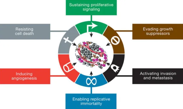
Hanahan and Weinberg (2000) described that tumors acquire six biological principles during multistep development (Figure 2):10 sustaining proliferative signaling; evading growth suppressors; resisting cell death; enabling replicative immortality;
inducing angiogenesis and activating invasion and metastasis.10,13
After a decade of conceptual progress The Hallmarks of Cancer “continue to provide a solid foundation for understanding the biology of cancer”; even though they reevaluated new findings and added two emerging and two enabling characteristics to hallmarks (traits) list, revealing that tumorigenesis cannot be just defined by self traits but also regarding tumor microenvironment (Figure 3).10
5 Traditional view of cancer describes it as an accumulation of genetic alteration/mutation, but several studies regarding epigenetic14 (study of heritable changes in gene function that not entail a change in DNA sequence15,16) showed that alterations in the epigenome have an important role in gene activation and repression regulation related to cancer. These heritable changes are disseminated as “covalent chemical changes to the cytosine bases and are referred to as DNA methylation”.16
Epigenetic regulation and chromatin compaction also understand histone tail modifications, ATP-dependent chromatin remodeling or non-coding RNA (ribonucleic acid) play, yet heritability is less clear. 16
DNA Methylation
Involved in several epigenetic processes like “gene expression, imprinting, X chromosome inactivation, silencing of retroviral and transposable DNA elements, and chromatin organization,”16 assuring proper regulation of gene expression and stable gene silencing.17 In cancer DNA is usually hipomethylated.18
Methylation is a covalent addition of a methyl group (-CH3) that occurs “exclusively at
the 5 position of the cytosine moiety” catalyzed by DNMT (DNA methyltransferase), this occurs within CpG islandsa dinucleotides (Figure 4).17 An alteration in gene expression caused by methylation could lead to an oncogene activation or inactivation of a tumor suppressor gene, increasing cancer risk.
a CpG islands are short interspersed DNA sequences rich in guanidine and cytosine (GC).103 Figure 3 | Emerging Hallmarks and Enabling Characteristics. Adapted from Hanahan and Weinberg (2011)
6 Epidemiology
Cancer is considered a rising epidemic disease19. Each year, worldwide appears approximately 14 million new cases of cancer and 8.2 million people die from cancer; this represents 13% of all death worldwide.19,20 In United States as in Portugal cancer is the second leading cause of death, but is expected to overcame heart diseases in the next few years.20–22 In 2016 around 595 690 Americans are expected to die due cancer, this represents an average of 1 630 death per day.21
Using trends, in cancer death rates to measure the improvement of the battle against cancer, is possible to see that in the 20th century, the total cancer death rate was high, reaching a peak in 1991 (Figure 5), this peak is often explained by the tobacco epidemic.21 Tobacco intake is associated as a risk factor for numerous tumors like “lung, larynx, oesophagus, oral cavity and pharynx, bladder, pancreas, kidney, liver, stomach, bowel, cervix, leukemia, and ovarian cancers”.21 So was expected that a decrease of its consumption – happened in the 70’s in United Kingdom (UK)23 and United States of America (USA)24, e.g. – would lead to a decrease of cancer death, but
Figure 4 | Cytosine methylation. A) Methylation occurs at position 5 of cytosine moiety (marked with an [*], the
process is catalyzed by DNMT. B) Within the promoter regulation region, unmethylated CpGs (dark brown) allow gene expression; when methylation occurs gene becomes silenced. Adapted from Kulis, M. and Esteller, M. (2010).
17
7
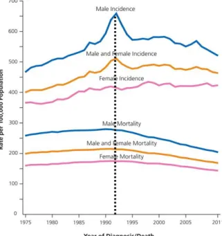
Figure 5 | Trends in cancer incidence and death rates by sex,
in USA between 1975 and 2011 – adapted from Siegel et al (2015)20
Figure 6 | Ten leading cancer types by sex in American citizens. A) Estimated new cases; B) Estimated
deaths. Estimates are rounded to the nearest 10 and excludes cases of basal cell and squamous cell skin cancers and in situ carcinoma except urinary bladder – adapted from Cancer Statistics, Siegel (2015)
that just happened between 1991 and 2012 (rate lowed in 23%).20 This is explained by the fact that usually is necessary many years, or decades, for the damages in DNA caused by smoking lead to cancer. The improvement in early detection and treatment is pointed as well as a cause of the reduction of cancer death.20
In USA, in spite of in both males and females the leading cause of death by cancer is lung and bronchus
cancer, incidence is gender specific – prostate cancer in men and breast cancer (BC) in women (Figure 6).20
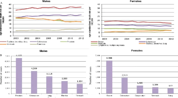
8 In Portugal mortality rates are quite different (Figure 7), in males lung and bronchus tumors are still the leading cause of death, but in females the first position is taken by breast cancer (lung and bronchus are in fifth place). Colorectal cancer is the second major cause of cancer in Portugal, in both genders. Although, cancer incidence in Portugal as is USA is gender specific. 25
Breast Cancer
EpidemiologyBreast cancer is a disease that affects many people worldwide, in specially women. In United States, 29% of the new cases of cancer in women are breast cancer, it is the second leading cause of death in women with cancer, representing 15% of the deaths20, in Portugal that number rises to 17%.25 Cancer statistics 2015 show that new cases and deaths in men represents only 1% of all cases,20 the same was assessed to Portuguese population.26 Comparatively to worldwide the incidence of BC in Western Europe is higher, although, Portugal have the smallest rate of mortality (in comparison with other European state members). In part this could be a result of early detection, promoted by mammographies screenings, that in 2016 is expected to reach 60% of coverage of women (between 45 and 69 years old).27
A B Numbe r of c as es Numbe r of c as es
Figure 7 |A) Mortality by gender in Portugal (2000-2012); B) Incidence by gender in Portugal. Adapted
9 Risk factors
There are some risk factors associated with breast cancer and they are commonly seen in about 50% of woman diagnosed. Those risk factors could be:
– Age – above 50 years old28,29
;
– Gender – occurs about 150 times more in females than in males; – Lifestyle, diet and environment factors;
– Personal or family history of breast cancer or benign disease;
– Hormonal and reproductive factors – females with no pregnancies – nulliparity – or pregnancies in an age greater than 30 years old, early menarche and late menopause;
– Endogenous estrogen exposure/reproductive factors; – Genetic predisposition.28
Hormonal risk
As we saw above, hormones play a tremendous role in breast cancer, and it have been demonstrated that an increased estrogen exposure is correlated with an increasing risk of cancer, these suggests a hormone dependence.28,30 Although the cancer risk, estrogen is essential for breast development and for the reproductive system. The number of menstrual cycles a woman goes throw in her life will be determinant to define her cancer risk because natural estrogen (produced in self organism) is “released from ovaries during every menstrual cycle”30. This way: women with earlier menarche and late menopause are exposed to more natural estrogen28,30; for each year earlier from the average menarche (12 years old) the risk increases by 5% ,30,31 and for late menopauses the risk increases 3% per year.30 In other hand having children is benefic to women due to a lack of periods during the pregnancy, this could be a risk factor for western societies where women delayed giving birth and have less children; each pregnancy is thought to lower the risk in 7%. 30 The risk could be lowered even further by breastfeeding [4.3% for every 12 months], studies suggest that breast cells could be changed in the process, making them less prone to develop cancer. 30,32,33
Women use hormones commonly as a contraceptive (the pill) or as a post-menopausal Hormone Replacement Therapy (HRT), this increases slightly breast cancer risk30,34–36. Notwithstanding, the risk vanishes slowly after quitting, being null after 10 years quitting the pill 30,37 and after 5 years after stopping HRT.28,30 The risk, is higher using combined therapy of estrogen and progestin compared with using estrogen
10 alone.28 In spite of the risk, big number of menopausal patients use HRT to relieve biological changes like hot flashes, vaginal dryness or as a way of protection against osteoporosis or other diseases of bone.28,38
Genetic risk
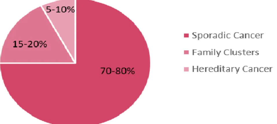
Between 70-80% (Figure 8) of breast cancer cases are due to sporadic cases (mix of genetic damages obtained over a lifetime) and a small percentage due to hereditary causes (genetic alterations passed by one generation to another) and family clusters (bigger percentage of breast cancer in a same family considering rate of sporadic cancer; putting in evidence a combination of risk factors like inherited susceptibility and environmental factors).39
Breast cancer 1 and 2 genes (BRCA1 and BRCA2) are the cause of most hereditary breast cancer cases, but mutations can occur also in PTEN, p53, MLH1, MLH2 and STK11.28 BRCAs are autosomal dominant genes that encode tumor suppressor protein, which help repair damaged DNA, playing an essential role in cell stability. When either of one is mutated or altered causing a malfunction of the protein, DNA may not be repaired properly and additional genetic alteration could lead to cancer. Specific mutations in these genes not only increase female risk of breast cancer but also ovarian cancers. Is known that 12% of woman in general population will develop breast cancer during their lives; recent studies show that 55 to 65% women with harmful mutations in BRCA1 and 45% with harmful mutations in BRCA2 will
Figure 8 | Distribution of breast cancers cases by sporadic vs hereditary cancer. Adapted
11 develop breast cancer by the age of 70 years old. 40
Staging
For staging it is mandatory doing a microscopic confirmation where the histologic type and grade should be recorded. Staging for carcinoma of the breast applies to infiltrating (including microinvasive) and in situ carcinomas. It is usually used TNM (Tumor, Node and Metastasis) classification of malignant tumors, published by the American Joint Committee on Cancer (AJCC) as staging system.28,41
Physicians use results from scans and diagnostic tests to answer questions like:
Anatomy
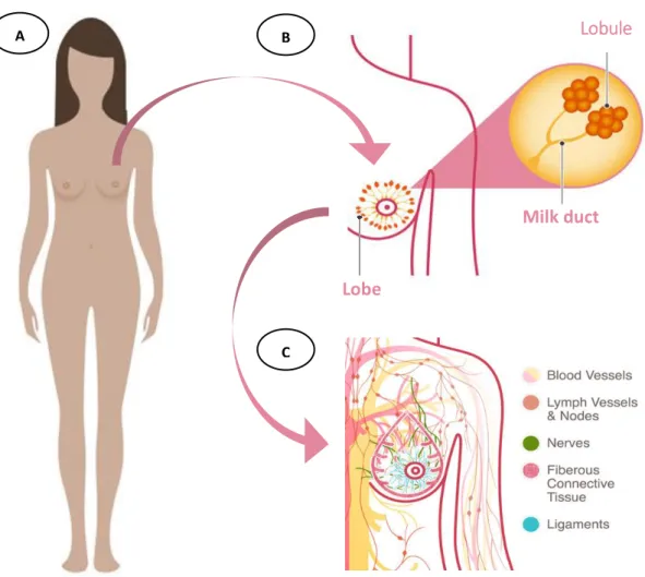
Female breast is mostly composed by adipose tissue. Each breast is divided in lobes, lobules and milk ducts. A healthy breast have between 12 to 20 lobes (which one is formed by many smaller lobules28 – gland that produces milk in nursing woman). Lobules and lobes are connected by milk ducts that when necessary carry milk to the nipple. In adipose tissue a network of ligaments, fibrous connective tissue, nerves, lymph vessels, lymph nodes and blood vessels (Figure 9).42
Lymph system
The lymph is part of the immune system and work in a similar way of the circulatory system, where lymph vessels and nodes are running throughout the entire body to transport disease-fighting cells and fluids. Bean-shaped lymph nodes clusters “are fixed in areas throughout the lymph system act as filters by carrying abnormal cells away from healthy tissue”.
“T (Tumor) – How large is the primary tumor? Where is it located?
N (Node) – Has the tumor spread to the lymph nodes? If so, where and how
many?
M (Metastasis) – Has the cancer metastasized to other parts of the body? If so
12 Usually the type of breast cancer is determined by the origin of the growth of cancer cells (usually in the lobes, lobules and ducts). Cancer found in nearby lymph nodes (LN) can help physicians determine if the tumor has spread; some additional distal nodes can be tested to understand how far the cancer has spread.
Regional lymph nodes
Axillary, transpectoral and internal mammary nodes are drained by the breast lymphatic vessels. For staging purposes intrammamary lymph nodes are coded as axillary, and supraclavicular lymph nodes are coded as regional lymph nodes. Metastasis to other lymph nodes (as cervical or contralateral internal mammary lymph nodes) are classified as distant (M1) (Figure 10)28
Figure 9 | Female and breast anatomy. A) Female anatomy; B) Localization of lobules, lobes and milk ducts in
breasts; C) Scheme of intrinsic systems in breast tissue and neighborhood. Adapted from National Breast Cancer Foundation (2015). Milk duct Lobe B C A Lobule
13
Figure 10 | Diagram of breast and regional lymph nodes.
1 – Low axillary, Level I; 2 – Mid-axillary, Level II; 3 – High-axillary, apical, Level III; 4 – Supraclavicular; 5 – Internal mammary nodes.
1. Axillary (ipsilateral): interpectoral (Rotter’s) nodes and lymph nodes along axillary vein and its tributaries that may (but not required) divided into:
a. Level I – low axilla b. Level II – mid-axilla
c. Level III – apical axilla (more anatomical information in Appendix table 3)
2. Internal mammary (ipsilateral): LN in the intercostal spaces along the edge of the sternum in the endothoracic fascia.
3. Supraclavicular: LN in the supraclavicular fossa, a triangle defined by the omohyod muscle and tendon, the internal jugular vein, and the clavicle and subclavicle vein. Adjacent LN outside of this triangle are considered to be lower cervical nodes (M1).
Classification
1. Clinical staging
The first step is a physical examination of the area, including observation and palpation of the skin, mammary gland and lymph nodes (axillary, supraclavicular and cervical), imaging and an examination of breast tissues and/or other tissues. 28,41
14
2. Pathologic staging
Includes all previous information and data from “surgical exploration and resection as well as pathologic examination of the primary carcinoma, regional lymph nodes and, metastatic sites (if applicable) including not less than excision of the primary carcinoma with no macroscopic tumor in any margin of resection by pathologic examination.”41
The pathologic stage (pT) only can be assessed if the marge involvement is microscopic and not macroscopic. If there is a macroscopic involvement the cancer is coded as pTX since the total extent of the primary tumor could not be measured. If the primary tumor is invasive and not just microinvasive it is recommended at least the resection of the low axillary lymph nodes (Level 1) (Figure 10) this is used for pathologic classification (pN). If the surgery occurs after “neoadjuvant therapy, hormonal therapy, immunotherapy, or radiation therapy, the prefix “y” should be used with the TNM classification” (e.g. ypTNM)41
TNM Classification
A. Primary Tumor (T)
For each case, it is used a specific kind of measurement to classify the primary tumor; it could be obtained by physical examination or mammographies and ultrasounds. Only the invasive component is measured, after that it is removed tissue to prosecute some specific studies, for instance, to evaluate estrogen and progesterone receptors. Some patients have to do multiple core biopsies, this could lead to an under classifying of the T component, in that cases tumor size should be reconstructed based on imaging and histological findings. 41
Applying TNM for staging the letter “T” is grouped to a number or letter to define the size and location. Some stages have some smaller groups – sub stages – to describe the tumor in more detail43
TX: As said previously, is used when the primary tumor cannot be
evaluated.28,41,43
TO: There is no evidence of breast cancer. 28,41,43
Tis – Carcinoma in situ, with no evidence of an invasive component41, its restricted within ducts or lobules of the breast tissue. 41,43 Tis is sub divided in three types:
15
Tis (DCIS) – Intraductal carcinoma in situ, is a noninvasive cancer, that can
evolve to an invasive breast cancer if not removed. 28,41,43
Tis (LCIS) – Lobular carcinoma in situ, called to abnormal cells found in the
lobules or glands of the breast – in spite of not being cancer increases the risk of progress to invasive breast cancer. 28,41,43
Tis (Paget’s) – Paget’s disease of the nipple28,41,43
, “rare form of early, noninvasive cancer”43 that only affect skin cells of the nipple. Simultaneously can occur an invasive breast cancer, staging in this case is only given by the invasive tumor.28,43
T1 – Tumor is no bigger than 20 millimeters (mm) in its widest area. It is
subdivided usually in substages:
T1mic – Microinvasion no larger than 1mm in the widest area.28,41
T1a – Tumor is larger than 1mm but smaller than 5mm in the widest
area.28,41,43
T1b – Tumor larger than 5mm but not more than 10mm in the widest
area.28,41,43
T1c – Tumor larger than 10mm but not more than 20mm in the widest area.
28,41,43
T2 – Tumor between 20mm and 50mm in the widest area. 28,41,43 T3 – Tumor greater than 50mm. 28,41,43
T4 – Tumor of any size with direct extension to: 28,41,43 T4a – Chest wall, not including pectoralis muscle. 28,41,43
T4b – Skin (Edema “[including peau d’orange] or ulceration of the skin of the
breast, or satellite skin nodules confined to the same breast)”. 41
T4c – Chest wall and skin. 28,41,43 T4d – Inflammatory carcinoma. 28,41,43
Notes:
i. In case of bilateral breast carcinoma, each breast is considered an independent organ, this way both will have an independent stage. ii. Inflammatory carcinoma is characterized by “diffuse erythema and
edema (peau d’orange) of the breast, often without an underlying palpable mass.”41
The symptoms are common to mastitis (inflammation of the breast). In this type of cancer, cancer cells block lymph vessels, causing the visual symptoms.44
16 iii. Carcinoma in situ – cancer located in epithelial cells is designated carcinoma. It occurs in glands and ducts, that is why most breast cancers are carcinomas. Benign (non-cancerous) cells do not invade beyond epithelial tissue, in carcinoma in situ abnormal cells look similar to invasive carcinoma cells (when analyzed under microscope). It was assumed that these cells could become invasive if not treated, but recent studies showed that the transition to invasive carcinoma is more “complex and subtle” than the previous idea based on microscopic resemblance. Long term follow-up of patients with carcinomas in situ established that not all of them progress to invasive cancer. 45
B. Regional Lymph Nodes (N)
Definitions for classifying the regional lymph nodes (N) are different for Clinical and for Pathologic classification. 28,41,43,46
B.1. Clinical Classification28,41
NX – Regional lymph node cannot be assessed (it could be removed previously,
or not removed, e.g.).
NO - No regional lymph node metastasis.
N1 – Metastasis to movable ipsilateral axillary lymph nodes
N2 - Metastasis in ipsilateral axillary lymph nodes fixed or matted (axillary
lymph nodes that are fixed to each other), or in clinically apparent ipsilateral internal mammary nodes in the absence of clinically evident axillary node metastasis.
N2a – Metastasis in ipsilateral axillary lymph nodes fixed to one another
or to other structures
N2b – Metastasis only in clinically apparent ipsilateral internal mammary
nodes in the absence of clinically evident axillary lymph node metastasis.
N3 – Metastasis to ipsilateral infraclavicular lymph node(s) with or without
clinically evident axillary lymph nodes, or in clinically apparent ipsilateral internal mammary lymph node(s) and in the presence of clinically evident axillary lymph node metastasis, or metastasis in ipsilateral supraclavicular lymph nodes with or without axillary or internal mammary nodal involvement.
17
N3b – Metastasis to ipsilateral internal mammary lymph node(s) and
clinically apparent axillary lymph nodes
N3c – Metastasis in ipsilateral supraclavicular lymph nodes with or
without axillary or internal mammary nodal involvement
B.2. Pathologic Classification 28,41,46
Classification based on axillary lymph node dissection (ALND) with or without sentinel lymph node dissection (SLND). Classification based merely in SLND should be designated sn.
NX – Regional lymph node cannot be assessed (they could be removed
previously, or not removed, e.g.).
NO - No regional lymph node metastasis.
N1 – Metastasis to movable ipsilateral axillary lymph nodes.
pNX – Regional lymph nodes cannot be assessed (e.g., previously removed, or
not removed for pathologic study).
pNO - No regional lymph node metastasis; no additional examination for
isolated tumor cells (ITCs, defined as single tumor cells or small lusters not greater than 0.2 mm, usually detected only by immunohistochemistry (IHC) or molecular methods but which may be verified on hematoxylin and eosin stains. ITCs do not usually show evidence of malignant activity [e.g., proliferation or stromal reaction])
pNO (i-) – No histological nodal metastasis, and negative by IHC
pNO (i+) – No histological nodal metastasis but positive by IHC, with no
cluster greater than 0.2 mm in diameter
pNO (mol-) – No histological nodal metastasis and negative molecular
findings (by reverse transcriptase polymerase chain reaction, RT-PCR)
pNO (mol+) – No histological nodal metastasis, but positive molecular
findings (by RT-PCR)
pN1 – Metastasis in 1-3 ipsilateral axillary lymph node(s) and/or in internal
mammary nodes with microscopic disease detected by SLND but not clinically apparent
pN1mi – Metastasis (greater than 0.2 mm, none greater than 2.0mm) pN1a – Metastasis in 1-3 axillary lymph nodes
pN1b – Metastasis to internal mammary lymph nodes with microscopic
18
pN1c – Metastasis in 1-3 ipsilateral axillary lymph node(s) and in
internal mammary nodes with microscopic disease detected by SLND but not clinically apparent. If associated with more than three positive axillary nodes, the internal mammary nodes are classified as N3b to reflect increased tumor burden.
pN2 – Metastasis in 4-9 axillary lymph nodes or in clinically apparent internal
mammary lump nodes in the absence of axillary lymph nodes
pN2a – Metastasis in 4-9 axillary lymph nodes (at least one tumor
deposit >2 mm)
pN2b – Metastasis in clinically apparent internal mammary lymph nodes
in the absence of axillary nodes
pN3 – Metastasis in 10 or more axillary lymph nodes, or in infraclavicular
lymph nodes, or in clinically apparent ipsilateral internal mammary lymph nodes in the presence of one or more positive axillary nodes; or in more than three axillary lymph nodes with clinically negative microscopic metastasis in internal mammary lymph nodes; or in ipsilateral supraclavicular lymph node(s)
pN3a – Metastasis in 10 or more axillary lymph nodes (at least one
tumor deposit greater than 2.0 mm), or metastasis to the infraclavicular lymph nodes
pN3b – Metastasis in clinically apparent ipsilateral internal mammary
lymph nodes in the presence of one or more positive axillary nodes: or in more than three axillary lymph nodes detected by SLND but not clinically apparent
pN3c – Metastasis in ipsilateral supraclavicular lymph node(s).
C. Metastasis
MX - Distant metastasis cannot be assessed. M0 – No distant metastasis
M1 - Distant metastasis
D. TNM stage grouping for breast cancer
Cancer stage is a combination of T, N and M. Staging is usually confirmed after a surgery, but for neoadjuvant therapyb staging is chiefly determined clinically. Physicians may use stage I to stage IIA to define early stages, and stages IIB to stage II
19 for locally advanced. Furthermore, it is imperative to retain the idea that tumor biology, like specific markers (e.g. estrogen receptors) have a huge impact on the advised treatment as well as in the prognosis. 43
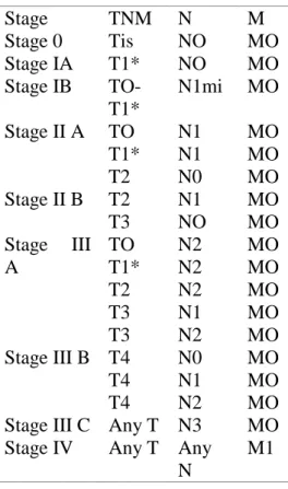
Table 1 | TMN stage groupingc for breast cancer, as in the 7thEdition of AJCC Cancer Staging Manual (2010).43,46 (* – T1 includes T1mi)
Stage TNM N M Stage 0 Tis NO MO Stage IA T1* NO MO Stage IB TO-T1* N1mi MO Stage II A TO T1* T2 N1 N1 N0 MO MO MO Stage II B T2 T3 N1 NO MO MO Stage III A TO T1* T2 T3 T3 N2 N2 N2 N1 N2 MO MO MO MO MO Stage III B T4 T4 T4 N0 N1 N2 MO MO MO Stage III C Any T N3 MO Stage IV Any T Any
N
M1
Histopathologic Type
Histological classification has been a valuable tool in past decades, and divide in situ carcinomas and invasive carcinomas;41,47 Relies on histology and put apart molecular markers that have a significant weight on prognosis.47
Table 2 | Histopathologic classification of breast cancers - as in the 7thEdition of AJCC Cancer Staging Manual (2010)48
Tumor Location Histologic subtype
Carcinoma, NOS (Not otherwise specified)
Ductal Intraductal (in situ)
Invasive with predominant component
c TMN stage grouping used in TCGA (The Cancer Genome Atlas, a database used in the thesis)
is from the7th Edition of AJCC Cancer Staging Manual, in order to even out the results it was decided to place the 7th edition classification here; 6th edition can be found in Appendix table 1.
20 Invasive, NOS
Comedo Inflammatory
Medullary with lymphocytic infiltrate Mucinous (colloid)
Papillary Scirrhous Tubular Other
Lobular Invasive with predominant in situ component
Invasive
Nipple Paget disease, NOS
Paget disease with intraductal carcinoma Paget disease with invasive ductal carcinoma
Other Undifferentiated carcinoma Metaplastic
The following tumor subtypes occur in the breast but are not considered typical breast cancers:
Phyllodes tumor.
Angiosarcoma.
Primary lymphoma Types of tumor
Commonly breast cancer is organized in subtypes, aiming the division in some (1) special receptors or in a (2) specific cluster of genes.
1. Hormone receptors (routinely assays are performed by pathologists on
tumor samples) 28
a. Hormone receptor positive – Tumors express estrogen receptors (ER), progesterone receptors (PR),44 ER responsive genes and other genes that encode typical proteins of luminal epithelial cells.49 They may depend on hormones (estrogen and progesterone) to grow.44
b. HER2 positive – Represents 20% - to 25% of breast cancers, these tumors are dependent of the gene HER2 (epidermal growth factor receptor 2) to grow and have a high abnormal number of HER2 receptors or copies of the gene. 44
21 c. Triple negative – if the tumor do not express any of the previous hormone receptors (ER, PR and/or HER2). This type may grow faster than the hormone positive and be more sensitive to chemotherapy. 44 2. Molecular classification of breast cancer subtypes
Molecular classification was described first by Perou et al in 200050, and gradually emerged as an answer to predict a response to targeted therapies and represents a high success in the design of individualized therapies in breast cancer, promoting an improvement in disease-specific survival.51,52Altough, studies show that combining both histological and molecular classifications result in significantly better predictive value. 47,51 After the first classification given by Perou et al (2000) several adjustments were made and now tumors are commonly classified into four major subtypes: luminal A, luminal B, HER2+ and basal like.50–54 This classification is a result of gene expression profiling given by “microarray datasets and progressed to a PCR-based test with a curated list of 50 genes (the PAM50 gene signature).”54 Each subtype has a particular set of “risk factors for incidence, response to treatment, risk of disease progression and preferential organ sites of metastases.”51
Luminal A – most common (50 to 60% of all BC); is characterized by an
increase in estrogen receptor 1 (ESR1) and/or PR+/Her2- status47,53,55 and lower levels of proliferation related genes, as Ki 67.d49,56 It is also associated to low-grade tumors and good prognosis; 47,53,55 they will likely have a good response to hormonal treatments; with relapse rate lower than other subtypes. Recurrence is common in bone, but in tissues like liver, lung and central nervous system occur in a little less than 10% of patients. Include special histological types (tubular, invasive cribriform, mucinous and lobular).51
Luminal B – Less common than luminal A (about 20% of all BC) although
approximately 30% of HER2-positive tumors defined by IHC are assigned to the luminal B subtype. Demonstrate increase expression of growth receptor signaling genes and have a more aggressive49 phenotype with bad prognosis.47,53
As an IHC point of view luminal B is defined by:
ER positive; HER2 negative; high Ki 67 or;
ER positive; HER2 positive.
d Ki 67 protein expression is strictly associated with cell proliferation,104 high levels are
22 But in fact, this definition does not englobe a small percentage (6%) of luminal B that are ER and HER negative. 49,56
Note:
Main difference between both luminal subgroups is increased expression of proliferation-related genes. Ki67 index is pointed as a potential marker to differentiate luminal A and B in clinical practice.49 Cheang et al (2009) stablished Ki67 index of 14% or more Ki67-positive tumor nuclei as the best cut point.
To summarize: Ki67 < 14% (low Ki67) – Luminal A Ki67 ≥ 14% (high Ki67 – Luminal B
However, Ki67 immunohistochemistry is not optimized and standardized as well the Ki67 cut off. 49,56
HER 2+ – About 15-20% of BC. Characterized by high expression of HER2 gene and other HER2 pathway related genes.49
HER2 is a member of four membrane tyrosine kinases. “Upon ligand binding to their extracellular domains, HER proteins undergo dimerization and transphosphorylation of their extracellular domains.” Due to HER2 lack of ligand, HER2 relies on “heterodimerization with another family member or homodimerization with itself to be activated” at high levels. These “phosphorylated tyrosine residues interact with numerous intracellular signaling molecules” provoking downstream activation of second messenger pathways and crosstalk with other membrane signaling pathways, activating many genes involved in cell proliferation, differentiation, survival, angiogenesis, invasion and metastasis, 49,57–60 conferring more aggressive biological and clinical behavior. In the absence of treatment, HER2+ have a poor diagnosis and a susceptibility to metastasize to brain and visceral organs.49
Basal-like – Low frequency (3-15% of all BC); frequently lack hormone
receptors (e.g. ER and PR) and HER2, hence are called triple negative breast cancer (TNBC), but not all basal like are TNBC and not all TNBC are basal-like.49 Either characterized for the expression of keratin 5, 6 and 17, integrin beta, fatty acids and laminin.50,51,53,61,62 Currently there is “no molecular-based targeted therapies”51 and only 20% have a good response to standard chemotherapy, nevertheless numerous agents are in clinical trials that unfortunately seem to fail treating TNBC (as a result
23 have a poor outcome) further studies point to a subdivision of TNBC in five or six classes, with specific molecular features, that can explain the low response to drugs;
51,63–65
Basal like cancers undergo frequent mutations in TP53e (tumor protein 53) gene; approximately ¾ of basal-like tumors are BRCA1 (breast cancer 1).
MGP – Matrix Gla Protein
Matrix Gla Protein (MGP) was first isolated from urea extracts of demineralized bovine bone, being the second vitamin K-dependent (VKD)f to be discovered in bone, after osteocalcin (also known BGP or bone Gla protein).66,67 Different tissues (e.g. heart, lung, kidney and breast) express MGP.66,68,69
Human MGP has a single copy gene66 located in the small arm of chromosome 12 (12p12.3) and encodes a 10-kD (kilo Dalton) skeletal extracellular matrix (ECM) protein.70
MGP primary structure consists in a signal peptide, a phosphorylation domain and a γ-carboxylase recognition site. MGP has also five Gla residues (γ-carboxylated glutamic acid) (Figure 11), which are converted by γ-glutamyl carboxylase enzyme (dependent of vitamin K66,71,72 – using it as a cofactor)70 through a posttranslational modification; this alteration gives high affinity for mineral and mineral ions70,73,74 like calcium, phosphate and hydroxyapatite crystals (mineral components of skeletal ECM).70
MGP variants
For several years MGP was known as a four exon gene (with a total of 1398 base pairs [bp]) but recently a specific MGP variant was identified in human fetal tissue, with an insertion of a new protein domain, resulting in the synthesis of a longer protein
e
TP53 – evidence of genomic instability and inactivation of the retinoblastoma (Rb) pathway.49
f VKD proteins have several functions, including: hemostasis, apoptosis and growth control.74 Figure 11 | Schematic representation of matrix Gla protein (MGP), pointing out the primary structure and Gla
domains.
Primary structure: Signal peptide – SP; Phosphorylation domain – P, in a green box (phosphoserine residues are
represented by green dots; γ-carboxylase recognition site – γ, in a blue box.
24 isoform (with 1473bp). This novel isoform was labeled E5 (contain five exons) while the previous isoform was labeled E4 (contain four exons) (Figure 12). Isoform E5 have three putative additional sites of gamma carboxylation,71,72 this feature could increase MGP binding capacity, resulting in a more powerful inhibitory function.
Alternative splicing
Humans produce around 90 000 different proteins, and scientific community was expecting that human genome project would reveal a similar number of genes (“one gene to one protein”) so it was a huge surprise that the real number of genes is almost four times smaller than expected – about 25000 genes. Insights in this paradigm resulted in the concept of alternative splicing – a mechanism where a single gene is the origin of multiple proteins, tallying protein diversity.75 In average, one human gene has eight exons and seven introns, that gives rise in average to three or more alternatively spliced mRNA (messenger RNA). Recent high-throughput sequencing studies point that 100% of human genes produce at least two or three isoforms.76
The first transcript of the gene is pre-messenger RNA (pre-mRNA) – copy of the gene including both introns (intended to be removed during pre-mRNA processing) and exons. In RNA splicing exons are retained and exons targeted for removal in order to create different mRNA from the same primordial information (pre-mRNA). The same nucleotide sequence can be an exon or an intron, depending on the splicing target.77
Characterization of the protein and its functions
Fraser and Price in 198869 described MGP as the first well characterized substrate of VKD found in discrete tissue-specific cells (kidney, lung, heart and spleen
Figure 12 | MGP schematic representation. Variant E5 (5 exons) and variant E4 (4exons) – novel isoform
(E5) have one extra exon (75bp) due to alternative splicing in MGP gene. Exon at red (Ex2) is only present in isoform E5.
Isoform E5
Isoform E4
25 cells), showing that MGP function is beyond specific connective tissues, thus, is unlikely that MGP acts just for accumulation in an extracellular matrix.
In 1997, Luo et al73 developed homozygous mice without Mgp expression (MGPm1/ MGPm1). Those mice until the second week were similar to control mice but then they became shorter and with a faster heart beat; within two months they died due hemorrhage triggered by thoracic or abdominal aortic rupture, showing signals of prominent calcification. Findings suggested:
Mgp inhibits calcification of certain ECMs, probably as a result of its capacity of mineral-ion-binding.
In early development other gene products play an important role to prevent earlier calcification, otherwise newborn or one-week-old MGPm1/ MGPm1 mice must have showed defects. 73
Studies show also the involvement of MGP in cell differentiation71,78 and proliferation.71,72
Mutations (with loss of function) in this gene were related with: atherosclerosis (vascular calcification), cancer68,79,80 and were proven to lead Keutel syndrome whom is characterized by abnormal formation of cartilage calcifications (in auricles, nose, larynxes, trachea and ribs), multiple peripheral pulmonary stenosis, neural hearing loss and short terminal phalanges. 70
MGP and cancer
As stated above, MGP was showed a role in cell differentiation and proliferation, and was suggested a role also in cell migration, differentiation, onset of angiogenesis81 and tumorigenesis.72,82,83 However, MGP role in oncogenesis is unclear as well as the correlation between its expression and tumor type.68 Some studies were performed and in some tumor types expression was higher and in other types lower than in normal tissue, giving in some cases prognostic value.
Regarding MGP mRNA expression analyses we can see:
Ovarian cancer – MGP mRNA expression in tumor was higher
compared to normal tissue. 68,84
Renal cell carcinoma – higher expression in tumor tissue with a
significant inverse correlation between mRNA expression and tumor size, lymph node metastasis and grade.68,85
26
Colorectal cancer – Fan et al (2001) 86 found a down-regulation of MGP mRNA in human adenocarcinomas but results obtained in our laboratory comparing cancer tissue and paired adjacent normal tissue showed an up-regulation.71
Astrocytic gliomas (AG) – comparing high and low grade AG, mRNA
levels were significantly increased in high-grade tumors. 68,83
Regarding protein expression, The Human Protein Atlas87 stained twenty different types of cancers and controls, the results (Table 3) in brief, show that malignant cells exhibited moderate to strong cytoplasmic and nuclear staining. Samples vary from 4 to 12, and the most exciting result regard MGP protein expression is in gliomas and liver cancers, where it could be observed a small upregulation of MGP relatively to normal tissue. Analyses of more samples could give more reliable results.
MGP and Breast Cancer
A study using cDNA hybridization revealed that MGP expression in breast cancer is 20-fold higher in metastatic cancer (cell line 600PEI) than in normal epithelium, 68,88 and Yoshimura et al (2009) presented MGP mRNA expression as a potential prognostic factor in BC patients, being overexpressed in poor prognostic patients (using microarray analyses). However, immunohistostaining of breast tissue microarrays did not revealed a correlation between overall survival or ER status metastasis with protein expression,68 this information is also supported by results in Table 3, where MGP is highly to medium expressed in cancer staining and normal tissue is also highly expressed.
Table 3 | MGP protein expression in tumoral vs normal tissue staining. Colors represent different levels of
27 Cell culture
Is unethical starting research in humans or other animals before testing it before, for this reason a variety of models were and are being developed. For instance, cancer cell lines which are used currently in several biomedical research laboratories.89,90
Cell lines are obtained by primary cultures (growing cells from tissue taken directly from an individual growing in a flask or petri dish. They provide an almost unlimited supply of cells (with similar genotypes and phenotypes). There are various observations that reveals cell lines as a good model for cancer research, as a first approach, for instance:90
Histopathology: most human cancer cell lines transplanted to
immunedeficient mice lead to tumors; histopathologic analysis of 127 human cell lines that formed tumors in mice reveled that they are all correlated with tumor origin. 90
Molecular genetics and receptor expression: comparison between lung
cancer and breast cancer cell line with origin cancers revealed that phenotypic (as ER receptors expression) and genotypic properties are retained for long periods of time. 90
28
Aims
More and more cancer research is focused on target therapies and biomarkers. This kind of approach is imperative to refine strategies of screening and diagnosis leading to early detection, that is correlated with better prognosis. Furthermore, target therapies are known as less toxic and with reduce side effects, improving the wellbeing of cancer patients. To improve this approach is necessary to enlighten the role of genes related with tumorigenesis. Knowing that MGP role in breast cancer (and cancer in general) is unclear, our biggest aim is to try to clarify it, searching for correlations between its expression in normal vs tumoral samples of breast cancer, as well as in particular subsets of patients. Alongside our main aims were either:
Clarify isoforms expression in breast cancer cell lines, as well as the result of isoforms overexpression in cellular proliferation and migration.
Identify some regulatory mechanisms of MGP expression, in particular epigenetic (methylation) and posttranscriptional regulation (mir155 analyze, even as, its correlation in normal and tumoral tissue.
29

![Figure 4 | Cytosine methylation. A) Methylation occurs at position 5 of cytosine moiety (marked with an [*], the process is catalyzed by DNMT](https://thumb-eu.123doks.com/thumbv2/123dok_br/18628499.910871/22.892.137.761.191.604/figure-cytosine-methylation-methylation-position-cytosine-process-catalyzed.webp)