Stanković Ana
Dissertation presented to obtain the Ph.D degree in Molecular Biology
Instituto de Tecnologia Química e Biológica António Xavier | Universidade Nova de Lisboa
Insert here an image
with rounded corners
epigenetic centromere propagation
Oeiras, April, 2017
Драги мамаи тата, надам се дасте поносни намене коликосамја на вас. Данас сам овде захваљујући вама и вашој подршци. Волим вас пуно ивамапосвећујемовутезу.
Dissertation presented to obtain the Ph.D degree
in Molecular Biology
Instituto de Tecnologia Química e Biológica António Xavier | Universidade Nova de Lisboa
Oeiras, April, 2017
epigenetic centromere propagation
Research work coordinated by:
Declaração ... i
Summary ... iii
Sumário ... v
Acknowledgments ... ix
List of Publications ... xi
Chapter 1 - General introduction: Cell cycle and Centromeres ... 1
1.1 The cell cycle ... 3
1.2 Cell-cycle control system and Cyclin-dependent kinases ... 4
1.4 Centromere function ... 7
1.5 Centromere organization across different species ... 9
1.6 Centromeric DNA ... 10
1.7 CENP-A is epigenetically marking active centromeres ... 13
1.8 CENP-A nucleosomes are stably propagated at centromeres through mitotic and meiotic divisions ... 16
1.9 Determinants of CENP-A stability ... 19
1.10 The Constitutive Centromere-Associated Network (CCAN) ... 21
1.11 The modularity of CENP-A dependent kinetochore assembly ... 27
1.12 Propagation of centromeric chromatin across cell divisions ... 32
1.13 Centromere as a paradigm for epigenetic inheritance ... 38
Introduction ... 67
2.1 The Mis18 complex centromere targeting is required for CENP-A propagation across species ... 67
2.2 The role of the Mis18 complex at the centromere ... 70
2.3 Mechanisms of centromere recognition by the Mis18 complex .... 75
2.4 Material and Methods ... 77
2.5 Results ... 82
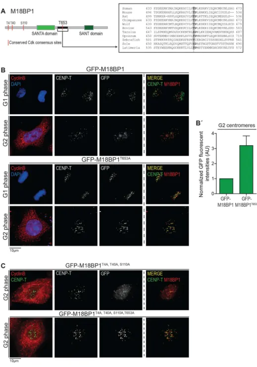
Recruitment of the Mis18 complex to the centromere is controlled by phosphorylation of M18BP1T653 ... 82
Mis18α can form a complex with M18BP1 or Mis18β in G2 phase of the cell cycle ... 88
Mis18β cannot form a complex with M18BP1 in G2 phase of the cell cycle ... 90
2.6 Discussion and Conclusions ... 95
Acknowledgements ... 100
References ... 101
Chapter 3 - Cdk1/2 dependent regulation of HJURP ... 109
Abstract ... 111
Introduction ... 111
3.3 Molecular basis of HJURP CENP-A selectivity ... 117
3.4 Additional factors involved in the efficient HJURP/CAL-1-mediated CENP-A deposition ... 119
3.5 Molecular mechanism of HJURP centromere targeting ... 122
3.6 Material and Methods ... 124
3.7 Results ... 130
HJURP is phosphorylated in a cell cycle dependent manner ... 130
The HJURP conserved domain interacts with Cyclin A and controls timing of CENP-A assembly ... 133
HJURP Serine 210/211 is functionally phosphorylated in G2 phase cells ... 141
Cdk activity controls HJURP localization not its chaperoning activity148 3.8 Discussion ... 148
A role for NPM-1 in regulating CENP-A assembly? ... 151
Acknowledgements ... 154
References ... 155
Chapter 4 - Full reconstitution of CENP-A assembly pathway under high Cdk1/2 activities ... 169
Abstract ... 171
Introduction ... 171
tight cell cycle timing of centromere propagation ... 178
Efficient CENP-A assembly requires displacement of M18BP1 from the centromere ... 181
4.2 Discussion ... 185
Acknowledgements ... 190
References ... 191
Chapter 5 – General Discussion: Histone-based Inheritance, Centromeres and Evolution ... 195
Abstract ... 197
5.1 The phenomenon of epigenetic inheritance... 197
5.2 Histone based epigenetic inheritance... 200
5.3 Maintenance of CENP-A nucleosomes across the cell cycle ... 205
5.4 Centromeric DNA, CENP-A and centromere ... 210
5.5 Seeding the centromere ... 215
References ... 222
Appendix 1 - Reductionism at the vertebrate kinetochore ... 235
Appendix 2 - A Dual Inhibitory Mechanism Sufficient to Maintain Cell-Cycle-Restricted CENP-A Assembly ... 239
i
own work, as developed between 2012 and 2017 in the laboratory of Dr. Lars Jansen at the Instituto Gulbenkian de Ciência in Oeiras, Portugal. Specific author contributions are indicated in each chapter, in the Acknowledgements section.
Financial support was granted by Fundação para a Ciência e a Tecnologia, doctoral fellowship SFRH/BD/51878/2012 and ERC-2013-CoG-615638– Epimechanism.
Declaração
Declaro que esta dissertação de doutoramento e os dados nela apresentados são o resultado do meu trabalho, desenvolvido entre 2012 e 2017 no laboratório do Dr. Lars Jansen no Instituto Gulbenkian de Ciência em Oeiras, Portugal. As contribuições de cada autor são indicadas em cada capítulo na secção dos Agradecimentos/Acknowledgements.
O apoio financeiro foi concedido pela Fundação para a Ciência e Tecnologia, através da bolsa de doutoramento SFRH/BD/51878/2012 e por fundos do Conselho Europeu de Investigação (ERC) ERC-2013-CoG-615638–Epimechanism.
iii
structure called the kinetochore during mitosis. This structure serves as a binding platform for microtubules during mitosis, enabling segregation of genetic material to two daughter cells. Although fascinating, the function of the centromere was not the focus of my PhD work. Rather, it was the epigenetic nature of its propagation. The centromere is epigenetically marked and inherited through incorporation of a specialized H3 variant called CENP-A. CENP-A is necessary and sufficient for specification of centromere genomic location and its inheritance. Incorporation of CENP-A into centromeric nucleosomes is orchestrated by several factors, and occurs in a cell cycle dependent manner, upon mitotic exit. Previous work from our laboratory demonstrated that the key molecular switch is driven by Cyclin-dependent kinases 1 and 2 (Cdk1/2) that negatively control the timing of CENP-A incorporation. Brief inhibition of these kinases resulted in precocious centromeric incorporation of CENP-A. This led to a proposal where the CENP-A loading machinery is present and poised for CENP-A assembly, but is held inactive due to Cdk1/2 activities. Key proteins necessary for the process of CENP-A deposition include the Mis18 complex and the A specific chaperone HJURP, which bears CENP-A-specific nucleosome assembly activity.
In Chapter 2, I describe how negative Cdk1/2 control is exerted upon members of the Mis18 complex. By identifying a key phosho-residue on the largest member of this Mis18 complex, M18BP1, we demonstrate that Cdk1/2 are controlling M18BP1 centromere localization, rather than its activity. Mutating key phospho-residue in M18BP1 resulted in premature centromere recruitment of this protein, thus demonstrating a direct
iv
result in complete alleviation of inhibition of CENP-A assembly.
In Chapter 3, I focus on the Cdk1/2 dependent regulation of the HJURP chaperone. As in the case of the Mis18 complex centromere localization, Cdk1/2 are acting directly upon HJURP centromeric targeting. Functional inhibition of HJURP is exerted at the level of its centromeric localization, rather than activity, given that mutations in key Cdk-dependent phosho-residues of HJURP result in premature localization of HJURP. Furthermore, these mutations result in a low level of precocious nascent CENP-A assembly.
In Chapter 4, we show that simultaneous uncoupling of the CENP-A loading factors, M18BP1 (chapter 2) and HJURP (chapter 3) from cell cycle control results in a full recapitulation of CENP-A assembly under high Cdk activities, indistinguishable from G1 assembly. This indicates that these two assembly factors are the main targets of the cell cycle control mechanism, restricting CENP-A assembly to G1 phase.
In summary, this work expands and provides direct evidence for the previously recognized role of Cdk1/2 in regulation of inheritance of epigenetic centromere. We define a dual inhibitory mechanism that is sufficient to maintain cell cycle restricted centromere propagation and characterize the molecular mechanism of how CENP-A assembly is turned on and subsequently turned off.
v
pela agregação do cinetocóro. Esta estrutura actua como uma plataforma na qual os microtúbulos se ligam durante o processo de mitose, permitindo a distribuição do material genético pelas duas células-filhas. No entanto, embora fascinante, o assunto central desta tese não é a função do centrómero, mas sim a natureza epigenética da sua propagação.
O centrómero é epigeneticamente definido e herdado através da incorporação de uma variante especializada da histona H3 designada por CENP-A. Esta proteína é necessária e suficiente não só para a determinação da localização genómica do centrómero, como também para sua propagação às células-filhas. A incorporação da CENP-A nos nucleossomas centroméricos é organizada por diversos factores e depende da fase do ciclo celular, ocorrendo especificamente durante a saída da mitose. Resultados prévios do laboratório tinham já mostrado que o controlo da incorporação de CENP-A é regulado negativamente pelas Cinases Dependentes de Ciclina 1 e 2 (Cdk1/2). Dado que a inibição temporária destas cinases resultou numa deposição precoce de CENP-A no centrómero, foi proposto que a maquinaria responsável por esta incorporação estaria já presente e pronta para funcionar, mas que seria mantida inactiva devido à actividade das Cdk1/2. Outras proteínas essenciais no processo de deposição da CENP-A incluiriam o complexo Mis18 e a chaperona HJURP, esta última que demonstrou ter uma actividade específica para a deposição da CENP-A durante a formação dos nucleossomas do centrómero.
O Capítulo 2 descreve a forma como o controlo negativo das Cdk1/2 é exercido sobre os membros do complexo Mis18. Através da identificação
vi
localização centromérica de M18BP1 - e não a sua actividade. A mutação deste fosfo-resíduo em M18BP1 resultou no recrutamento prematuro desta proteína, o que revelou que as Cdk1/2 têm um efeito directo na regulação da localização de M18BP1 para o centrómero. No entanto, este recrutamento prematuro do complexo Mis18 não resultou na atenuação completa da inibição deste sobre a incorporação de CENP-A no centrómero.
No Capítulo 3, a regulação da chaperona HJURP pelas Cdk1/2 é discutida. De forma semelhante ao controlo da localização centromérica do complexo Mis18, as Cdk1/2 actuam directamente sobre a localização centromérica de HJURP. A inibição da função desta chaperona, mais uma vez, ocorre ao nível da sua localização - e não da sua actividade - dado que mutações em fosfo-resíduos dependentes de Cdk1/2 em HJURP são suficientes para induzir baixos níveis de incorporação precoce de CENP-A.
No Capítulo 4 é demonstrado que o desacoplamento simultâneo dos factores que regulam a acumulação de CENP-A, M18BP1 (Capítulo 2) and HJURP (Capítulo 3), do ciclo celular resulta numa recapítulação completa da incorporação de CENP-A sob altos níveis de actividade de Cdk, o que é idêntica à deposição desta histona na fase G1.
Em resumo, este trabalho aumenta e reforça o importante papel já previamente reconhecido das Cdk1/2 na regulação da propagação epigenética do centrómero. Este estudo não só define um mecanismo inibitório duplo que revelou ser suficiente para restringir a propagação do centrómero à fase apropriada do ciclo celular, como também caracteriza o
ix
exquisite individuals. Although I cannot mention all of them here, they all deserve my utmost gratitude.
Lars, you are an amazing and inspiring supervisor. Thank you for patiently sitting next to me and teaching me everything from how to clone to how to write a scientific paper. Thank you for putting so much trust in me and letting me develop my own ideas as well as represent our lab on multiple conferences. My journey as a scientist doesn’t end here, but the foundation that you have set will always be a part of my scientific endeavors. I am privileged to have you as my role model not only for on how the science should be done, but also how to deal with ups and downs of everyday life. I could not have asked for a better supervisor. Thank you!
João (JoMão), Mihailo and I are always saying that the world needs more of Joãos. I am thankful to have you not only as a lab colleague, but also as a friend. Thank you for all the conversations, all advices, all the comforting and support. Thank you for birthday cakes and (much needed) coffee in the morning. Thank you for being there for me, always. I will truly miss you. Dani, it was great to have you as a lab buddy! We were always getting along, and continued to do so. From coffee breaks to gin bar in Lisbon, I always had an amazing time with you. My dear labbies: Dragan (Drogan), Wojtek (Aruba, Jamaica), Marina (Spanish inquisition), Sreyoshi (Lord of the grapes), Bas (the youngster) and Inês (a person with a house)-you guys are fantastic! Thank you all for motivating me, all the fun and laughter! I would also like to thank Ben Black and members of his lab for hosting me for few weeks in their group. Especially, I would like to thank Lucie, for saving the day by preforming a critical mass-spec analysis during the time
x
committee. Thank you for your selfless friendship and off course, the infamous μουσακάς. Jörg, thank you for being part of my thesis committee and for all great input I received from you. Raquel, I truly appreciate professional and personal connection that we have developed over the years (including Lia, off course). I would also like to thank IGC and IGC PhD program for being an amazing an inspiring place to develop my work. A special thanks goes to past and present members of Zheng-Ho wing and to the cell cycle club community. I would especially love to thank members of Mónica Bettencourt Dias lab, since I have indulged in their generous support, starting from reagents and protocols to developing awesome friendships. I would love to give a big thank you to the members of my PhD class, PIBS 2011/2012, together with former and present program directors, Thiago Carvalho and Elio Sucena. In particular, I have to say ´´obrigada`` to my friend Rita. Rita, I am so happy that we have this great bond. I will always cherish our friendship and all the great memories that we shared (#v.activeOeiras). And now, Serbian acknowledgments. Branka, toliko toga smo prošle zajedno u poslednjih deset i više (!) godina. Nikada nisam imala sumnje u tebe, niti ću ikada, jer znam da si mi istinski prijatelj. Znam da, dokle god se držimo zajedno, postoji šansa da sve bude u redu. Hvala ti na svemu! Sad imaš to i napismeno. Dušice, nikada neću zaboraviti kako si me dočekala na lisabonskom aerodromu, uzneverenu i poprilično uplašenu. Hvala ti na svoj podršci, savetima i naravno, izlascima! Moj dragi Mihailo, ti već sve znaš. Hvala ti na ljubavi, podršci, razumevanju, šetnjama i uspomenama. Ti ćes uvek biti moj broj jedan.
xi
In chronological order:
Ana Stankovic and Lars E.T. Jansen (2013). Reductionism at the
vertebrate kinetochore, J. Cell Biol. 200, 7–8.
Ana Stankovic, Lucie Y. Guo, João F. Mata, Dani L. Bodor, Xing-Jun Cao,
Aaron O. Bailey, Jeffrey Shabanowitz, Donald F. Hunt, Benjamin A. Garcia, Ben E. Black and Lars E.T. Jansen (2017). A Dual Inhibitory Mechanism Sufficient to Maintain Cell-Cycle-Restricted CENP-A Assembly. Molecular Cell, 65, 231–246.
Ana Stankovic and Lars E.T. Jansen (2017). Quantitative microscopy
reveals centromeric chromatin stability, size and cell cycle mechanisms to maintain centromere homeostasis,Springer series, ´´Progress in Molecular and Subcellular Biology``, In press
1
This chapter contains sections of the publication: Ana Stankovic and Lars E.T. Jansen (2017). Quantitative microscopy reveals centromeric chromatin stability, size and cell cycle mechanisms to maintain centromere homeostasis. Springer series, ´´Progress in Molecular and Subcellular Biology``, In press
CHAPTER 1
General Introduction:
Cell cycle and Centromeres
2
3
1.1 The cell cycle
Cell reproduction is conveyed by an orderly sequence of events in which cell duplicates its contents and divides in two. This cycle of duplication and divisions is known as the cell cycle.
The process of duplication occurs in in S or synthesis phase, which is followed by equal partition of the duplicated material between two daughter cells during mitosis (M phase). S phase is flanked by two phases in which the cell continues to grow. The G1 phase (Gap1) is the time period between the completion of M phase and the beginning of S phase (Figure 1.1). During G1 phase, initial steps for DNA replication are taken in form of DNA ´´licencing´´, consisting of the formation of a specific protein-DNA complex called the pre-replicative complex (pre-RC) (Blow and Dutta, 2005; Machida et al., 2005). When cells are committed for division they enter the S phase (Synthesis phase). At this stage, the initiation step of DNA replication occurs, and consists of activation of the pre-RC complex (assembled at G1 phase) and in establishment of bidirectional replication forks, leading to DNA replication and chromosome duplication (Bell and Dutta, 2002; Tanaka and Araki, 2010). Once DNA is replicated, cells enter the second Gap (or G2) phase. In this phase, the cell increases in size and activates regulatory mechanisms that will ensure the cell´s competence for mitotic entry (Pollard et al., 2017) (Figure 1.1). In Mitosis, the duplicated chromosomes along with organelles are segregated and equally divided to two daughter cells. The first stage of mitosis, prophase, is characterized by nuclear envelope breakdown, followed by the onset of DNA condensation together with centrosome-mediated microtubule nucleation that will form the mitotic spindle. As microtubules emanate from the spindle poles, they contact the kinetochore on each chromosome. Simultaneously, continuous
4
growth of microtubules at their plus-end positions chromosomes close to the centre of the mitotic spindle. Once all chromosomes are aligned with sister chromatids facing opposite poles (bi-orientation), cell will progress from metaphase into anaphase. During anaphase, two sister chromatids are separated towards the opposite poles of the cells. Finally, in telophase, the nuclear envelope is reformed together with formation of the contractile ring which constricts the cell equator during cytokinesis to give rise to two daughter cells (Pines, 2006; Pollard et al., 2017).
1.2 Cell-cycle control system and Cyclin-dependent kinases
To ensue faithful and one-directional flow of the cell cycle, eukaryotic cells possess a complex network of regulatory proteins known as the cell-cycle control system (Crosby, 2007; Morgan, 1997; Nurse, 2000; Pollard et al., 2017). This system guarantees that the events of the cell cycle will occur in a sequential manner and that each process has been completed before the next one begins. The central components of the cell cycle control system are a family of enzymes called cyclin-dependent kinases (Cdks) that catalyze the covalent attachment of ATP-derived phosphate groups to Serine or Threonine of their protein substrates. This phosphorylation modulates substrate’s enzymatic activity and interaction with other protein complexes. Importantly, Cdk activities cyclically rise and fall as cell progress through cell cycle. These oscillations result in cyclical changes in phosphorylation of components of the cell-cycle machinery, driving the transition between cell cycle stages. Switching Cdk activities on and off at the appropriate times is partly the responsibility of another set of proteins in the control system—the cyclins. The binding of the enzymatic kinase with the corresponding cyclin results in the formation of a cyclin-Cdk complex,
5
which is followed by phosphorylation of Cdk by an activating protein kinase (CAK) at a conserved threonine residue (Krek and Nigg, 1992; Morgan, 2007; Solomon et al., 1993). Without cyclins, Cdk bear little enzymatic activity. Cyclins can be classified into four categories depending on the timing of their accumulation: the G1 phase cyclin (cyclin D in vertebrates), the G1/S phase cyclin (cyclin E in vertebrates), the S phase cyclin (cyclin A in vertebrates) and the mitotic cyclin (cyclin B in vertebrates) (Morgan, 2007) (Figure 1.1). The concentration of cyclins oscillates during the cell cycle and their abundance is regulated at several levels: transcriptional, translational and at the level of protein stability. One of the common ways of regulating cyclin accumulation is achieved via control of their nuclear import and export; cyclin E and A are present in the nucleus during interphase (import is favoured over export) whereas cyclin B is cytoplasmic in interphase and enters the nucleus only upon mitotic entry (Murray, 2004). At the end of mitosis, ubiquitin-mediated proteolysis drives the destruction of mitotic Cyclin B, which results in inactivation of Cdks activities (Morgan, 2007; Zachariae et al., 1998). This inactivation allows cell to re-enter interphase. Upon mitotic exit, low Cdk activities allow the recruitment of components of pre-replicative (pre-RC) complex that will serve as a landing pad for the assembly of other proteins known to be essential for the initiation of DNA replication. Formation of pre-replicative complex (pre-RC) allows chromatin to be ´´licenced`` for replication in subsequent S phase. As cell advances from G1 into S phase, the activities of S phase-specific Cdks are raising, and together with cdc7 kinase, activate pre-RCs that recruit DNA replicating enzymes on sites of DNA replication. Therefore, differential levels of Cdk activities are essential for temporal disconnection between licencing and initiation of DNA replication; while the licensing step requires low Cdk activity, the initiation step needs
6
high Cdk activity (Arias and Walter, 2007; Pollard et al., 2017; Tanaka and Araki, 2010).
Figure 1.1 Overview of eukaryotic cell cycle stages together with principal (human) cyclin-Cdk complexes that are active at a specific phase. There are the four stages of
the cell cycle, the G1 (Gap 1), S, G2 (Gap 2) and M (Mitosis) phases (Nurse, 2000; Pollard et al., 2017). Processes and transitions between cell cycle phases are controlled by cyclin-Cdk complexes. In G1 phase the major complex is formed between cyclin-Cdk4-cyclin D and Cdk6-cyclin D, S phase entry is controlled by Cdk2-cyclin E, Cdk2-cyclin A and Cdk1-cyclin A regulate the completion of S and G2 phases, and Cdk1-cylin B control mitosis.
1.3 Cyclin-dependent kinases and their choice of substrates
Once activated through binding to its regulatory Cyclin subunit, the active site of Cdks recognize and phosphorylate Serine or Threonine present on a substrate, which are embedded within a typical [S/T*]-P-X-[K/R] consensus motif (where X is any amino acid), and K/R amino acid is not absolutely required) (Errico et al., 2010; Morgan, 2007; Murray, 2004). However, due to the ubiquitous occurrence of Ser/Thr–Pro sequences in both substrates and non-substrates, Cdk targets can harbour an additional cyclin docking ´´Cy`` motif (Morgan, 2007). For example, both cyclin A and cyclin E
7
contain a hydrophobic binding pocket on their surface, that recognizes an RXL motif (where X is any amino acid) on a Cdk substrate (Brown et al., 2007). This motif is important to increase substrate specificity, especially when the substrate contains a truncated Cdk consensus motif (S/T-P). RXL motifs are prevalent in S phase substrates of cyclin A and Cdk2-cyclin E and in some CKI proteins (Errico et al., 2010; Morgan, 2007). Whether there is an equivalent, different motif for cyclin B is unknown, however, Cyclin B shows little affinity towards RXL motifs due to the different sequence in its hydrophobic patch compared to the one present in Cyclin A/ Cyclin E (Brown 2007). The most pervasive mechanism by which cyclin B confers substrate specificity is most likely through its subcellular localization.
1.4 Centromere function
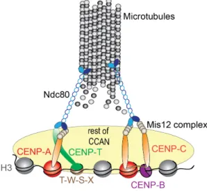
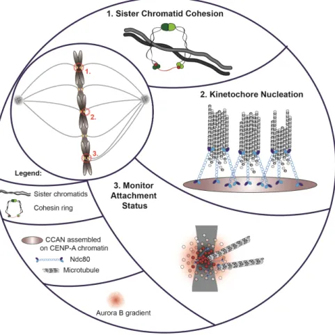
Centromeres are specialized genomic loci that drive accurate genome segregation across cell divisions. The core region of the centromere provides a structural platform for formation of the kinetochore, a protein complex that links chromosomes to spindle microtubules during mitosis (Figure 1.2) (Cheeseman and Desai, 2008; Foltz et al., 2006; Okada et al., 2006). The centromere nucleates the kinetochore via the assembly of a large group of proteins, the centromere-associated network (CCAN) (Cheeseman and Desai, 2008; Foltz et al., 2006; Izuta et al., 2006). Members of the CCAN network are constitutively present at the centromere throughout the cell cycle. During mitosis, they recruit a secondary protein complex known as the kinetochore. The core microtubule binding site of the kinetochore is comprised of the conserved microtubule-binding KMN network, consisting of the protein KNL1 as well as the Mis12 and Ndc80 complexes (Cheeseman et al., 2004, 2006; DeLuca et al., 2006).
8
The KMN network serves as an initial scaffold for the recruitment of Spindle assembly checkpoint proteins (SAC). SAC is the mitotic checkpoint ensuring that anaphase does not take place before chromosomes are bi-oriented on the spindle (facing opposite poles of the mitotic spindle) (Musacchio and Salmon, 2007). The SAC achieves this by generating a signal at unattached kinetochores, known as the MCC complex (MAD2, BUBR1, BUBR3 and CDC20). The MCC complex directly inhibits the anaphase promoting complex (APC/C), which is the key regulator of anaphase onset. In this way, unattached kinetochores generate a stop signal allowing for the spindle to attach. Upon spindle attachment, the MCC signal generation is inhibited, in part, by stripping off the MCC components from the kinetochore (Musacchio and Salmon, 2007). While the checkpoint blocks anaphase as long as kinetochores are unattached and not bi-oriented, regulation of such proper kinetochore-microtubule attachments is achieved by an error-correction mechanism and its key effector, Aurora B (Carmena et al., 2012). Inter-centromere localized Aurora B acts by phosphorylating its kinetochore targets and de-stabilizing attached microtubules (Figure 1.2). Upon correct attachment, kinetochore geometry changes due to the exerted tension. This shift removes Aurora B form its substrates, thus stabilizing the microtubule attachments that generate sufficient tension. As bi-orientation is a state in which the highest amount of tension will be exerted on kinetochore pairs, this state is preferably stabilized (Dewar et al., 2004; Watanabe, 2012).
The broader centromeric domain, termed pericentric heterochromatin, is the site which keeps the chromosomes together during mitosis. This is enabled by a ring-like molecule called cohesin which encompasses replicated DNA strands upon their duplication in S phase (Figure 1.2) (Haarhuis et al., 2014). Cohesin is dynamically associated along the length
9
of the chromosome; however most of the cohesin is unloaded during prometaphase, leaving only centromeric cohesin to keep the chromosomes together (Waizenegger et al., 2000). Cohesin degradation along with Cyclin B degradation marks the onset of anaphase. This simultaneous degradation is orchestrated by the APC/C. APC directly targets Cyclin B for degradation (Pines, 2006). Additionally, APC degrades securing (Uhlmann et al., 1999), which is an inhibitor of Separase. Separase, in turn, is a protease that cleaves cohesion at the metaphase to anaphase transition (Nasmyth and Haering, 2009; Peters et al., 2008), ensuring rapid segregation of chromosomes, coinciding with Cyclin B degradation and mitotic exit.
1.5 Centromere organization across different species
The role of the centromere in driving chromosome segregation is highly conserved, yet centromere size and genomic localization is remarkably different across eukaryotes (Malik and Henikoff, 2009). Depending on the size and localization of the centromere, eukaryotic chromosomes can be classified as monocentric or holocentric. The most prevalent monocentric chromosomes assemble centromeres on a single defined region, whereas holocentric ones do so along the whole length of the chromosome (nematodes, arachnids, and insects (Schvarzstein et al., 2010). Monocentric chromosomes can have two defined varieties of centromeres; the point centromere, as the one present in Saccharomyces cerevisiae, which is formed on a small stretch of centromeric DNA (~125 bp in budding yeast). The second type is a regional centromere that is nucleated on a larger chromosomal domain, ranging from a few kilobases in fungi such as Schizosaccharomyces pombe and Candida albicans, to hundreds of kilobases in most plants and animals (Malik and Henikoff, 2009).
10
Figure 1.2 Centromeres control chromosome segregation and mitotic progression. (1)
Sister chromatid cohesion is maintained at pericentric heterochromatin in mitosis to prevent premature chromosome separation (image adapted from: Mirkovic and Oliveira, 2017). (2) Centromeres form a structural platform for kinetochore nucleation, during mitosis. The latter includes the microtubule binding protein Ndc80 which allows chromosome segregation. (3) An Aurora B gradient originating from the inter-centromere region destabilizes proximal kinetochore-microtubule interactions to prevent asymmetric chromosome segregation.
1.6 Centromeric DNA
Since centromeres are directly associated with centromeric DNA, early models postulated that specific features of centromeric DNA are directing nucleation of the functional centromere. This is indeed true in the case of
11
the point centromere of S. cerevisiae that is defined by a specific DNA sequence found on all chromosomes (Clarke and Carbon, 1983). This centromeric DNA sequence is comprised of three functional elements termed centromere DNA element I (CDEI), CDEII and CDEIII. Combined they form a sequence of approximately 125 bp that is sufficient to confer mitotic stability when introduced into plasmids (Clarke and Carbon, 1980; Fitzgerald-Hayes et al., 1982; Hieter et al., 1985). The sequences of CDEI and CDEIII are conserved among all S. cerevisiae chromosomes, with CDEII organizing a single centromeric nucleosome containing the S. cerevisiae homologue of CENP-A called Cse4 (Meluh et al., 1998; Stoler et al., 1995).
Centromeres in most other organisms appear to be determined in DNA sequence independent fashion, insofar as that specific DNA sequences driving centromere assembly have not been identified. Instead of assembling on the specific DNA sequence, the majority of regional centromeres associate with highly repetitive tandem sequence repeats (Choo, 2001; Tyler-Smith and Floridia, 2000). In humans, the best characterized repeat unit is a 171 bp monomer known as α-satellite (or alphoid) DNA (Willard, 1985, 1990). These repeats exist in two distinct subtypes, type I and type II. Type I repeats, also known as α-I satellite DNA contain a 17 bp sequence termed the CENP-B box that recruits the conserved centromere protein B (CENP-B) (Earnshaw et al., 1987; Ikeno et al., 1994; Masumoto et al., 1989). α-I satellite repeats are flanked by α-II satellite DNA which contains divergent repetitive sequences and retrotransposons. Whereas repeat unit length tends to be similar between different organisms (e.g. 171 bp for primates, 186 bp in fish, 155 bp in insects) (Henikoff et al., 2001), the nucleotide sequence of these repeats displays high variability even between closely related species. Additionally,
12
multiple eukaryotic subfamilies display variable genomic localization of mitotically active centromeres, which shifted along the chromosome, independently of the surrounding sequences or structural rearrangements (Montefalcone et al., 1999; Rocchi et al., 2012; chapter 5 of this thesis). One of the most compelling evidence arguing against the role of DNA sequence as principal determinant of centromere localization came with the discovery of centromeres on atypical loci. These so-called neocentromeres, initially identified in 1993 on a mitotically stable derivative of chromosome 10, lacking typical centromeric sequence as well as the CENP-B protein that binds to those sequences (Voullaire et al., 1993). Up to this date, more than 130 unique human neocentromeres, spanning all chromosomes except 22, have been identified (Liehr, 2014; Marshall et al., 2008). Neocentromere genomic location is stably maintained throughout cell divisions where they confer mitotic stability to carrier chromosomes and, in some cases, neocentromeres are inherited through human generations (Amor et al., 2004; Capozzi et al., 2009; Knegt et al., 2003; Tyler-Smith et al., 1999; Ventura et al., 2004; Wandall et al., 1998), pointing to their meiotic stability as well. Importantly, large arrays of vacated α-satellite sequences do not display any centromeric function and can be retained on neocentric chromosomes, including meiotically stable ones (Bukvic et al., 1996; Hasson et al., 2011; Liehr et al., 2010; Tyler-Smith et al., 1999). Therefore, the case of the neocentromere argues that centromeric sequences are neither necessary nor sufficient for centromere specification in human cells.
Although not strictly required for establishment of an active centromere, specific features of centromeric DNA may have a contributory role in centromere specification. One well known feature of mammalian
13
centromeric DNA is the recruitment of CENP-B, a sequence specific DNA binding protein that recognizes a sequence, named CENP-B box, found within a proportion of α-satellite monomers (Masumoto et al., 1989). Although the absence of CENP-B protein doesn’t impair viability (Hudson et al., 1998), the presence of CENP-B boxes together with α-satellite DNA was found to be essential for formation of functional de novo centromere on artificial human chromosomes (Ohzeki et al., 2002). Additionally, the presence of CENP-B was found to enhance mitotic fidelity of human centromeres through stabilization of the kinetochore nucleating component of CCAN, CENP-C (Fachinetti et al., 2015; Hoffmann et al., 2016). The intricate relationship between repetitive centromeric DNA and centromere evolution is further discussed in the chapter 5 of this thesis.
1.7 CENP-A is epigenetically marking active centromeres
Epigenetic traits are heritable features whose propagation is not solely driven by underlying DNA sequences. As outlined above, centromeric DNA appears not to have a critical role in driving formation of a functional centromere. The current consensus in the centromere field is that the centromere-specific histone H3 variant CENP-A lies at the core of a positive epigenetic feedback loop and is sufficient to initiate and propagate centromeres. CENP-A, along with CENP-B and CENP-C were among the first centromere proteins to be identified using antibodies isolated from auto-immune sera from human scleroderma patients (CREST) (Earnshaw and Rothfield, 1985). These sera stained proteins at all active centromeres but, importantly, they are absent from an inactive centromere, suggesting a ´´chromatin based regulation´´ of the centromere (Earnshaw and Migeon, 1985). Soon after its initial discovery CENP-A was found to copurify with core histone proteins (Palmer et al., 1987) and have histone-like properties
14
(Sullivan et al., 1994). In addition, centromere specific CENP-A homologues exist in nearly all species analyzed so far (Malik and Henikoff, 2003; Talbert et al., 2012), with the exception of kinetoplastids and some holocentric insects that do not appear to contain a recognizable CENP-A homologue (Akiyoshi and Gull, 2013; Drinnenberg et al., 2014). A remarkable feature of centromeric chromatin is its requirement for the maintenance of centromeric chromatin across the germline in several, but not all organisms analyzed thus far. In mammals, early work has shown that CENP-A is present in mature bovine sperm, evading protamine deposition (Palmer et al., 1990), suggesting CENP-A may play a transgenerational role in mammals. Indeed, stable paternal transmissions of neocentormeres within human families demonstrate that the position of the centromere is inherited epigenetically at least through the male germline (Amor et al., 2004; Tyler-Smith et al., 1999). Sperm retained CENP-A was also found in X.laevis and D. melanogaster (Dunleavy et al., 2012; Milks et al., 2009; (Dunleavy et al., 2012; Milks et al., 2009; Raychaudhuri et al., 2012). In Drosophila, a causative role for CENP-A in germline centromere maintenance has been shown. Selective removal of the CENP-A homologue [known as CID or cenH3 (Talbert and Henikoff, 2013)] from paternal centromeres resulted in successful fertilization but in the selective failure to segregate paternal chromosomes in the zygote, despite normal segregation of maternal chromosomes and the availability of a maternal pool of CID (Raychaudhuri et al., 2012). The transgenerational necessity of CENP-A is not universal in life. C. elegans, sperm is devoid of CENP-A which is provided de novo through the maternally deposited pool of CENP-A (Gassmann et al., 2012). Further, during oogenesis, pre-existing CENP-A is removed, and is de novo deposited (Monen et al., 2005).
15
In proliferating somatic cells, loss of CENP-A is lethal due to the severe defects in chromosome segregation in all species analyzed (Black et al., 2007; Blower and Karpen, 2001; Buchwitz et al., 1999; Fachinetti et al., 2013; Henikoff et al., 2000; Howman et al., 2000; Régnier et al., 2005; Stoler et al., 1995; Talbert et al., 2002). Additionally, CENP-A is sufficient for the recruitment of virtually all known centromere and kinetochore proteins (Barnhart et al., 2011; Carroll et al., 2009; Foltz et al., 2006; Guse et al., 2011; Heun et al., 2006; Liu et al., 2006; Mendiburo et al., 2011; Okada et al., 2006), with the exception of the sequence specific DNA binding protein CENP-B (Pluta et al., 1992; Voullaire et al., 1993). In a groundbreaking study, (Mendiburo et al., 2011) used Drosophila S2 cells to tether CENP-A to a naïve chromatin domain containing Lac operator sequences (using a LacI DNA binding domain), not previously associated with centromere function. Once tethered, CENP-ACID-LacI creates a local nucleosome pool that is able to recruit virtually all known downstream centromere and kinetochore proteins allowing stable binding of microtubules. Importantly, once formed, this nascent centromere recruited naïve CENP-ACID, not previously associated with this region, even after the initial tether had been lost, indicative of self-propagation of CENP-ACID. Analogous experiments were performed with the CENP-A loading factor HJURP. In this case, not only de novo centromere formation was observed (Barnhart et al., 2011; Hori et al., 2013) but this centromere was shown to rescue chromosome stability and cell viability after deletion of the endogenous centromere in chicken DT40 cells (Hori et al., 2013). Exploiting conditional endogenous centromere deletion combined with artificial genomic targeting of LacI-fused CCAN components to LacO array, (Hori et al., 2013) demonstrated that CENP-C and CENP-I are sufficient to initiate a heritable centromere at the LacO array, as observed in the case of
LacI-16
HJURP tethering. This ectopic centromere functionally replaced the endogenous one and faithfully maintained chromosome Z ploidy in the cell population. Although these two CCAN components have been shown to be required for CENP-A assembly at endogenous centromeres (Okada et al., 2006; Erhardt et al., 2008; Carroll et al., 2010), these results show they can also be sufficient for de novo recruitment of CENP-A on naive chromatin. Importantly, this implies that although CENP-A chromatin provides a stable heritable core, its propagation involves a positive epigenetic feedback mechanism in which other CCAN components, themselves dependent on CENP-A, play an active role in CENP-A recruitment.
1.8 CENP-A nucleosomes are stably propagated at centromeres through mitotic and meiotic divisions
Early work indicates that total cellular CENP-A protein exhibits a remarkably long half-life and lives as long as the cell itself, equating ~50% decrease per cell generation (Shelby et al., 1997). The apparent slow turnover required the employment of specific tools to assess protein dynamics. Fluorescence recovery after photobleaching (FRAP) which relies on local, irreversible photo-bleaching of a fluorophore, followed by subsequent repopulation of a bleached area with unbleached molecules provides information of the local rate of protein turnover. FRAP experiments on budding yeast kinetochores (containing a single microtubule attachment site), revealed that the yeast CENP-A homologue, Cse4 displays very low turnover rates at centromeres except during S phase where all of the preexisting Cse4 nucleosomes are exchanged (Pearson et al., 2004). Cse4 was found to be stable specifically at the centromere, whereas the non-centromeric Cse4 is degraded via ubiquitin-mediated proteolysis (Collins et al., 2004). Stable binding of Cse4 at centromeres was recently confirmed in
17
elegant experiments using a photoconvertible Cse4-tdEos (Wisniewski et al., 2014). Eos, green in the unconverted state can be stably switched to red emission upon short wavelength excitation. Following conversion, Cse4 molecules were found to be stably associated with centromeres until their turnover during DNA replication.
Stability of the fission yeast, kinetochore-bound, CENP-A homologue was demonstrated using photobleaching of Cnp1-GFP (Coffman et al., 2011), which displayed a similar dynamics as previously described for Cse4 (Pearson et al., 2004). Interestingly, in contrast to the yeasts, holocentric C. elegans embryos, characterized by extremely short division times (~15min), photobleaching of embryonic CeCENP-A-GFP in anaphase in the one-cell embryo results in the complete fluorescence recovery in the next cell division, indicative of complete loss of pre-existing CeCENP-A nucleosomes (Gassmann et al., 2012). Here, sites for CeCENP-A deposition appear to be based on other genomic features rather than pre-existing CENP-A. These regions include those with low transcriptional activity in the parental germline (Gassmann et al., 2012) and sites of high DNA accessibility (Steiner and Henikoff, 2014).
In vertebrate cells, following the initial determination of CENP-A stability with a tagged but overexpressed shut-off allele in human cells (Shelby et al., 1997), a shut-off in the context of a full deletion of the CENP-A gene in chicken DT40 cells (Régnier et al., 2005) revealed that the loss rate of the cellular CENP-A pool is very slow indeed, with the first mitotic defects occurring only after 7-8 cell cycles. Similar results were obtained in human cells after conditional deletion of CENP-A (Fachinetti et al., 2013). The fact that these cells can survive for extended amount of time without continuous supply of fresh CENP-A, strongly suggests that pre-existing CENP-A, once
18
assembled into nucleosomes, remains stably bound to centromeric chromatin. While these studies determined that CENP-A turns over slowly, establishing the actual turnover rate proved difficult to determine. The FRAP methodology is suitable for determining protein dynamics at short time scales such as in organisms which have a short cell division time, but proofs limited for dissecting protein turnover and replenishment rates at long time intervals. This limitation was surmounted by the use of a fluorescent pulse labeling strategy such as SNAP-tag technology, which allows for pulse labeling and visualization of different cohorts of the same protein within whole cell populations. SNAP is a derivative of a human DNA repair enzyme, O6-alkylguanine-DNA alkyltransferase (AGT). The endogenous AGT enzyme recognizes O6-alkylated guanine in DNA, and transfers the alkyl group to a reactive cysteine residue. This self-labeling capacity is exploited in a mutant version of AGT (named SNAP) which has a high affinity towards synthetically engineered small, cell permeable molecules, such as benzylguanine (BG) (Keppler et al., 2003). The enzymatic reaction between SNAP and its substrate is irreversible, highly efficient and specific. Combining serial labeling of SNAP-tagged proteins with different SNAP substrates enables visualization and fate determination of pre-existing versus newly synthesized pools of the same protein (Bodor et al., 2012). Following a pulse labeled cohort of CENP-A-SNAP molecules over the course of 48-72 hours, demonstrated the stable transmission of CENP-A through mitotic divisions (Bodor et al., 2013; Jansen et al., 2007). The loss rate of this pool was found to equate ~50% during each cell division, consistent with quantitative recycling of old CENP-A during S phase, with no additional turnover (Bodor et al., 2013; Dunleavy et al., 2011; Jansen et al., 2007). This high rate of retention appears to be unique to CENP-A nucleosomes. Similar pulse labeling experiments on H3.1 and
19
H3.3 did not reveal such retention at centromeric chromatin (Bodor et al., 2013; Falk et al., 2016), indicating that the property of stable transmission is linked to CENP-A itself, not the centromeric chromatin environment as a whole. However, histone H4 shows a striking differential stability. In the genome overall its turnover rates are similar to that of H3.1, but at the centromere H4 is retained to the extent of CENP-A (Bodor et al., 2013). CENP-A directly contacts H4 in the prenucleosomal complex as well as within the nucleosome, forming a highly rigid structure (Black et al., 2004, 2007), likely directly stabilizing H4 at the centromere. The other remaining nucleosome partners, H2A and H2B, like H3.1 and H3.3 do not display any elevated retention at the centromere (Bodor et al., 2013). Hence, CENP-A/H4 forms a stable subnucleosomal complex that represents the epigenetic core of the centromere which is quantitatively maintained throughout multiple cell divisions.
The most striking example showcasing extreme stability of CENP-A nucleosomes is recent work in female mouse meiosis (Smoak et al., 2016). Like in humans, mouse oocytes are arrested in meiotic prophase I for an extended period of time. CENP-A is readily detected in arrested mouse oocytes. However, no assembly occurs at any appreciable rate. Consistent with that observation, deletion of the CENP-A in early oogenesis has no impact on long term (~1 yr) retention of centromeric CENP-A despite the lack of a nascent pool.
1.9 Determinants of CENP-A stability
The molecular underpinnings enabling the remarkable CENP-stability are, at least in part, embedded within CENP-A nucleosome itself. H3 and its isoform CENP-A, share substantial degree of common features, including ~75% sequence similarity in their histone fold domain (HFD). On the other
20
hand, a very low level of homology exists between the N-terminal histone tails of H3 and CENP-A, suggesting that this could be a differential feature functionally separating these two histones (Palmer et al., 1991; Sullivan et al., 1994). Unexpectedly, using chimeric proteins of H3 and CENP-A, it was shown that the HFD rather than the tail of CENP-A is responsible for its centromere targeting (Sullivan et al., 1994). The portion of CENP-A that confers its centromere targeting lies within this domain (HFD), in a subdomain termed CENP-A targeting domain (CATD), consisting of loop1 and the α2-helix (Black et al., 2004) (Figure 1.3A). Replacement of the equivalent domain in H3 with that of CENP-A is sufficient to target an H3CATD chimera to centromeres (Black et al., 2004, 2007) and neocentromeres (Bassett et al., 2010). Importantly, the CATD confers increased conformational rigidity to (CENP-A/H4)2 tetramers as well as CENP-A nucleosomes (Black et al., 2004). In addition, the CATD is directly recognized by its specific chaperone and assembly factor, HJURP (albeit different residues participate in HJURP recognition from those that are responsible for increased rigidity) (Bassett et al., 2012) (Figure 1.3B), which targets and deposits nascent CENP-A to centromeres (Dunleavy et al., 2009; Foltz et al., 2009; Shuaib et al., 2010). Remarkably, complete genetic substitution of endogenous CENP-A with H3CATD showed that this chimera retained the capacity to maintain its own centromeric levels over multiple cell cycles, suggesting that CATD is the critical subdomain responsible for longevity of CENP-A nucleosome in vivo (Fachinetti et al., 2013). Importantly, H3CATD chimeras display identical loading dynamics as wild type CENP-A, and crucially, induce increased stability of a direct binding partner of CENP-A-histone H4 (Bodor et al., 2013).
Taken together, these results indicate that long-term stability of CENP-A nucleosomes is conferred by its CATD, which allows for centromeric
21
maintenance of not only CENP-A, but (CENP-A/H4)2 subnucleosome core
as well. Interestingly, a C terminal LEEGLG motif of CENP-A (absent from H3), is responsible for the recruitment of the majority of downstream centromere and kinetochore proteins (Carroll et al., 2010; Fachinetti et al., 2013; Guse et al., 2011). This appears to be the main reason why a H3CATD chimera could, for a limited amount of time, reside at the centromere but was not sufficient to rescue a complete depletion of endogenous CENP-A. Thus, the CATD seems to be mostly dispensable for CENP-A function (recruitment of kinetochore proteins); rather it is responsible for maintenance of centromere identity.
In summary, the CATD emerges as a differential feature of CENP-A nucleosomes functionally separating CENP-A and H3 histones and implies that the extreme stability of CENP-A nucleosomes is encoded within CENP-A molecule itself. Recent work however defined CENP-C, a member of CCAN network, as an additional extrinsic factor contributing to CENP-A stability. CENP-C binds directly to chromatin-bound CENP-A, and as a consequence, induces structural changes in conformation of CENP-A nucleosomes. This results in increased rigidity of CENP-A nucleosomes, a feature likely contributing to its stable maintenance at centromeres, since CENP-C depletion causes a rapid loss of CENP-A from the chromatin (Falk et al., 2015).
1.10 The Constitutive Centromere-Associated Network (CCAN)
CENP-A acts as the most upstream component in kinetochore assembly by specifying the point of contact between the DNA and mitotic spindle. CENP-A directs the formation of the constitutive centromere associated network (CCAN) which in turn, during mitosis, recruits a secondary protein complex known as the kinetochore (Fukagawa and Earnshaw, 2014). The
22
constitutive centromere-associated network (CCAN) is composed of 16 proteins (Amano et al., 2009; Foltz et al., 2006; Hori et al., 2008; Obuse et al., 2004; Perpelescu and Fukagawa, 2011) among which only two directly recognize chromatin-incorporated molecules: C and CENP-N. CENP-C and CENP-N recognize distinct domains in the CENP-A nucleosome; CENP-N recognizes the CATD domain embedded within the HFD (Figure 1.3C) of CENP-A whereas CENP-C recognizes the C-terminal LEEGLG motif of CENP-A (Carroll et al., 2009, 2010; Logsdon et al., 2015). Recently, it has been shown that both the CATD and the N-terminal portion of CENP-A, together with its C terminal tail are contributing to the efficient recruitment of CENP-C to an ectopic genomic locus (LacO array in this case) (Logsdon et al., 2015) (Figure 1.3D)
CENP-N is required for mitotic progression and accurate chromosome segregation (Carroll et al., 2009; Foltz et al., 2006; McClelland et al., 2007). Centromeric localization of CENP-N is necessary for centromere localization of several CCAN components, including CENP-H, CENP-I, CENP-K, CENP-C and CENP-O (Carroll et al., 2009; McClelland et al., 2007). However, CENP-N doesn’t appear to be involved in recruitment of kinetochore proteins since depletion of CENP-N doesn’t affect the levels of the Nnf1 component of the Mis12 complex (McClelland et al., 2007). CENP-N and CENP-A centromere presence appear to be mutually dependent since depletion of CENP-N causes defective loading of nascent CENP-A (Carroll et al., 2009), and rapid destruction of CENP-A using an auxin-degron system leads to diminished centromere deposition of new CENP-N molecules in S phase (Hoffmann et al., 2016).
CENP-C is a DNA binding protein that localizes to inner centromere in a CENP-A dependent manner (Saitoh et al., 1992). CENP-C homologues
23
have been identified in virtually all model organisms, including yeast, flies, plants and mammals, and have been shown to be required for proper chromosome segregation and mitotic progression (Dawe et al., 1999; Erhardt et al., 2008; Fukagawa et al., 2001; Moore and Roth, 2001; Oegema et al., 2001; Schuh et al., 2007; Tomkiel et al., 1994). Interestingly, CENP-C is the only CCAN component thus far identified in Drosophila, which may explain why centromeric localization of CENP-ACID is absolutely dependent on CENP-C (Erhardt et al., 2008). CENP-C is required for the centromere localization of several kinetochore proteins, including Knl1, the Mis12 complex and the Ndc80 complex that together are known as the KMN network, which forms the principal microtubule binding complex in the kinetochore (Cheeseman et al., 2006; Klare et al., 2015; Milks et al., 2009; Screpanti et al., 2011). CENP-C is also required for the recruitment of checkpoint proteins, for mitotic checkpoint function (Kwon et al., 2007; Przewloka et al., 2011; Screpanti et al., 2011), and for the centromere localization of other CCAN components such as CENP-H, CENP-I, CENP-K and CENP-T (Carroll et al., 2010). In humans, CENP-C deposition occurs a few hours after CENP-A assembly, and the presence of CENP-A nucleosomes is absolutely required for centromeric targeting of C (Hoffmann et al., 2016). In accordance to direct binding of CENP-C to CENP-CENP-A nucleosomes, excision of endogenous CENP-CENP-A, its CENP-C terminal domain or acute degradation of CENP-A using and auxin-degron system, leads to the proportional reduction in the amount of centromeric CENP-C (Fachinetti et al., 2013; Hoffmann et al., 2016). However, after the initial reduction, the levels of CENP-C become stabilized even when total amount of CENP-A is reduced to ~1% of its initial level. This retention of CENP-C at the centromeres is enhanced by the DNA binding CENP-B protein that recognizes the amino tail of CENP-A nucleosomes and
24
adjacent alphoid DNA (Figure 1.3E); once at the centromeres, CENP-B further stabilizes the levels of centromeric CENP-C through a direct protein-protein interaction, thereby providing an additional parallel pathway for CENP-C dependent kinetochore assembly (Fachinetti et al., 2015; Hoffmann et al., 2016) (Figure 1.4). Therefore, at centromeres, CENP-C has a dual role: on one hand it stabilizes centromeric CENP-A nucleosomes, thus contributing to its maintenance across cell cycle; and on the other, forms a critical link between inner centromere domain and the kinetochore proteins that bind plus ends of spindle microtubule.
Another member of the CCAN is CENP-T, which together with its binding partners CENP-S, CENP-W and CENP-X forms a platform for binding of proteins that in turn interact with microtubules (Amano et al., 2009; Foltz et al., 2006; Hori et al., 2008, 2013; Obuse et al., 2004) (Figure 1.4). CENP-T and -W form a hetero-tetramer with CENP-S and –X and shows DNA binding activity. It protects a ~100 bp region of nucleosome-free DNA forming a nucleosome-like structure (Nishino et al., 2012). The CENP-T-W complex does not directly associate with CENP-A, but with histone H3 in the centromere region (Hori et al., 2008). Consistently, deposition of nascent CENP-T does not solely depend on CENP-A nucleosomes (Nishino et al., 2012). However, recently it has been shown that both N and C-terminal tails of CENP-A, together with CATD promote recruitment of CENP-T to ectopic centromeres (Logsdon et al., 2015). Whether this A driven recruitment of T resulted in the formation of CENP-T/W/S/X complex is not known. However, it is likely that another adaptor protein (possibly a member of CCAN) is mediating the interaction between CENP-A and CENP-T, given that CENP-T remains mostly unaffected by rapid CENP-A degradation, which is a sharp contrast to the members of
25
CCAN that are directly interaction with CENP-A nucleosomes (Hoffmann et al., 2016).
Figure 1.3 CENP-A protein sequence along with all relevant domains [alpha N helix (αN), alpha-1 helix (α1), Loop-1 (L1), alpha-2 helix (α2), Loop-2 (L2), alpha-3 helix (α3)] are depicted (yellow). Domains that are responsible for specific features of CENP-A are highlighted in red. (A) Centromere targeting domain (CATD) is allowing differentiation between CENP-A and H3, and is responsible for centromeric targeting of CENP-A. (B) HJURP recognizes CATD within CENP-A/H4/HJURP prenucleosomal complex. Serine 68 located outside of CATD further stabilizes this interaction. (C) CENP-N directly recognizes CATD in CENP-A nucleosomes. (D) CENP-C directly recognizes the C-terminal LEEGLG sequence of CENP-A nucleosomes. Additionally, CENP-C recognizes N terminal portion of
26
Figure 1.3 Continued: CENP-A nucleosomes, where it interacts with CENP-B. (E) CENP-B
binds to CENP-B boxes in alpha satellites and recognizes N terminal domain of CENP-A.
The N terminus of CENP-T proteins interacts directly with the Ndc80 complex in the outer kinetochore (Gascoigne et al., 2011; Nishino et al., 2013). The CENP-T/-W complex is not maintained at centromeres through cell divisions, and new deposition is absolutely required at each cell cycle for kinetochore function. Consistently, CENP-T and CENP-W are loaded and become enriched at centromeres during late S and G2 phases, just before the recruitment of the KMN network to the kinetochores (Prendergast et al., 2011).
Figure 1.4 The CCAN connects centromeric DNA and microtubule plus ends.
CENP-T/W/S/X complex forms a bridge between centromeric chromatin and the mitotic kinetochore through a direct interaction between CENP-T and the Ndc80 microtubule-binding complex. CENP-C (which directly recognizes CENP-A nucleosomes) binds the remaining of CCAN and the Mis12 complex, which in turns directly interacts with Ndc80 complex. CENP-C forms a dimer and interacts with the DNA sequence specific protein CENP-B. CENP-B functions as a dimer and binds CENP-B boxes at alpha satellites. Simultaneously, CENP-B directly
27
Figure 1.4 Continued: interacts with CENP-C, resulting in increased mitotic fidelity of a
CENP-C dependent branch of kinetochore nucleation.
Remarkably, CENP-A per se is expendable for proper mitotic division given that acute destruction of CENP-A (employing an auxin-degron system), 4h prior to mitosis doesn’t significantly influence proper chromosomes segregation (Hoffmann et al., 2016). However if CENP-A is depleted prior to critical time window when CCAN components are targeted to the centromere, the integrity of whole kinetochore is diminished, thus leading to higher frequency of mitotic errors. Taken together, these results indicate that CENP-A containing nucleosomes provide the initial structural platform for the assembly of the kinetochore. Once assembled, building blocks of kinetochore are autonomous in regard to CENP-A, as exemplified by the stable microtubule-kinetochore attachment during mitosis and faithful genome segregation occurring in the absence of CENP-A.
1.11 The modularity of CENP-A dependent kinetochore assembly
As outlined above, CENP-A nucleosomes actively participate in the nucleation of kinetochore, while being dispensable for its maintenance after the initial establishment. Current models for centromere and kinetochore architecture are based on repeated individual subunits, in which the amount of centromere components directly dictates the number of downstream kinetochore proteins, and ultimately the number of microtubule attachment sites. This form of organization was initially proposed in 1991, when islets of proteins recognized via CREST antibodies were identified in a stretched centromeric DNA fiber (Zinkowski et al., 1991). Evidence for such a modular organization is found at the S. cerevisiae point centromere in which the proteins forming the interface between centromeric chromatin and the microtubule plus end exist in specific stoichiometries (Joglekar et
28
al., 2006). Regional centromeres tend to assemble on large stretches of centromeric DNA compared to the budding yeast point centromeres and they are bound by multiple spindle microtubules [ranging from 2-4 in fission yeast to ~17 in the case of humans (McEwen et al., 2001; Sagolla et al., 2003). Initial studies, focused on the centromeres of fission yeast and C. albicans (Joglekar et al., 2008), found a strikingly constant ratio between the amount of centromeric CENP-A nucleosomes, structural components of kinetochore and number of microtubules attached during mitosis. In these cases, while absolute numbers differ, the number of kinetochore proteins per microtubule attachment are very similar between budding and fission yeast. For both yeasts there are 6-8 molecules of KMN network per kinetochore-microtubule attachment. These findings strongly argue that the regional centromeres of fission yeast are composed of repeated structures reminiscent of the ones existing in budding yeast. This apparent kinetochore architecture extends to certain metazoan species, such as chicken DT40 cells, in which the copy number of CCAN network members (namely CENP-C, CENP-H, CENP-I and CENP-T) is in nearly stoichiometric relation to KMN network members (Mis12, Knl1 and Ndc80), which, once again, assemble at ~8 molecules per microtubule (Johnston et al., 2010).
However, a direct relationship between the number of centromeric CENP-A nucleosomes and amount of downstream kinetochore components is incompatible with the fact that constitutive overexpression of Cnp1 does not lead to significant changes in the copy number of kinetochore protein (Joglekar et al., 2008). Consistently, in C. albicans, the number of CaCse4 nucleosomes is larger than the number of microtubule attachment sites (Joglekar et al., 2008), indicating that the relationship between centromeric chromatin and microtubule attachment sites is less defined. This notion is
29
further supported by the fact that CENP-A depletion in human cells resulting in ~7% of total centromeric (Fachinetti et al., 2013) or ~10% of cellular pool (Liu et al., 2006) had no effect on centromere integrity at least in the short term. Upon partial loss of CENP-A, proteins such as CENP-C and CENP-T remain largely unaffected (Fachinetti et al., 2013). In an extreme case, upon complete acute complete loss of CENP-A, the centromere remains mitotically functional at least initially, after which failure to propagate the centromere in the next division results in gradual loss of centromere components (Hoffmann et al., 2016). In agreement with the stoichiometric disconnect between centromeric chromatin and the rest of the centromere, altering CENP-A levels in human RPE cell line between 40% and 240% relative to wild type, showed no significant effect on the amount of critical kinetochore proteins (Bodor et al., 2014). These included CENP-C and CENP-T, which are responsible for mitotic recruitment of the KMN network (Gascoigne et al., 2011), as well as the key microtubule binding protein Hec1/NDC80 (Cheeseman et al., 2006; DeLuca et al., 2006). Taken together, these results argue that on a typical human centromere the amount of CENP-A nucleosomes is in excess compared to the critical number necessary to maintain the centromere, which could in part be facilitated through semi-stable self-regulated recruitment of downstream CCAN proteins.
Another insight into the relationship between CENP-A chromatin and the kinetochore comes from overexpression studies. Excess CENP-A results in its mislocalization to non-centromeric sites (Athwal et al., 2015; Heun et al., 2006; Lacoste et al., 2014). Mistargeted CENP-A is not randomly distributed; rather it is enriched at sites of high histone turnover (Athwal et al., 2015; Lacoste et al., 2014). Even at physiological expression levels, CENP-A is present outside the centromere in a surprisingly high amount

![Figure 1.3 CENP-A protein sequence along with all relevant domains [alpha N helix (αN), alpha-1 helix (α1), Loop-1 (L1), alpha-2 helix (α2), Loop-2 (L2), alpha-3 helix (α3)] are depicted (yellow)](https://thumb-eu.123doks.com/thumbv2/123dok_br/19194704.951292/46.765.107.665.211.688/figure-cenp-protein-sequence-relevant-domains-depicted-yellow.webp)