ANTICANCER ACTIVITY OF
MARINE-DERIVED FUNGI
EXTRACTS AND ISOLATED
COMPOUNDS IN HUMAN
CANCER CELL LINES
MARIA PRATA E CASTRO DE SENA ESTEVES DISSERTAÇÃO DE MESTRADO APRESENTADA
AO INSTITUTO DE CIÊNCIAS BIOMÉDICAS ABEL SALAZAR DA UNIVERSIDADE DO PORTO EM
MARIA PRATA E CASTRO DE SENA ESTEVES
ANTICANCER ACTIVITY OF MARINE-DERIVED FUNGI EXTRACTS
AND ISOLATED COMPOUNDS IN HUMAN CANCER CELL LINES
Dissertação de Candidatura ao grau de Mestre em Ciências do Mar – Recursos Marinhos submetida ao Instituto de Ciências Biomédicas de Abel Salazar da Universidade do Porto
Orientadora – Doutora Alice Fernanda Abreu Ramos Categoria – Investigadora Post-Doc
Afiliação – Centro Interdisciplinar de Investigação Marinha e Ambiental
Co-Orientador – Professor Doutor Eduardo Jorge Sousa Rocha Categoria – Professor Catedrático Afiliação – Instituto de Ciências Biomédicas Abel Salazar
Acknowledgements
I would like to thank the following people who made this Master thesis possible:
Professor Doctor Eduardo Rocha, my co-supervisor, for entrusting me this work and for all the support and guidance provided during my Masters training.
Doctor Alice Ramos, my supervisor, I am very thankful for the limitless support, advise, cooperation and friendship.
Professor Doctor Anake Kijjoa and his research group for providing both the extracts and compounds used in this study, which would otherwise have been impossible.
Professor Doctor Daniela Gargiulo, for all the thoughtfulness, help and for explaining to me stress relief in neurological terms.
Pedro Marques and Márcia Moreira for their invaluable assistance in the practical work.
The staff at the LECEMA group at CIIMAR, who were always kind and supportive when answering my queries.
The staff at the Laboratory of Histology and Embryology at ICBAS, in particular to Dr. Fernanda Malhão, for the assistance and support.
To my family. To Bruno.
This work was partially funded by the ICBAS and by the Project MARBIOTECH (reference NORTE-07-0124-FEDER-000047), co-financed by the North Portugal Regional Operational Programme (ON.2 – O Novo Norte), under the National Strategic Reference Framework (NSRF), through the European Regional Development Fund (ERDF).
Abstract
The marine environment is a prolific and largely unexplored source for the prospection of bioactive compounds. The isolation of such substances from marine microorganisms has gained increasing interest over the last decade. Marine fungi are known to produce a great variety of secondary metabolites with unique chemical structures that have appealing bioactivities.
Cancer is currently one of the most globally prevalent diseases. Furthermore, the incidence of cancer is steadily increasing, accompanying the ageing and growth of populations, as well as imbalanced life-styles and declining environmental conditions. The treatment of cancer is commonly (partially or solely) based on chemotherapy, however, resistance to common chemotherapeutic agents and hazardous side effects highlight the importance of the search for safer, more efficient and tumour-specific anticancer drugs.
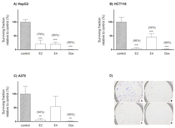
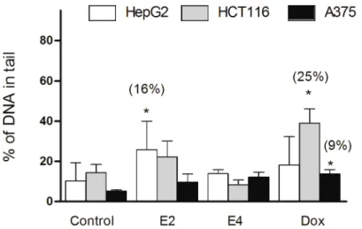
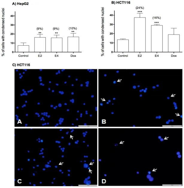
The first part of this study aimed to assess the in vitro anticancer activity of the crude ethyl extracts of three marine sponge-derived fungi, namely, Aspergillus similanensis KUFA 0013 (E1), Neosartorya paulistensis KUFC 7897 (E2) and Talaromyces trachsypermus KUFC 0021 (E3), and of one sea fan-derived fungi Neosartorya siamensis KUFA 0017 (E4) in a panel of seven cancer cell lines, glioblastoma (U251), malignant melanoma (A375), non-small cell lung cancer (A549), hepatocellular carcinoma (HepG2), colon carcinoma (HCT116 and HT29) and breast adenocarcinoma (MCF-7). Extracts E2 and E4 significantly decreased cell proliferation in HepG2, A375 and HCT116 cancer cells, while extract E2 also decreased long-term cell proliferation in all three cell lines, and extract E4 in HepG2 and HCT116 cell lines, as observed by the clonogenic assay. Both extracts also managed to induce cell death in HCT116 and HepG2 cells. No genotoxic effect was observed, thus the observed cell death induction does not seem related to the induction of DNA damage, namely by the induction of DNA strand breaks.
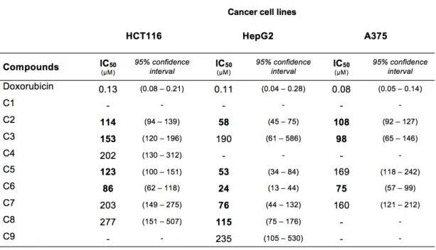
The second part of this study focused on nine compounds isolated from Neosartorya siamensis KUFA 0017 (E4), namely, 2,4-dihydroxy-3-methylacetophenon (C1), nortryptoquivaline (C2), chevalone C (C3), tryptoquivaline H (C4), fiscalin A (C5), epi-fiscalin C (C6), epi-neoepi-fiscalin A (C7), epi-epi-fiscalin A (C8) and tryptoquivaline F (C9), which were also screened for in vitro anticancer activity in the three cell lines in which extract E4 presented most activity, namely in HepG2, A375 and HCT116 cells. Results showed that compounds C2, C3, C5, C6, C7 and C8 presented a significant anti-proliferative effect against the selected cell lines, with IC50 values ranging from 24 to 153 µM. The induction
of cell death was observed in HepG2 cells by compounds C2, C5, C7 and C8, and in HCT116 cells by compounds C2, C3 and C5. The induction of cell death is once more possibly unrelated to induction of DNA damage — at least to a point of induction of DNA strand breaks — as none of the compounds exhibited genotoxic activity assessed by the comet assay.
In summary, crude ethyl extracts of the marine-derived fungi Neosartorya paulistensis and Neosartorya siamensis were shown to have anticancer activity against hepatocellular carcinoma, colon carcinoma and malignant melanoma cell lines. The activity of compounds isolated from N. siamensis was in most cases greater than that of the extract, presenting appreciable IC50 values, ranging from 24 to 153 µM, and considerable induction of cell
death, with compounds exhibiting a range of 5 to 12% increase of cells presenting nuclear condensation. Further studies should be undertaken to elucidate the underlying mechanisms of action and molecular targets.
Resumo
O meio marinho é um ambiente altamente prolífico e ainda parcamente explorado na procura de compostos bioactivos. O isolamento de compostos bioactivos provenientes de microorganismos marinhos tem adquirido particular relevância na última década. Os fungos marinhos são já reconhecidos pela produção de uma grande variedade de metabolitos secundários com estruturas químicas singulares, apresentando bioactividade de interesse.
O cancro é actualmente uma das doenças com maior prevalência a nível global. A incidência do cancro tem aumentado consistentemente, acompanhando o envelhecimento e crescimento das populações, bem como estilos de vida pouco equilibrados e condições ambientais prejudiciais. O tratamento do cancro é habitualmente baseado, em parte ou exclusivamente, na quimioterapia, contudo, o aumento da resistência associada a agentes quimioterapêuticos de uso comum, acompanhado de efeitos secundários nefastos, revela a importância na pesquisa de drogas anticarcinogénicas mais seguras, eficientes e específicas.
A primeira parte deste estudo baseou-se na avaliação da actividade anticarcinogénica in vitro de extractos de acetato de etilo de três fungos marinhos associados a esponjas, nomeadamente, Aspergillus similanensis KUFA 0013 (E1), Neosartorya paulistensis KUFC 7897 (E2) e Talaromyces trachsypermus KUFC 0021 (E3), e de um fungo associado a gorgónias, Neosartorya siamensis KUFA 0017 (E4), num painel de sete linhas celulares de cancro, nomeadamente, glioblastoma (U251), melanoma maligno (A375), carcinoma de pulmão de não-pequenas células (A549), carcinoma hepatocelular (HepG2), carcinoma do cólon (HCT116 e HT29) e adenocarcinoma da mama (MCF-7). Os extractos E2 e E4 levaram a uma diminuição significativa da proliferação celular em células HepG2, A375 e HCT116, enquanto que o extracto E2 diminuiu também a proliferação celular a longo termo nas mesmas três linhas celulares, sendo que o extracto
E4 teve efeito similar em HepG2 e HCT116, tal como observado pelo ensaio clonogénico.
Ambos os extractos induziram morte celular em células HCT116 e HepG2. Não foi observado qualquer efeito genotóxico, consequentemente é possível sugerir que a indução de morte celular observada não aparenta estar relacionada com a indução de danos no ADN, nomeadamente indução de quebras nas cadeias de ADN.
A segunda parte deste estudo focou-se em nove compostos isolados de Neosartorya siamensis KUFA 0017, nomeadamente, 2,4-dihydroxy-3-methylacetophenon (C1),
nortryptoquivaline (C2), chevalone C (C3), tryptoquivaline H (C4), fiscalin A (C5), epi-fiscalin C (C6), epi-neoepi-fiscalin A (C7), epi-epi-fiscalin A (C8) e tryptoquivaline F (C9), que foram também analisados em termos de actividade anticarcinogénica in vitro nas três linhas celulares onde o extracto E4 apresentou maior actividade, especificamente em células HepG2, A375 e HCT116. Os resultados demonstraram que os compostos C2, C3,
C5, C6, C7 e C8 apresentaram um efeito anti-proliferativo significativo contra as linhas
celulares seleccionadas, com valores de IC50 a variar entre 24 a 153 µM. A indução de
morte celular foi observada em HepG2 por via dos compostos C2, C5, C7 e C8, enquanto que em HCT116, por via dos compostos C2, C3 e C5. Mais uma vez, a indução de morte celular parece não estar relacionada com a indução de danos no ADN nomeadamente indução de quebras nas cadeias do ADN, uma vez que nenhum dos compostos exibiu actividade genotóxica observável pelo ensaio cometa.
Em suma, os extractos de acetato de etilo dos fungos marinhos Neosartorya paulistensis e Neosartorya siamensis demonstraram possuir uma actividade anticarcinogénica relevante contra linhas celulares de carcinoma hepatocelular, carcinoma do cólon e melanoma maligno. A actividade dos compostos isolados de N. siamensis suplantou na sua maioria a actividade do extracto, apresentando valores de IC50 apreciáveis, numa gama de valores de IC50 entre 24 a 153 µM, e uma indução de
morte celular considerável, onde os compostos induziram um aumento de 5 a 12% de células com núcleos condensados. Estudos futuros deverão procurar elucidar quais os mecanismos e alvos moleculares associados.
Index
Acknowledgments ... i Abstract ... iii Resumo ... v Index ... vii Abbreviations ... ix Preface ... xiii List of publications ... xv1. Chapter I – General introduction ... 1
1.1. Cancer ... 3
1.1.1. Demographics of cancer ... 3
1.1.1.1. Colorectal carcinoma ... 4
1.1.1.2. Hepatocellular carcinoma ... 4
1.1.1.3. Malignant Melanoma ... 5
1.1.2. The biology of cancer ... 5
1.1.2.1. Carcinogenesis ... 5
1.1.2.2. Programmed cell death and cancer ... 8
1.1.2.2.1. Apoptosis in cancer ... 9
1.1.3. Therapeutic approaches in cancer ... 11
1.2. Marine systems as a source of bioactive compounds ... 13
1.2.1. Marine fungi ... 14
1.2.2. Fungi associate relationships as a source of bioactive compounds ... 17
1.2.3. Anticancer compounds of sponge and sea fan derived fungi origin ... 19
1.2.4. Neosartorya genus ... 21
1.2.5. References ... 23
2. Chapter II – Testing the potential of four marine-derived fungi extracts as anti-proliferative and cell death-inducing agents in seven human cancer cell lines ... 31
3. Chapter III – Cytotoxic activity of compounds isolated from the marine fungi
Neosartorya siamensis in human cancer cells ... 59
3.1. Manuscript ... 63
4. Chapter IV – Conclusions and future perspectives ... 83
4.1. Conclusions and future perspectives ... 85
5. Appendix – Experimental protocols ... 87
P1. Cell culture ... 89
P2. MTT colorimetric assay ... 93
P3. Comet assay ... 97
P4. Nuclear condensation assay ... 103
Abbreviations
A375 – Human Malignant Melanoma Cell Line A549 – Human Non-small Lung Cancer Cell Line Apaf-1 – Apoptosis Protease Activating Factor-1 BGC823 – Human Gastric Cell Line
CSC – Cancer Stem Cell CTC – Cancer Transitioning Cell DAPI – 4',6-diamidino-2-phenylindole DISC – Death Inducing Signaling Complex DMEM – Dulbecco’s Modified Eagle Medium DMSO – Dimethyl sulfoxide
DNA – Deoxyribonucleic acid
ED50 – Half Maximal Effective Concentration
EDTA – Ethylenediaminetetraacetic acid EMA – European Medicines Agency EU – European Union
Fas – First Apoptosis Signal FBS – Fetal Bovine Serum
FDA – Food and Drug Administration
GI50 – Half Maximal Growth Inhibitory Concentration
IAP – Inihibitor of Apotosis Proteins NCI-H1975 – Non-small cell lung cancer
HEPES – 4-(2-hydroxyethyl)-1-piperazineethanesulfonic acid HBV – Hepatitis B Virus
HCT116 – Human Colorectal Carcinoma Cell Line HCV – Hepatitis C Virus
HepG2 – Human Hepatocellular Carcinoma Cell Line HL-60 – Human Promyelocytic Leukemia Cell Line Hs683 – Human Oligodendroglioma Cell Line Huh-7 – Human Hepatocellular Carcinoma Cell Line
HT29 – Human Caucasian Colon Adenocarcinoma Grade II Cell Line IC50 – Half Maximal Inhibitory Concentration
K562 – Human Erythromyeloblastoid Leukemia Cell Line LMP – Low Melting Point Agarose
MCF-7 – Human Breast Adenocarcinoma Cell Line MEM – Minimum Essential Medium Eagle
MOLT4 – Human Acute Lymphoblastic Leukemia Cell Line
MTT - 3-(4,5-dimethylthiazol-2-yl)-2,5-diphenyltetrazolium bromide NCI-H460 – Human Non-small Lung Cancer Cell Line
NMP – Normal Melting Point Agarose
P388 – Murine Lymphocytic Leukemia Cell Line PBS - Phosphate Buffered Saline
PFA – Paraformaldehyde PLC – Primary Liver Cancer ROS – Reactive Oxygen Species
RPMI – Roswell Park Memorial Institute Medium SK-MEL-28 – Human Malignant Melanoma Cell Line SMAC – Small Mitochondria-derived Activator of Caspases TNF – Tumour Necrosis Factor
U373 – Human Glioblastoma Cell Line
Preface
In the last few decades, there has been an increasing demand for novel compounds with pharmaceutical applications. The marine environment has become a leading hotspot for the bio-prospection of bioactive compounds, and due to the notable diversity and unique chemical structures, marine sourced natural products possess interesting properties that deserve further research and development into biotechnological applications.
This work was developed in the context of the project MARBIOTECH in the Interdisciplinary Centre of Marine and Environmental Research (CIIMAR) of the University of Porto, which aims to obtain bioactive compounds from marine organisms, with the objective of screening these compounds for a diversity of biological activities, such as allelopathic, antimicrobial, antifouling and anticancer activity.
In this context, the aim of this study was to screen several extracts obtained from marine invertebrate-associated fungi for in vitro anticancer activity in a panel of cancer cell lines. The screening of anticancer activity was based on the observation of anti-proliferative activity, cell death induction and genotoxic activity by induction of DNA damage (single strand breaks and alkali-labile sites). The following goal was to select extracts with demonstrated bioactivity and proceed to the subsequent isolation of compounds, and to screen these compounds for in vitro anticancer activity.
In summary, this work is organized in four chapters. Chapter I is a brief review to the themes of cancer and bioactive compounds of marine origin. Chapter II and III are comprised by original manuscripts, of which the first has been submitted and the latter is to be submitted to a peer-reviewed international journal. Chapter IV presents an overall conclusion of the work and future perspectives, while the Appendix displays the protocols used during this work to a more extensive detail.
List of publications
The elaboration of this Master thesis, and cooperation with other ongoing related works, rendered data sets that were included both in presentations in international meetings, with published abstracts, and in original papers, as follows:
1. Articles under submission or to be submitted to international peer-reviewed journals − Ramos A. A.*, Prata-Sena M.*, Castro-Carvalho B., Dethoup T., Buttachon S., Kijjoa A. &
Rocha E. (2014) Testing the potential of four marine-derived fungi extracts as anti-proliferative and cell death-inducing agents in seven human cancer cell lines. Submitted.
− Ramos A. A.*, Castro-Carvalho B.*, Prata-Sena M., Dethoup T., Buttachon S., Kijjoa A. & Rocha E. (2014) Extracts from Neosartorya (fungi) species exhibit anti-proliferative activity with cell death induction in colon, breast and skin cancer cell lines. Submitted.
− Prata-Sena M., Ramos A. A., Castro-Carvalho B., Dethoup T., Buttachon S., Kijjoa A. & Rocha E. (2014) Cytotoxic activity of compounds isolated from marine-derived fungi
Neosartorya siamensis in human cancer cells. To be submitted.
− Castro-Carvalho B., Ramos A. A., Prata-Sena M., Dethoup T., Buttachon S., Kijjoa A. & Rocha E. (2014) Marine-derived fungi extracts and isolated compounds enhance the anticancer activity of doxorubicin in non-small cell lung cancer cells. To be submitted.
2. Abstracts published in international peer-reviewed journals
− Prata-Sena, M., Ramos, A., Castro-Carvalho, B., Dethoup, T., Buttachon, S., Kijjoa, A., & Rocha, E. (2014). Anti-proliferative and pro-apoptotic activities of two marine sponge-derived fungi extracts in HepG2, HCT116 and A375 cancer cell lines. Planta Medica,
80(16), P1N3. doi: 10.1055/s-0034-1394594
− Castro-Carvalho, B., Ramos, A., Prata-Sena, M., Dethoup, T., Buttachon, S., Kijjoa, A., & Rocha, E. (2014). Extracts from the marine fungus Neosartorya tsunodae and the soil fungus Neosartorya fischeri exhibit anti-proliferative and pro-apoptotic effects in human cancer cell lines. Planta Medica, 80(16), P1N32. doi: 10.1055/s-0034-1394622
− Ramos, A., Moreira, M., Castro-Carvalho, B., Prata-Sena, M., Dethoup, T., Buttachon, S., Kijjoa, A., & Rocha, E. (2014). Marine-derived fungi extracts increase doxorubicin's cytotoxic effect in lung cancer cells. Planta Medica, 80(16), P1N25. doi: 10.1055/s-0034-1394615
− Ramos, A., Malhão, F., Ferreira, A., Alves, Â., Castro-Carvalho, B., Prata-Sena, M., Gargiulo, D., Dethoup, T., Buttachon, S., Lobo-da-Cunha, A., Kijjoa, A., Rocha, E. (2014) Marine and soil fungi extracts with anti-proliferative activity induce morphological alterations in breast cancer cells. Microscopy and Microanalysis (in press)
Note: 1) Abstracts published in Planta Medica concern posters presented at the 62nd Interna-tional Congress and Annual Meeting of the Society for Medicinal Plant and Natural Product Re-search – GA2014, between the 31st of August and 4th of September 2014, in Guimarães; and 2) extended abstract to appear in Microscopy and Microanalysis was derived from a poster presented at the INCOMAM'14 — International Conference on Microscopy and Microanalysis, XLVIII Con-gress of the Portuguese Microscopy Society, from the 6th to the 7th of November 2014, in Porto.
CHAPTER I
GENERAL INTRODUCTION
1.1. CANCER
Cancer is the general denomination for a series of diseases associated to regulatory abnormalities in cell growth and homeostasis. Overall, more than 100 distinct types of cancer have been described (Hanahan & Weinberg, 2000).
The etymology of the term “cancer” originates from the greek word karkinos, and its coinage is attributed to Hippocrates (460-370 B.C.), who made an analogy of the disease and its lesions to a moving, clasping crab. This early observation and appellation comes as no surprise, considering that in spite of cancer usually being considered a disease of the ‘modern ages’, it has accompanied both animals and mankind since time immemorial. The oldest description of cancerous lesions, in this case of breast cancer, dates back to Egypt in 3000 B.C. (Hajdu, 2011).
Cancer currently represents a high economic and societal burden for both developed and developing countries. Several factors have been pinpointed as causative factors of cancer. Environmental factors account for around 90-95% of all cancers, while inherited genetic factors are involved in 5-10% of the cases (Anand et al., 2008). Examples of environmental factors that have been correlated with the onset of cancer are, for example, smoking tobacco, alcohol consumption, diet, obesity, infectious agents, environmental pollution, radiation and physical inactivity (Jemal et al., 2011). Ultimately, this disease is best fought by prevention, where prophylactic measures such as annual cancer screenings in critical age-groups, vaccination for viral-induced cancers, the consumption of fruit, vegetables, wholegrain foods, teas and spices, vitamins, physical exercise and caloric restrictions are deemed essential for the maintenance of good health and lowering of cancer risk factors (Anand et al., 2008).
1.1.1. Demographics of cancer
In the 2012 GLOBOCAN project, it was estimated that there was a worldwide incidence of 14.1 million new cancer cases, 32.6 million people living with cancer and 8.2 million deaths due to cancer. Over half of all cancer cases and cancer-related deaths occurred in developing countries. The five most common cancers are lung, breast, colorectal, prostate
including liver cancer, are considered to be the deadliest cancers, with the highest annual mortality rates. Considering the ageing and growth of the global populations, projections based on this study suggest an increase of 19.3 million new cases of cancer per annum by the year 2025 (Ferlay et al., 2013).
1.1.1.1. Colorectal carcinoma
Colorectal cancer is one of the most commonly diagnosed cancers on a global scale, arising each year 1.23 million new cases (Ferlay et al., 2010). Around 55% of the total colorectal cancer cases arise in more developed regions (Ferlay et al., 2013). Notwithstanding its decreasing tendency in developed countries due to increased awareness and anticipated detection, shifts in key risk factors such as smoking, alcohol abuse, obesity, high caloric intake, low vegetable and fruit intake and physical inactivity have lead to an increase in the number of cases (Ferlay et al., 2010; LeMarchand et al., 1997). An important factor, however, is the hereditary factor involved in the development of colorectal cancer (Jasperson et al., 2010).
1.1.1.2. Hepatocellular carcinoma
The most current annual estimate for the incidence of primary liver cancers (PLC) is a total of approximately 780.000 cases diagnosed worldwide (Ferlay et al., 2013). The annual worldwide mortality rate is just as high, and in 2004 it was estimated that PLC was the cause of 1% of all deaths (Blachier et al., 2013; Nordenstedt et al., 2010). Consequently, in 2011 primary liver cancer held fifth and second place in the list of worldwide most common cancers and cause of cancer death, respectively (Jemal et al., 2011). Hepatocellular cancer (HCC) represents around 85-90% of all PLC cases. This type of cancer is most prevalent amongst men, and its distribution is most centered in South-East and East Asia and West and Middle Africa populations (Jemal et al., 2011). This may be in part explained by the risk factors associated to hepatocellular carcinoma, which are mainly attributed to chronic infection by hepatitis B virus (HBV) and hepatitis C virus (HCV), accounting for 80 to 90% of all cases. Other risk factors that have been suggested as influential in the development of HCC carcinogenesis are alcohol,
non-alcoholic fatty liver disease, diabetes, tobacco, oral contraceptives, obesity and exposure to dietary aflatoxin (El-Serag & Rudolph, 2007; Nordenstedt et al., 2010).
1.1.1.3. Malignant melanoma
Malignant melanoma is a type of skin cancer that origins from pigment-producing epidermal melanocytes. Common risk factors include exposure to ultraviolet light in sunbeds and sunlamps, history of sunburn, chronic sun exposure and familial genetic factors (Choi & Fisher, 2014; Ferlay et al., 2010; Shi et al., 2014). Melanoma of the skin was estimated to have caused 232.130 new cases and 55.488 deaths worldwide in 2012 (Ferlay et al., 2013). Fortunately, primary melanomas have a high cure rate, with 98.3% of patients presenting a 5-year rate survival, however, when the metastatic process is initiated, the cancer becomes highly aggressive and seriously undermines the 5-year survival rate to about 16% (Garraway & Chin, 2011).
1.1.2. The biology of cancer
1.1.2.1. Carcinogenesis
The process of carcinogenesis is defined as that by which normal cells gradually acquire a malignant, invasive profile. Cell division is an essential process that ensures the renovation and repopulation of tissues and organs. The cells involved in this proliferative activity are stem cells, which are able to divide and differentiate. Although every individual goes through the process of cell renewal daily, the total amount of cells in the body is maintained (Bertram, 2000). This occurs due to intricate control mechanisms that determine the extent of cell proliferation, but also govern cell death. The programmed death of cells is vital for the renewal of important tissues and removal of defective or damaged cells. However, the process of cell proliferation is dependent on several factors, such as the microenvironment cells are surrounded by, influence of exogenous factors and the accurate functioning of DNA control and repair mechanisms. The DNA molecule is inherently instable, so damage to the DNA can occur spontaneously due to replicative errors, errors in repair, or even by chemical induction (Cohen & Arnold, 2008). DNA
damage may also occur when induced by environmental carcinogens, which can be of chemical or physical nature (Bertram, 2000). Normal cells constantly ensure the maintenance of their DNA, by sensing and responding to DNA damage when it occurs and adequately repairing it. However, when a cell suffers a critical alteration in its DNA, which it is unable to repair, the cell usually enters a process of programmed cell death by apoptosis. Notwithstanding, when this does not occur and the damage to the DNA is maintained, the potential beginning of carcinogenesis is observed (Kryston et al., 2011). If this damage remains unrepaired and several other specific mutations appear, which may vary from a few dozen to thousands, remaining unchecked and allowed to accumulate, then the cell acquires a malignant profile (Greaves & Maley, 2012). These changes may take several years to take place. Mutations that promote oncogene expression and inhibit tumor suppressor gene expression are of critical importance (Babashah & Soleimani, 2011). In addition, alterations in epigenetic mechanisms can lead to altered gene function and the onset of malignancy (Sharma et al., 2010).
The most critical point that defines whether these genetic alterations are successful is the ability to proliferate and generate multiple clones bearing the same mutations. These accumulated mutations may confer cancer cell clones a selective advantage over other clones. The reproductive success of a malignant cell generates clone cells that no longer obey the strict homeostatic protocol of normal cells, ignoring the cooperative agenda of their regular counterparts, focusing instead on unrestrained multiplication and demonstrating phenotypic alterations that have been defined as the hallmarks of cancer (Greaves & Maley, 2012). Hanahan and Weinberg (2000) proposed the groundbreaking six hallmarks of cancer, which have more recently been updated to ten (Hanahan & Weinberg, 2000). These hallmarks represent distinctive features that are common to all types of cancer and are those responsible for tumour growth and dissemination of metastasis. The ten hallmarks are: sustained proliferative signaling, inducing angiogenesis, enabling replicative immortality, resisting cell death, evading growth suppressors, activating invasion and metastasis, tumour-promoting inflammation, avoiding immune destruction, deregulating cellular energetics and genome instability and mutation (Hanahan & Weinberg, 2011).
The proliferation of cancer cells leads to a neoplasm, also known as a tumour. Not all tumours are malignant, benign tumours (e.g., melanocytic nevi) are not invasive or metastatic and do not cause significant health threats, thus they are not cancerous in nature. Malignant tumours display high heterogeneity in terms of phenotypic expression, which result of the influence of both genetic and non-genetic factors. This heterogeneity may complicate therapeutic approach (Marusyk et al., 2012).
The development of metastasis is in fact the main cause of death in cancer patients. Metastasis are formed when a cancer cell originated from a primary tumour develops an invasive phenotype and invades surrounding tissue, eventually penetrating the microvasculature of either blood or lymphatic systems, i.e. intravasation. These transitioning cancer cells (CTC) circulate through the bloodstream until reaching small vessels in a distant tissue, where they then exit, i.e. extravasion. These cells must then evade the innate immune response in order to survive, and when successful, they must adapt to the new microenvironment and thus proliferate, forming a secondary tumour, i.e. colonization (Chaffer & Weinberg, 2011). Metastatic sources for common cancers are exemplified in Figure 1.
It is widely accepted that tumours possess cancer cells that are heterogenous in terms of phenotype and function. Recent studies have shown that cancer cells in tumours obey to a hierarchy in terms of cells with tumorigenic and invasive potential. This hierarchy is consisted by stem cells, progenitor cells and differentiated cells. Cancer stem cells (CSC) lie on the top of this hierarchy, and they have been labeled as self-renewing and as the source of the heterogenous lineages of cancer cells contained in the tumour, as they are able to differentiate, much like normal stem cells (Sugihara & Saya, 2013). CSCs possess a high clonogenic potential, and are thought to be responsible for the enhancement tumour growth, local tissue invasion and the formation of distant metastasis (Sampieri &
Figure 1 – Sources of metastasis for common cancers. Source: Mikael Häggström, via Wikimedia Commons, Creative Commons CC0 1.0.
Fodde, 2012). Recent research has tried to solidify the hypothesis of CSCs being the main culprits in the phenomenon of migration and metastasis. In fact, the latest studies suggest that CSCs may very well be the root of cancer, and highly responsible not only for the invasion by metastasis, but also for relapses in patients who have been in remission for several years, for CSCs can survive in dormancy for extended periods of time (Tirino et al., 2013). Considering that CSCs have also been found to be highly resistant to both chemotherapy and radiotherapy, efforts are being made to produce targeted therapy towards CSCs in hopes of limiting the aggressiveness of the cancer and its invasive potential, as well as eliminating residual cancer stem cells and mitigating relapses of the disease (Shiozawa et al., 2013).
1.1.2.2. Programmed cell death and cancer
Programmed cell death (PCD) is an essential mechanism involved in cell death and survival. There are three known types of PCD, namely apoptosis, autophagy and programmed necrosis. All these types of PCD are involved to some extent in the pathological process of cell death, and more specifically in this approach, in the development of cancer (Ouyang et al., 2012).
Apoptosis is mediated by a strict intracellular program, which ultimately leads a cell exposed to a certain stimulus to pursue a path that leads to its own death (Kerr et al., 1972). It is involved in several physiological processes, such as lymphocyte development and homeostasis, sculpting of tissue during embryonic development, destruction of cells with consequent proliferative replacement and physiologic involution (Taylor et al., 2008; Zhang et al., 2005). Disorders in apoptosis play a major role in pathogenesis, namely in cancer, neurological and cardiovascular disorders and autoimmune diseases (Favaloro et al., 2012).
Autophagy is a highly regulated process, which plays a role in the elimination of dysfunctional or unnecessary cellular components. Autophagy is not only involved in normal cell homeostasis, development, fight against infection and disease, but also in the response to metabolic stress (Mizushima et al., 2008). Its course of action is followed when the unnecessary cellular components are trapped by autophagic vacuoles and their degradation is enforced by lysosomes. In fact, autophagy may play a pro-survival role or a pro-death role. This pro-death role has been suggested as being triggered in situations where apoptosis is inhibited or impeded, acting as a last resort. Autophagic cell death is proposed to occur when the autophagic process eliminates such a large portion of cellular
components that the cells’ function and viability are mortally compromised (Levine & Yuan, 2005). Nonetheless, autophagic induced cell death is still a controversial subject, as there is still no consensual evidence that autophagy can effectively kill a cell (Shen et al., 2012). Moreover, autophagy may also trigger apoptosis and vice-versa. The exact mechanisms involved in the cross-talk between apoptosis and autophagy remain largely unclear, however, both seem to be regulated by Bcl-2 family proteins, and autophagic proteins (Atg) have been proposed as pro-apoptotic effectors and regulators of caspase induction (Ryter et al., 2014). Autophagy plays a dual role in cancer, as it can be both tumour-suppressing by removing damaged organelles, toxic unfolded proteins and oncogenic proteins, or be tumour-promoting, by providing the cancer cells with substrates for metabolism, maintaining mitochondria and thus inducing stress-tolerance (White, 2012).
Necrosis is most commonly referred as uncontrolled cell death. In contrast, new forms of necrotic cell death have been described, such as necroptosis, are considered to have regulated signaling pathways, which in turn may be linked to cross-talk with apoptosis and autophagy (Feoktistova & Leverkus, 2014). Necrosis occurs when a cell is exposed to trauma, infection and toxins that cause damage beyond a threshold of feasible repair, and ultimately leads to cell and organelle swelling, loss of membrane integrity and the leakage of intracellular components to the extracellular medium, which in turn causes an aggravated inflammatory response by the immune system. This type of immune response is not only costly to the organism, but can also lead to the development of further damage to surrounding cells and tissues. This localized induction of inflammation may promote tumour growth (Vakkila & Lotze, 2004). Nonetheless, the exploitation of necrotic cell death in cancer therapy may be of interest (Ouyang et al., 2012).
1.1.2.2.1. Apoptosis in cancer
Apoptosis is characterized by a series of morphological and biochemical hallmarks, such as membrane blebbing, cell shrinkage, nuclear condensation and fragmentation, phosphatidylserine externalization, detachment from the cellular matrix, mitochondrial fragmentation amongst others (Elmore, 2007). In contrast with necrosis, where the leaking of cell content stimulates inflammation, apoptotic cells trigger direct chemotactic signaling to phagocytes, thus avoiding inflammation and damage to neighboring cells (Taylor et al., 2008).
Apoptosis is triggered when an exogenous or endogenous stressor stimulates the cell to produce apoptotic signals, which cause regulatory proteins to initiate the apoptotic pathway. This process usually occurs either by the extrinsic or death receptor pathway or the intrinsic or mitochondrial pathway, which ultimately activate executioner caspases and induce cell death. Briefly, the extrinsic pathway initiates apoptosis by involving transmembrane receptor-mediated pathways, where cytokine ligands (e.g., TNF) bind to the death receptors (Fas), which will aggregate to the cell surface and form a Death Inducing Signaling Complex (DISC) that will then activate the caspase cascade (Elmore, 2007). The intrinsic pathway occurs with the involvement of mitochondria, the Bcl-2 family proteins, which regulate mitochondrial permeability, and the p53 tumour suppressor protein, which regulates the Bcl-2 proteins. The initial response is based on a response to a stress stimulus, which is then sensed by cytosolic or intra-membrane molecules, that consequently send the signal to the mitochondria. This results in changes in the mitochondrial membrane, such as loss of mitochondrial membrane potential that increases protein permeability, the SMAC proteins (small mitochondria-derived activator of caspases) then exit the mitochondria and diffuse in the cytosol. SMAC proteins will bind to and deactivate inhibitor of apoptosis proteins (IAP), thus avoiding the arrest of the apoptotic pathway. The mitochondrial cytochrome c will bind to the apoptosis protease activating factor-1 (Apaf-1), thus a series of subsequent processes will create the apoptosome and activate the caspase cascade (Elmore, 2007; Khosravi-Far & Esposti, 2004).
Cancer cells manage to avoid the induction of apoptosis due to three main factors: reduced caspase function, imbalance of anti-apoptotic and pro-apoptotic proteins and impaired death receptor signaling. The reduction of apoptotic caspase activity, which comprises initiator caspases (e.g., caspase-2) and effector caspases (e.g., caspase-3), may lead to impaired apoptosis and carcinogenesis. Abnormalities in pathways involved in the signaling of death, such as the downregulation or impairment of receptors, lead to deficient signaling, thus the stimulus of apoptosis is reduced and may lead to carcinogenesis (Wong, 2011). Several proteins are involved in promoting or inhibiting apoptotic activity. The main proteins of interest involved in this process are proteins of the Bcl-2 family, p53 and IAPs.
The Bcl-2 family is comprised of pro-apoptotic proteins (e.g. Bax, Bak, Bad, Bcl-XS, Bid, Bik ad Bim) and anti-apoptotic proteins (e.g., Bcl-2, Bcl-xL and Mcl-1), which are located in the outer mitochondrial membrane (Ouyang et al., 2012). An imbalance of expression of these proteins can cause a dysregulation of apoptosis, in particular when overexpressing anti-apoptotic proteins and underexpressing pro-apoptotic proteins.
Mutations in genes encoding these proteins are common to many cancers and may even lead to multi-drug resistance (Kelly & Strasser, 2011; Wong, 2011).
The protein p53 is the most notorious tumor suppressor protein, encoded by the gene TP53. It is regarded as the guardian of the genome, being responsible for apoptosis induction, autophagy modulation, cell cycle regulation, DNA repair, differentiation and development, DNA recombination, cell senescence, chromosomal segregation and gene amplification (Maiuri et al., 2010; Wong, 2011). Studies have shown that p53 is mutated in at least 50% of cancers (Bai & Zhu, 2006). The p53 tumour suppressor gene reacts when the cell suffers DNA damage, by arresting the cell cycle until the DNA is repaired. If p53 is mutated, cells will continue to divide disregarding the DNA damage, which may lead to the appearance of malignancy (Bai & Zhu, 2006).
The inhibitor of apoptosis proteins (IAP) are involved in apoptosis, signal transduction, cytokinesis immunity, and are also endogenous caspase inhibitors. Overexpression of these proteins may lead to inhibition of apoptosis and promotion of pro-survival signals that contribute towards tumour proliferation (De Almagro & Vucic, 2012).
1.1.3. Therapeutic approaches to cancer
Early cancer treatments were mostly based on exploratory surgery resulting in the resection of cancerous lesions, even so, many lesions were deemed unresectable and afflicted patients were treated only with palliative medication. At the present time, the surgical approach to cancer treatment has been much improved due to the diagnostic imaging techniques which are now available, such as computed tomography (CT), ultrasound sonography, positron emission tomography (PET) and magnetic resonance imaging (MRI). The development of less invasive surgical techniques with the aid of scopes, video cameras, lasers and other technological tools has greatly aided patient survival and quality of life (Fisher, 2008). However, the effectiveness of surgical treatment is undermined when the cancer has spread to other organs by metastasis.
The use of ionizing radiation for cancer treatment began in the 19th century, soon after the discovery of X-rays and radium, however, to a limited success, until the pioneering of fractioned radiotherapy in the early 20th century. Radiation therapy continued to evolve, in the light of technological advances in X-ray therapy, the development of intensity-modulated radiation therapy and extensive studies on the response of tumours and cells to radiation. It was later found that ionizing radiation acts through several pathways, such
as the activation of cell surface receptors, induction of double strand DNA breaks, production of ceramide (a pro-apoptotic molecule) by cell-membrane sphyngomyelin, activation of intracellular signaling pathways and by bystander effect to other neighboring cells (Connell & Hellman, 2009). Currently, radiation therapy remains a standard treatment for several cancers, and is also frequently used in combination with surgery or chemotherapy to potentiate results (Siegel et al., 2012).
In parallel, in beginning of the 20th century, efforts were made to develop chemotherapeutic agents with specificity towards cancer, by screening chemicals using transplantable tumour systems in rodents as models. The interest in screening for chemotherapeutical agents continued to rise, and major breakthroughs were achieved both during and in the aftermath of World War II, which lead to the use of many of the discovered chemotherapeutic agents in hematologic cancers. The discovery of truly successful chemotherapeutic agents only came to be in the 1960s, where the first cases of chemotherapy-induced remissions were observed. From hereon, the acceptance of this approach gave rise to the progress of more agents, as well as the development of adjuvant and combination chemotherapy (DeVita & Chu, 2008). Several types of chemotherapeutic drugs have been developed, such as alkylating agents, which damage the DNA (e.g., mechlorethamine and dacarbazine); antimetabolite agents, which induce cell death at S phase and inhibit enzymes responsible for RNA and DNA production (e.g., 5-fluorouracil and cytarabine); compounds that interfere with enzymes involved in DNA replication, for example, by inhibiting topoisomerase enzymes responsible for DNA strand separation (e.g., doxorubicin, etoposide and topotecan); mitotic inhibitors, which inhibit the progression of mitosis and associated enzymes (e.g., taxol and vinblastine) and corticosteroids with cytotoxic and cytostatic activity (e.g., prednisone and dexamethasone) (Skeel & Khleif, 2011). Nonetheless, the use of chemotherapy faces several setbacks, as for example, the non-specific cytotoxic effect with high toxicity and giving rise to resistance (Gordon & Nelson, 2012; Hedigan, 2010).
Common side effects related to current cancer therapies include cardiomyopathy, nausea, cognitive deficits, peripheral neuropathy, fatigue, infertility, osteopenia, osteoporosis and pulmonary dysfunction (Farrell et al., 2013; Saad et al., 2014; Siegel et al., 2012; Yahalom & Portlock, 2011).
More recently, efforts have been made in developing targeted therapy, which is a more selective and mechanism-based treatment. Targeted cancer therapy is based on molecules that specifically block vital biochemical pathways or abnormal proteins that are essential for the survival and proliferation of tumours (Vanneman & Dranoff, 2012). Such
examples are hormone therapies, apoptosis inducers, immunotherapies, gene expression modulators, angiogenesis inhibitors, signal transduction inhibitors, and delivery molecules (e.g. nanoparticles) (Brannon-Peppas & Blanchette, 2012; Garzon et al., 2010; Jordan, 2014; Vanneman & Dranoff, 2012; Wiezorek et al., 2010).
1.2. MARINE SYSTEMS AS A SOURCE OF NATURAL BIOACTIVE COMPOUNDS
The marine environment is home to an immensely vast and complex array of species and ecosystems, most of which remain undiscovered. This does not come as a surprise, considering that the ocean covers approximately 70% of the planet’s surface. This massive body of water encompasses different ecological niches, some of which are highly productive and prosperous in biodiversity, such as the sea-land interface and deep ocean thermal vent communities, others, such as the vast open ocean waters, possess limited production and are poor in biomass and diversity. Appeltans et al. (2012) enumerate a total of approximately 226.000 described marine eukaryotic species, and estimate that one-third to two-thirds of marine species are yet to describe (Appeltans et al., 2012).
The quest for novel compounds of natural origin has been a persistent ambition for pharmaceutical research. Natural compounds are the main source of active ingredients in medicines, and in spite of modern pharmaceutical synthesis techniques, natural products are still the basis of almost half of all approved drugs. This success is in part due to the fact that natural products usually display high bioavailability, high affinity to target, as well as a minor loss of entropy when binding to proteins (Harvey, 2008). Indeed, the discovery of natural products in terrestrial fauna and flora has produced a grand diversity of bioactive compounds with the most varied chemistry and effects, as diverse as anticancer, anti-inflammatory, anti-parasitic, antiviral, analgesic, immunomodulator, anti-diabetic activity, amongst many others (Newman & Cragg, 2012).
The focus is now turning also towards to the marine environment, where the broad and yet to explore biodiversity make promise of new chemical structures. In spite of this interest, the quest for marine natural products poses several challenges which limit its expansion; large and complex molecules, enhanced costs to collect and manipulate species, difficult culturability in laboratory conditions, the lack of technological tools and innovations and also environmental concerns (Bhatnagar & Kim, 2010). Approaches to
solving the problem of product availability may include the optimization of cultivation of target organisms with the goal of achieving mass-cultivation, either by adapting cultivation conditions (e.g., development of appropriate medium, adapting of growth conditions, novel technological tools) or by genetic engineering (Lang et al., 2005; C. Raghukumar, 2008). However, this is particularly hard to achieve with more complex organisms, such as many invertebrates, nonetheless, it is possibly more approachable when considering marine microorganisms. Marine microorganisms are solid candidates for the isolation of bioactive secondary metabolites, and as microbial cultivation and fermentation technologies advance in the future, there may be a substantial improvement in the availability of compounds from microbial origin and a lowering of the associated cost (Xiong et al.,, 2013).
The screening for novel bioactive compounds from marine sources can be undertaken by an array of procedures. These procedures must have as initial intent the choice of the target organism. Current screening strategies encompass conventional bioactivity guided screening, metagenomics, genomics, synthetic biology and combinatorial biosynthesis (Xiong et al., 2013).
As a result of several marine compound screening initiatives, there are already a few drugs originated from marine compounds that have been approved for pharmaceutical use in humans or are currently under clinical trials (Martins et al., 2014). ET743 (also known as Trabectedin or Yondelis®) is a drug originated from the sponge Ecteinascidia
turbinata and is currently approved in the EU for the treatment of advanced tissue sarcoma, as well as for the treatment of platinum-sensitive ovarian cancer in other countries (Newman & Cragg, 2014). Ziconotide (Prialt®) was approved by the FDA and
EMA for use in chronic pain management, and is derived from the peptide ω-conotoxin isolated from the venom of a cone snail (Schmidtko et al., 2010). Another example of a marine drug currently in clinical use is cytarabine (Cytosar-U®, also known as arabinosyl
cytosine), a synthetic pyramidine nucleoside based on spongothymidine, a nucleoside origined from the sponge Tethya crypta, is an FDA and EMA approved cytotoxic drug used in the treatment of several types of leukemia (Löwenberg et al., 2011; Mayer et al., 2010).
1.2.1. Marine fungi
In the last few years, marine fungi have gained a growing interest from the scientific community as sources of bioactive compounds of biotechnological interest. This interest
rises from the fact that fungi produce secondary metabolites with potential concern in pharmacological and biological studies (Rateb & Ebel, 2011).
Marine fungi differ from their terrestrial and freshwater counterparts in a critical factor. Fungi inhabiting the marine medium suffer a great influence from the seawater salinity, which is on average of 33 to 35 ppt. This poses several challenges for marine organisms, for they must adapt to several factors such as an increased pH, exposure to high sodium levels and alterations in internal water potential, and also low temperature, high hydrostatic pressure, oligotrophic nutrient conditions in deep-sea environments (Jennings, 1983; Raghukumar, 2008). The latter is of critical importance, for marine fungi must present efficient osmoregulatory mechanisms to counter high salinity environments, as is the case of the observation of greater production of osmolytes such as glycerol when exposed to such conditions (Blomberg & Adler, 1992). The tolerance of salinity is also highly dependent on temperature (Lorenz & Molitoris, 1992). In actual fact, these factors are of such vital consequence that marine fungi geographic distribution is mostly influenced by sea temperature and salinity (Jones, 2000).
Marine fungi can be effectively divided into two groups: obligate and facultative marine fungi. Those classified as obligate must by definition grow and sporulate exclusively in the marine environment, while those classified as facultative are commonly found in terrestrial or freshwater environments, however, are able to grow and sporulate in the marine medium due to a series of physiological adaptations (Kohlmeyer, 1974). However, the distinction between marine obligate and facultative is not always clear. Current studies have listed 530 obligate filamentous marine fungus species, inserted into 321 genera. A great majority of these species belong to the orders Ascomycota (424 species in 251 genera), Halosphaeriales (126 species in 53 genera) and Deuteromycota (94 species in 61 genera) and the remaining 12 species belonging to Basidiomycota (in 9 genera) (Jones et al., 2009). These numbers may however manifest a significant rise as research in this field increases and more recent molecular tools are applied. In spite of a potential escalation of the number of species, the actual number of marine fungi species culturable in laboratorial conditions is still extremely low (<1% of all estimated fungi biodiversity). This low culturability may be due to the artificial nature of the regularly used culture media for marine organisms, which may be lacking in essential nutrients (Alain & Querellou, 2009).
In an ecological perspective, marine fungi may be separated into various groups according to their habitat within the marine environment, such as oceanic, coastal, estuarine, deep-water, areniculous, or manglicolous marine fungi – different habitats in the
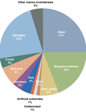
marine environment exert great influence on overall fungi diversity and adaptations (E. Jones, 2000). These fungi also happen to be associated to a large diversity of hosts, comprising several types of marine vertebrates and invertebrates, marine plants, algae, and marine microbial communities (Debbab et al., 2012; König et al., 2006; Raghukumar, 2006; Rateb & Ebel, 2011). The sources of marine-derived fungi from which compounds have been extracted are portrayed in Figure 2.
Fungi are known to produce secondary metabolites, which are small molecules that generally do not interfere in fungus’ normal development and growth (Fox & Howlett, 2008). Marine-derived fungi strains are known to mostly produce polyketides, peptides, terpenoids, prenylated polyketides, alkaloids, shikimates, lipids and mixed biosynthesis compounds, which are in agreement with the secondary metabolites commonly produced by fungi (Rateb & Ebel, 2011; Swathi et al., 2013).
There is currently one anticancer compound based on marine-derived fungi metabolites that has entered clinical trials. This alkaloid compound, plinabulin, is a modified structure based on (-)-phenylahistin, a metabolite of both terrestrial and marine fungi Aspergillus ustus (Hayashi et al., 2013; Kanoh et al., 1997).
Figure 2 – Sources of fungi from which bioactive compounds have been isolated. Adapted from Rateb & Ebel (2011).
1.2.2. Fungi associate relationships as a source of bioactive compounds
The total scale of marine fungi species is most probably greatly underrated. Current research has aimed at understanding marine fungi’s associations with other species, which has resulted in the collection of several novel species and strains (Passarini et al., 2013; Wang et al., 2012). As previously mentioned, marine fungi are associated to several organisms, most importantly invertebrates, algae and plants. While in the case of invertebrates (e.g. corals, ascidians, holothurians, gorgonians and sponges) and algae, fungi usually interact by association, when concerning marine plants, marine fungi habitually act as endophytes (Debbab et al., 2012). An endophyte is a fungal or bacterial microorganism that colonizes a plant or algae either at an intra-cellular or inter-cellular level, this colonization takes place without an apparent pathogenicity towards the host and may occur during a part or the whole of the host’s life cycle (Tan & Zou, 2001).
In the case of endophyte-host plant interaction, fungi secondary metabolites have been proven to contribute to the host’s survival and performance, by affecting factors such as chemical defense against predation, competitors and pathogens, growth rate, salt tolerance, photosynthesis and overall fitness, amongst others (Debbab et al., 2012). Whereas in fungal association to marine invertebrates, in particular sessile invertebrates, the role of fungi remains essentially unknown, however, fungi have been proposed as being able to improve the stability of the host skeleton, enable chemical defense against predators and competitors, parasitize pathogens, enhance nutrition and stimulate the host’s immune system (Selvin et al., 2010). This intricate relationship provided by association or endophytic behavior proves itself interesting for bioactive compound research since the colonization process and fungal-host interaction relies on chemical communication
Sponges (phylum Porifera) represent some of oldest metazoans in existence. They are simply structured sessile invertebrates, which inhabit mostly marine and seldom freshwater systems. They are habitually attached to benthic substrates of the intertidal or deeper zones of tropical, temperate and polar waters (Thomas et al., 2010). Considering their relatively simple body structure and function, they heavily rely on chemical defense mechanisms to avoid predation and combat pathogens and competitors. Intense competition is in fact one of the main drivers for the production of toxic compounds, as the concentration of biomass is often overwhelming and space is limited in many ecological niches. Consequently, these compounds must reveal a surprising efficiency to exert their
effect as they are rapidly diluted in the seawater (Haefner, 2003). As a result, a great deal of attention was guided towards sponge secondary metabolites, and an extensive number of compounds have been extracted from these organisms. Several studies have attributed numerous pharmacological attributes to sponge metabolites, such as neuroprotective, antifungal, antibacterial, cyotoxic and antiparasitic activity (Kossuga et al., 2008; Qaralleh et al., 2010; Sasaki et al., 2011).
Sponges are home to a massive microenvironment of associate microorganisms. These associated microorganisms are essentially of bacterial nature, however, recent research has shed some light into the actual diversity of symbiotic marine fungi attached to sponges. Höller et al. (2000) collected 16 sponge species from 6 different locations, from which they isolated 681 fungal strains. These fungal strains were predominantly representatives of ubiquitous genera, but marine genera were also observed (Holler et al., 2000). In parallel, Morrison-Gardiner (2002) described the isolation of 208 fungal strains from marine sponges of tropical waters of Australia (Morrison-Gardiner, 2002). In similarity to the previous case, both ubiquitous and marine fungi genera were observed. The persistent observation of ubiquitous genera suggests that these fungi may be highly similar to their terrestrial counterparts, and may be in fact contaminants to the sponge. In fact, the exact role of marine fungi in their relationship with sponges remains unclear. However, the adaptations needed to endure marine conditions, vast marine fungal biodiversity and a prospective bioactive potential comparable to that of terrestrial fungi, make these fungi an undoubtedly interesting focus for the search of bioactive compounds (Taylor et al., 2007). Table 1 shows examples of the bioactivity of several compounds isolated from sponge-derived fungi.
Gorgonian corals (phylum Cnidaria), also known as sea fans, are sessile colonial cnidarians which are mainly distributed in the tropics and sub-tropics. Much like sponges, due to their bodies and sessile nature, they are also exposed and vulnerable to predation and competition. While the need of sea fan for chemical defense and interaction makes these organisms themselves a prolific source of bioactive compounds (Rocha, Peixe, Gomes, & Calado, 2011), the focus on their microbial associates is beginning to arise. The interaction of fungi with sea fans is far less studied than that of other corals, in exception of the pathogenic relationship often observed, since sea fan communities are frequently attacked by violent fungal infections (Toledo-Hernández et al., 2013). However, there are a series of studies that have examined healthy sea fan populations and observed that healthy populations possess a more diverse array of fungi, which are considered residential (Toledo-Hernández et al., 2007). The association between
non-pathogenic fungi and sea fans has been proposed to be of saprophytic nature, where the fungus consumes dead tissue from the coral (Kanoh et al., 1997). Meanwhile, reports of novel compounds derived from these sea fan associate fungi are rapidly increasing, and descriptions of antifouling, antibacterial, cytotoxic and other activities are already available (Bao et al., 2012; C. Li et al., 2011).
Table 1 - Examples of compounds isolated from marine sponge-associated fungi and their bioactivity.
1.2.3. Anticancer compounds of sponge and sea fan derived fungi origin
Marine invertebrates have been on the spotlight of marine anticancer compound screening for a few decades. Sponges, gorgonians and soft corals have been the main targets for compound isolation (Bhatnagar & Kim, 2010), however, more recently, research has centered on discovering and isolating microorganism associates of these species and assessing compounds and extracts for anticancer activity.
Bioactivity Fungus Sponge Metabolite Reference
Antibacterial Aspergillus sp. Xestospongia testudinaria (−)-sydonic acid (D. Li et al., 2012)
Aspergillus sp. Tethya aurantium Austalide R (Zhou et al., 2014)
Exophiala sp. Halichondria panacea Chlorohydroaspyrones A and B
(D. Zhang, Yang, Kang, Choi, & Son,
2008)
Antifungal Penicillium spp. Tethya aurantium Nortryptoquivalin
(Wiese, Ohlendorf,
Blümel, Schmaljohann, &
Imhoff, 2011)
Aspergillus insuetus Psammocinia sp. Insuetolides A, B and C (E. Cohen et al., 2011)
Cytotoxic Clonostachys
sp. Unidentified sponge IB-01212
(Cruz et al., 2006)
Acremonium sp. Axinella sp. Efrapeptin E, F, Eα, G, H
(Claudia M Boot et al., 2007; C. M. Boot, Tenney, Valeriote, & Crews, 2006) Penicillium
auratiogriseum Mycale plumose
(S)-2,4-dihydroxy-1-
butyl(4-hydroxy)-benzoate
(Xin et al., 2005)
The screening of extracts is often the first route to examine the potential in investing in the isolation of compounds. As an example of a sponge-associated fungus extract possessing anti-proliferative activity, the ethyl acetate extract of the fungus Eurotium cristatum obtained from the sponge Mycale sp. was shown to possess activity against three cancer cell lines: MCF-7 (breast adenocarcinoma), NCI-H460 (lung cancer), and A375-C5 (melanoma). The IC50 values were, respectively, 44.3, 45.5 and 71.3 µg/ml.
Three compounds were isolated from this extract, of which 2-(2’, 3-epoxy-1’,3’-heptadienyl)-6-hydroxy-5-(3-methyl-2-butenyl) benzaldehyde exhibited the most interesting cytotoxic activity in all three cell lines, however, the overall cytotoxic effect displayed by the crude extract quite possibly is also due to other unidentified compounds (Almeida et al., 2010).
There are several examples of compounds isolated from sponge-associated fungi, as is the case of dankastatin C, a polyketide tyrosine derivative, isolated from the marine sponge-derived (Homaxinella sp.) fungus Gymnascella dankaliensis. This compound was a potent inhibitor of the P388 lymphocytic leukemia cell line, with an ED50 (half maximal
effective concentration) of 57 ng/ml, having obtained an equivalent potency to that of the common chemotherapeutic drug 5-fluorouracil (Amagata et al., 2013). Another example is of epoxyphomalin A, isolated from the sponge-derived (Ectyplasia perox) fungus Phoma sp., which revealed a cytotoxicity at nanomolar concentrations against 12 human tumour cell lines, with IC50 values ranging from 0.010 µg/ml to 0.038 µg/ml (Mohamed et al.,
2009).
While gorgonians or sea fans have been widely screened for cytotoxic compounds, their associate fungi counterparts have only very recently been approached in this matter. The following examples illustrate a few examples of compounds with different chemical composition and cytotoxic activity, originating from sea fan associate fungi. Nigrosporanene A, a cylohexene derivative isolated from the fungus Nigrospora sp. PSU-F11 derived from the gorgonian sea fan Annella sp. presented an IC50 of 9.37 µg/ml in
MCF-7 breast adenocarcinoma cells (Rukachaisirikul et al., 2010). Aspergillone A, a benzylazaphilone derivative isolated from the fungus Aspergillus sp. associated to the sea fan Dichotella gemmacea was found to demonstrate a potent cytotoxic effect against MCF-7 breast adenocarcinoma, A549 lung cancer and HL-60 human promyelocytic leukemia with the respective IC50 values of 25.0, 37.0 and 3.2 µg/ml (Shao et al., 2011).
Asperterrestide A, a cyclic tetrapeptide, was isolated from the fungus Aspergillus terreus that was isolated from the tissue of the sea fan Echinogorgia aurantiaca, exhibited a cytotoxic effect against two human leukemia cell lines, U937 leukemic monocyte
lymphoma and MOLT4 acute lymphoblastic leukemia (IC50 of 6.4 and 6.2 µM,
respectively) (He et al., 2013). Oxalicumones D and E, two dihydrothiophene-condensed chromones, were isolated from the fungus Penicillium oxalicum SCSGAF 0023 associated to the sea fan Muricella flexuosa. Oxalicumone E demonstrated a strong cytotoxic activity against a panel of eight cancer cell lines (H1975, U937, K562, BGC823, MOLT4, MCF-7, HL60 and Huh-7) with IC50 values ranging from 1.36 to 8.80 µM. Oxalicumone D,
however, exhibited a cytotoxic effect against BGC823 gastric cancer and MOLT4 acute lymphoblastic leukemia cell lines with an IC50 of 10.10 and 5.74 µM, respectively (Bao et
al., 2014).
1.2.4. Neosartorya genus
Neosartorya is a genus of fungi that is common in the terrestrial environment, where it has been described as occurring in the soil, organic materials, air, food and human habitations but also in the marine environment (Yaguchi et al., 2010). The Neosartorya genus belongs to the phylum Ascomycota and species of this genus are a telemorphic (i.e., sexual reproductive) stage of the Aspergillus section Fumigati. Neosartorya species are known to produce an asexual state with conidiospores and a sexual state with ascospores (Frisvad et al., 2008). Although some Neosartorya species have been identified as the cause of several human pathologies, such as invasive aspergillosis, endocarditis, and osteomyelitis (Peláez et al., 2013; Summerbell et al., 1992), recent studies have approached these species as a source of interesting bioactive compounds with potential application in human health (Eamvijarn et al., 2013; Gomes et al., 2014; Padhye et al., 1994).
Several terrestrial strains of Neosartorya have yielded interesting cytotoxic compounds, for instance, the pyrroloindole sesquiterpenoid fischerindoline, isolated from Neosartorya pseudofischeri CBS 404.67. This compound presented moderate cytotoxic activity in five human cancer cell lines (A549, Hs683, MCF-7, SKMEL-28 and U373) and one murine melanoma cell line (B16F10), with IC50 values ranging from 25 to 37 µM (Masi et al.,
2013). Another example is the indoloazepinone deriviative sartorymensin, isolated from Neosartorya siamensis KUFC 6349, which exhibited cytotoxic activity against five human cancer cell lines (U373, Hs683, A549, MCF-7 and SKMEL-28) with IC50 values that
More recently, marine strains of Neosartorya have been subjected to isolation of compounds and subsequent screening of cytotoxic activity. In a study by Eamvijarn et al. (2013) several compounds were isolated from Neosartorya tsunodae (KUFC 9213), Neosartorya fischeri (KUFC 6344) and Neosartorya laciniosa (KUFC 7896). The isolated compounds, 13-oxofumitremorgin B, sartorypyrone A, aszonapyrone A and sartorypyrone B, presented low to high cytotoxic activity (GI50 values ranging from 123.3 to 10.2 µM)