Doi: https://doi.org/10.25186/.v15i.1637
Coffee protects cardiovascular health by maintaining the structure of
coronary arterial wall intimal collagen
I Dewa Ayu Susilawati1 , Suryono Suryono2 , Purwanto Purwanto1 , Juris Burlakovs3 , Abubakar Yaro4
1Jember University, Department of Biomedical Science, Faculty of Dentistry, Jember, Indonesia 2Jember University, Department of Cardiology, Faculty of Medicine, Jember, Indonesia 3Linnaeus University, Faculty of Health and Life Sciences, Kalmar, Sweden
4Institute of Health Sciences & Research, AHRO Scientific Publishing Ltd, Glasgow, Scotland
Contact authors: dewasusi@unej.ac.id, suryonocardio@yahoo.com, purwanto@unej.ac.id, juris.burlakovs@lnu.se, afhereor@gmail.com Received in November 11, 2019 and approved in March 10, 2020
ABSTRACT
This study aimed to determine whether coffee consumption affects the structure of coronary arterial wall and protects against coronary artery disease (CAD) in atherosclerotic rat model induced by periodontitis. Rats (n=21) were divided into three groups (i) Coffee group (periodontitis + coffee), (ii) Peri-odontitis group (no coffee), and (iii) Control group (no periPeri-odontitis, no coffee). A single dose of coffee suspension (representing one cup) was given daily by stomach sondation to the rats in the coffee group. The experiment was conducted for 5 wk. At the completion of the experiment, all of rats were sac-rificed. Their hearts containing coronary arteries were removed and analyzed by histochemistry assay. In addition, the serum level of collagen degrading enzymes matrix metalloproteinase–2 (MMP–2) was also analyzed using Enzyme–link immunosorbent assay (Elisa). Results demonstrated that coronary atherosclerotic lesions including atheroma, stenosis, and vascular occlusion were rarely identified in the coffee group. The coronary arterial wall demon-strated relatively symmetrical intima-media thickness (IMT) and the lumen diameter remained adequate for blood flow. The intimal collagen was intact, dense and thick. MMP–2 level was significantly lower (P < 0.05) in the coffee group. In conclusion, coffee maintained the structure of coronary arterial wall particularly the intimal collagen, providing protection against CAD. This might also mediate the vascular resistance against rupture and thrombosis that might precipitate the occurrence of acute coronary syndrome (ACS).
Key words: Acute coronary syndrome (ACS); Coronary artery disease (CAD); Histochemistry; Intima-media thickness (IMT); Matrix metalloproinase-2 (MMP-2).
1 INTRODUCTION
The structure of the coronary arterial wall, particularly the intima (the innermost layer) is an important indicator of cardiovascular health. Changes in this structure might increase the risk of coronary artery disease (CAD). Intimal thickening and lipids accumulation in the intimal layer have been demonstrated in the occurrence of atherosclerosis (Aziz; Yadav, 2016). On the other hand, intimal thinning might promote the vessel vulnerability leading to rupture (Otsuka, 2016; Sakakura et al. 2013), followed by the luminal thrombosis. This is the currently accepted mechanism responsible for the evidence of the acute coronary syndromes (ACS) and sudden coronary death.
The intima is comprised mainly of collagen. Matrix-degrading enzyme (matrix metalloproteinases, MMPs) has been proposed to play a significant role in the intimal collagen rupture (Newby 2015; Tummer et al. 2010). These enzymes are produced by cells as an inactive enzyme pro–MMP. Its activation can be induced by proteases, oxidants, or free radicals (Kameda et al. 2003; Kamdasamy et al. 2010). This phenomenon has led the idea to inhibit MMP activation and collagen degradation using antioxidant.
Coffee is well–known as an antioxidant–rich beverage. It is therefore plausible to hypothesize that coffee might reduce
the activation of MMPs and collagen degradation as well as maintain the morphology of coronary artery and protect against CAD and ACS.
This study aimed to determine whether coffee consumption affects the structure of the coronary arterial wall in atherosclerotic rat model induced by periodontitis. In addition, histopathological analysis of the intima and serum MMP–2 level examinations were conducted to study the collagen integrity within the intima. The use of atherosclerotic model using periodontitis induction for this purpose has been studied previously (Brondala et al. 2005; Susilawati et al. 2020).
2 MATERIAL AND METHODS 2.1 Animals and groups
Male rats [Rattus norvegicus (Berkenhout, 1769)], 12 wk (± 0.2 kg body weight) were purchased from Faculty of Medicine, Universitas Brawijaya,Malang, Indonesia. Animals were maintained under standard laboratory conditions and in concordance with the guidelines established by the Institutional Animal Care and Ethics Committee. The animal treatment procedure was approved by The Ethical Committee of Faculty of Medicine Universitas Jember, Indonesia (Number, 1100/
H25.1.11/KE/2016). Dry pellet normocholesterol standard diet and water were given ad libitum. All rats were housed in pens in identical condition to minimize the possible effects of environmental factors. A total of 21 rats were separated into three groups randomly as follows: (i) Coffee group (periodontitis + coffee), (ii) Periodontitis group (no coffee), and (iii) Control group (no periodontitis, no coffee).
2.2 Preparation of coffee suspension
This study used commercial pure robusta (Coffea canephora Pierre ex A. Froehner) ground coffee produced by The State Plantation Company named PTPN XII Jember, East Java, Indonesia. To prepare one cup of coffee suspension, a total 3 g ground coffee was poured into 0.2 L boiled water and stirred for 1 min. The dose given to the rats was converted by comparing body weight of rat (0.2 kg) to adult man (70 kg). This resulted in a dose of 0.57 mL suspension (rounded to 0.6 mL) for each rat in each administration. This dose of coffee suspension was then given once daily per rat for 5 wk by means of stomach sondation.
2.3 Atherosclerotic rat model induced by periodontitis
The atherosclerotic model was created in the rats by injecting periodontitis bacteria Porphyromonas gingivalis (ATTC, 33277) in the buccal gingival sulcus of left mandibular teeth (as previously described by Susilawati et al. 2020). 2.4 Preparation of histopathological samples
At the end of the study, all rats were sacrificed using 0.02 mL Ketamine intra-peritoneal injection. The heart containing coronary arteries were subsequently removed and fixed in 10 % formalin (in PBS) and trimmed cross–sectionally (perpendicular to the direction of blood flow) in about one– half coronal area of the heart. These samples were prepared by frozen section. A serial cryo–sections (10 μm thickness) were made and then mounted on the microscope slides (three sections on each slide).
2.5 Histopathological samples analysis
To analyzethe coronary arterial wall thickness and examine its morphology, samples were stained using the collagen staining kit (Picro sirius Red, Scy Tek, USA). Collagen would appear red while elastin would appear yellow. The images were visualized with 400× light microscope magnification and documented with an optilab microscope camera. Although the histopathological samples were cross–sections of the heart, the analysis was focused on coronary arteries only. Each sample was represented by two coronary artery images. Therefore, each group would be represented by 14 coronary artery images.
2.6 Coronary arterial wall thickness and morphology
The coronary arterial wall thickness was measured as intima–media thickness (IMT) in the thickest area or atheroma. The measurement was conducted using the available software in optilab (m). IMT data was expressed as mean ± standard deviation and were statistically analyzed by one-way Anova and least significant difference (LSD). In addition, the symmetry of thickness was identified. Morphological analysis of atherosclerotic indicators (the existence of atheroma or arterial wall protrusion into the lumen, stenosis or luminal narrowing, and vascular occlusion) was analyzed qualitatively and denoted as present or absent.
2.7 Intimal collagen thickness and integrity This work also focused on coronary artery intimal collagen thickness and integrity. The presence of collagenous fibrous cap plays vital role in preserving the vascular resistance and atherosclerotic plaque against rupture and thrombosis. Thinning and disintegration of intimal collagen could cause thinning of fibrous cap that might increase the risk of vascular rupture. This study analyzed the integrity of intimal collagen qualitatively and denoted as intact or disintegrated.
2.8 Analysis of serum MMP–2
One of MMPs that plays an important role in cardiovascular disease is MMP-2 (Azevedo et al. 2014). This study analyzed the level of serum MMP–2 by means of Elisa. Serum sample of the rats was prepared according to the procedures described by Takahashi et al. (2014), with modification. Immediately after rats were sacrificed, intra-cardial blood was collected and centrifuged for 5 min at 1 200 ×g at room temperature (RT). The tubes were then kept at 4 °C for 1.5 hr to 2 hr. After collecting the fibrin clots, the tubes were then centrifuged for 5 min at 1 200× g at RT. The serum was carefully transferred into a 15 mL sterile tube using a sterile pipette in a laminar air-flow cabinet. The serum was stored at -20 °C until before being analyzed. MMP-2 was measured using the Elisa kit (Elabscience). Serum level of MMP–2 was expressed as mean ± standard deviation and statistically analyzed by one–way Anova and least significant difference (LSD).
3 RESULTS
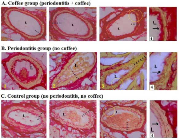
Coronary arterial wall in the coffee group demonstrated significantly lower IMT (P < 0.05) compared to periodontitis group (Table 1). Moreover, the IMT was relatively symmetrical around arterial wall. This finding was associated
with the absent of atheroma, minimum luminal narrowing/ stenosis and vascular occlusion (Figure 1A). In contrast, the periodontitis group demonstrated higher IMT due to the thickening of the elastic layer and the existence of atheroma in some areas in arterial wall (Figure 1B). Asymmetrical IMT, luminal narrowing and vascular occlusion were identified in the periodontitis group (Table 2).
Further analysis of the coronary arterial wall focused on the thickness and integrity of the intimal collagen layer. The intimal collagen appeared thick and intact in the samples from the coffee group while appearing thin and disintegrated in the periodontitis group and appearing as a thin layer in the control group. These findings were in concordance with the serum MMP–2 level which was significantly lower (P < 0.05) in the coffee group than other groups (Table 3).
4 DISCUSSION
Several studies have reported the beneficial effect of coffee on cardiovascular health including its effects
on subclinical inflammation and high density lipoprotein (HDL) cholesterol (Kempf et al. 2010) and resistance against coronary calcification (Van Woudenbergh et al. 2008). Coffee consumption has been found to be inversely associated with markers of inflammation and endothelial dysfunction (Lopez-Garcia 2006) and risk of heart failure (Mostofsky et al. 2012), increase the resistance of LDL to oxidative modification (Natella et al. 2007), and improve endothelial function (Shechter et al. 2011; Siasos et al. 2013).
Table 1: Histometrics of rat’s coronary artery Intima-Media Thickness (IMT).
Groups n Thickness (IMT)Intima-Media
X ± SD (µm)
Coffee (Periodontitis + coffee) 14 1.75 ±0.43
Periodontitis (no coffee) 14 4.11 ± 1.64 *
Control (no periodontitis, no coffee) 14 2.04 ± 0.50
n: number of coronary arteries images (seven samples, each represented by two images)
*: Significantly different from other groups (P < 0.05) Anova and LSD.
Figure 1: The Histomorphology of the rat’s coronary arteries stained with the Collagen Staining Kit (Picro Sirius Red). A.
The coffee group demonstrated more symmetrical intima–media thickness (IMT) and wider lumen. The intimal collagen appeared more intact, denser and thicker. B. The periodontitis group demonstrated signs of atherosclerosis including
atheroma (B1), intimal collagen disintegration (B2) vascular occlusion (B3). C. Intimal thickening was identified in the
control group. Figure in column 4 demonstrated the magnified view of the intimal collagen (arrow). The distance between bars: 1μm; L: lumen.
The mechanism of how coffee affects cardiovascular health, however, is still under debates. Some reports have concerned with the harmful effect of caffeine used in isolation (Adebayo et al., 2007; Cornelis; El-Sohemy, 2007). However, coffee has a very complex chemical composition. In addition to caffeine, coffee contains many other substances that might act as antioxidants (Damat et al., 2019; Halvorsen et al., 2002; Moriera et al., 2013; Natella et al., 2007; Suryono et al., 2020) and anti– inflammation (Cardenas; Quesada; Medina, 2011; Dewanti et al., 2019; Kempf et al., 2010; Moriera et al., 2013) that might counteract the adverse effect of caffeine. Due to its rich antioxidant and anti–inflammatory content, it has been hypothesized that coffee consumption might be of benefit to cardiovascular health since oxidation and inflammation represent the important mechanisms in coronary artery disease (Rafieian-Kopaei, 2014). The anti-atherosclerotic property of antioxidants in prevention and improvement atherosclerosis had been reviewed by Yalameha (2019).
Within the current study, atherosclerosis was induced by chronic periodontitis. Previous studies have demonstrated that challenge of intravenous administration of periodontitis bacteria (Porphyromonas gingivalis) promoted atherosclerosis (Brondala, 2005; Li et al., 2002). This study showed that periodontitis induced the morphological deterioration of coronary arterial wall leading to accelerated CAD. It was consistant with previous research (Susilawati et al., 2020).
The deleterious effect of chronic periodontitis could be counteracted by coffee consumption hence its protection against CAD. The most important finding of this research was its effect on the morphology of intimal collagen. In the coffee group, the coronary intimal collagen was intact, dense
and thick. This structure might mediate the resistance of the vasculature against injurious agents from blood stream,hence the reduced response to injury that would subsequently lead to reduced atherosclerosis. In addition, coffee consumption might also provide resistance to the vasculature and atherosclerotic plaque against rupture since rupture mostly occurred in the area of thin collagen fibrous cap (Gough, 2006). As atherosclerotic plaque rupture is the proximate event of clinical manifestation of ACS, it could therefore be proposed that coffee consumption might prevent ACS by maintaining the morphology of coronary arterial wall, particularly the intimal collagen.
The integrity of vascular intimal collagen is modulated by MMPs. During systemic infection, the entrance of injurious agents into blood circulation could stimulate inflammatory responses in the vessels including the activation of inflammatory cells neutrophils and monocytes to produce a large amount of pro-MMPs and reactive oxigen species, ROS (Newby, 2015). Interaction of these substances results in the activation of MMPs that degrade the vascular collagen and cause thinning of intimal collagen or fibrous cap of atherosclerotic plaque, leading to its increased vulnerability for rupture.
This study demonstrated an intact integrity of coronary intimal collagen in rats that consumed coffee. The hypothesis is the beneficial effect of coffee was due to its abundant antioxidant and anti-inflammatory content. Anti-inflammatory compounds might reduce inflammatory responses, while anti-oxidants might be scavenging ROS, and collectively these mechanisms result in the inhibition of MMPs activation leading to reduced collagen degradation and hence the preserved its integrity. Further studies are needed to elucidate the effect of coffee on MMPs and collagen metabolism.
The present study used a single dose of pure coffee suspension per day that represents one cup. The effect of the dose still needs further analysis since some studies have indicated that a high intake of coffee could adversely affect cardiovascular health. In addition to doses, coffee serving usually was mixed with other ingredients including milk and sugar. It is not yet to be elucidated whether the interaction of coffee and others nutrients might influence the effect of coffee on cardiovascular health.
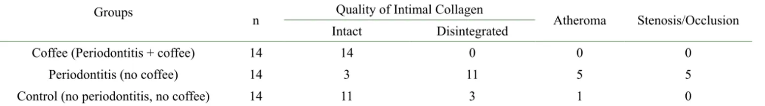
Table 2: Coronary artery disease parameters in rats*.
Groups n Quality of Intimal Collagen Atheroma Stenosis/Occlusion
Intact Disintegrated
Coffee (Periodontitis + coffee) 14 14 0 0 0
Periodontitis (no coffee) 14 3 11 5 5
Control (no periodontitis, no coffee) 14 11 3 1 0
n: number of coronary arteries images (seven samples, each represented by two images) *:Qualitative analysis (present or absent).
Table 3: Serum level of MMP–2 in rats.
Groups n Serum level of MMP–2 X ± SD (g L–1)
Coffee (Periodontitis + coffee) 7 3.88 ± 1.25 *
Periodontitis (no coffee) 7 5.52 ± 0.98
Control (no periodontitis, no coffee) 7 5.11 ± 1.96
n: number of samples
The beneficial effect of coffee to maintain the structure and morphology of vascular intimal collagen is a novel a perspective. This finding was the first study that reported the role of coffee on the vascular collagen integrity. Other important finding is the indication that coffee might affect blood composition and coagulation. The luminal space of coronary artery of the rats in the coffee group demonstrated a clearer image with minimum coagulation components. Further studies are needed to clarify the anticoagulation properties of coffee.
5 CONCLUSIONS
In conclusion, one cup of coffee consumption per day maintains the morphology of coronary arterial wall particularly the intimal collagen leading to protect against CAD. This protective effect might also explain the resistance mechanism of vasculature against rupture, thrombosis and ACS.
6 ACKNOWLEDGMENTS
This study was conducted under the grant from the Ministry of Research-Technology and Higher Education of the Republic of Indonesia (Grant number, 0481/UN25.3.1/ LT2017). The authors would like to thank to anonymous reviewers in the Scientific Committee of JICC (Jember International Coffee Conference) who have supported this paper to be published. The responsibility of paper’s contents is by writers. The authors also would like to thank Mrs. Wahyu, Mr. Agus and Mr. Pinardi for helping us conducting the laboratory works.
7 REFERENCES
ADEBAYO, J. O. et al. Effect of caffeine on the risk of coronary heart disease. A reevaluation. Indian Journal of Clinical Biochemistry, 22(1):29-32, 2007.
AZEVEDO, A. et al. Matrix metalloproteinases are involved in cardiovascular diseases. Basic & Clinical Pharmacology & Toxicology, 115(4):301-314, 2014.
AZIZ, M.; YADAV, K. S. Pathogenesis of atherosclerosis a review. iMedPub Journals - Medical & Clinical Reviews, 2(3):1-6, 2016.
BRODALA, N. et al. Porphyromonas gingivalis bacteremia induces coronary and aortic atherosclerotic in
normocholesterolemic and hipercholesterolemic pigs. Arteriosclerosis, Thrombosis, and Vascular Biology, 25(7):446-1451, 2005.
CÁRDENAS, C.; QUESADA, A. R.; MEDINA, M. A. Anti-angiogenic and anti-inflammatory properties of kahweol, a coffee diterpene. Plos One, 6(8):1-9, 2011.
CORNELIS, M. C.; EL-SOHEMY, A. Coffee, caffeine, and coronary heart disease. Current Opinion in Clinical Nutrion and Metabolic Care, 10(6):745-751, 2007. DAMAT, D. et al. Dietary fiber and antioxidant activity of
gluten-free cookies with coffee cherry flour addition. Coffee Science, 14(4):493-500, 2019.
DEWANTI, I. D. A. R. et al. The effect of steeping Robusta coffee beans on monocytes: Expression of IL-1β and TNF-α against Streptococcus mutans. Coffee Science, 14(4):477- 483, 2019.
GOUGH, P. J. et al. Macrophage expression of active MMP– 9 induces acute plaque disruption in apo E–deficient mice. Journal of Clinical Investigation, 116(1):59-69, 2006. HALVORSEN, B. L. et al. A systematic screening of total
antioxidants in dietary plants. Journal of Nutrition, 132(3):461-471, 2002.
KAMEDA, K. et al. Correlation of oxidative stress with activity of matrix metalloproteinase in patients with coronary artery disease. European Heart Journal, 24(24):2180-2185, 2003.
KANDASAMY, A. D. et al. Matrix metalloproteinase-2 and myocardial oxidative stress injury: Beyond the matrix. Cardiovascular Research, 85(3):413-423, 2010. KEMPF, K. et al. Effects of coffee consumption on
subclinical inflammation and other risk factors for type 2 diabetes. American Journal of Clinical Nutrition, 91(4):950-957, 2010.
LI, L. et al. Porphyromonas gingivalis infection accelerates the progression of atherosclerosis in a heterozygoes apolipoprote in e-deficient murine model. Circulation, 105(7):861-867, 2002.
LOPEZ-GARCIA, E. Coffee consumption and risk of chronic diseases: Changing our views. American Journal of Clinical Nutrition, 95(4):787-788, 2012.
MOREIRA, M. E. D. C. et al. Anti-inflammatory effect of aqueous extracts of roasted and green Coffea arabica. L. Journal of Functional Foods, 5(1):466-474, 2013.
MOSTOFSKY, E. et al. Habitual coffee consumption and risk of heart failure: A dose-response meta-analysis. Circulation Heart Failure, 5(4):401-405, 2012.
NATELLA, F. et al. Coffee drinking induces incorporation of phenolic acids into LDL and increases the resistance of LDL to ex vivo oxidation in humans. American Journal of Clinical Nutrition, 86(3):604-609, 2007.
NEWBY, A. C. Metalloproteinases promote plaque rupture and myocardial infarction: A persuasive concept waiting for clinical translation. Matrix Biology, 44-46:157-166, 2015. OTSUKA, F. et al. Pathology of coronary atherosclerosis and
thrombosis. Cardiovascular Diagnosis and Therapy, 6(4):396-408, 2016.
RAFIEIAN-KOPAEI, M. et al. Atherosclerosis: Process, indicators, risk factors and new hopes. International Journal of Preventive Medicine, 5(8):927-946, 2014. SAKAKURA, K. et al. Pathophysiology of atherosclerosis
plaque progression. Heart, Lung, and Circulation, 22(6):399-411, 2013.
SUSILAWATI, I. D. A. et al. Coronary artery disease in periodontitis rat model. Annals of Tropical Medicine and Public Health, 23(3A):34-43, 2020.
SHECHTER, M. et al. Impact of acute caffeine ingestion on endothelial function in subjects with and without coronary
artery disease. American Journal of Cardiology, 107(9):1255-1261, 2011.
SIASOS, G. et al. Consumption of a boiled greek type of coffee is associated with improved endothelial function. Vascular Medicine, 18(2):55-62, 2013.
SURYONO, S. et al. Elevated blood serum neutrophil collagenase and NADPH oxidase-1 (NOX-1) in acute coronary syndrome, Annals of Tropical Medicine and Public Health, 23(3A):171-179, 2020.
TAKAHASHI, M. et al. Preparation of rat serum suitable for mammalian whole embryo culture. Journal of Visualized Experiments, 90:e51969, 2014.
TUMMERS, A. M. et al. Serum levels of matrix
metalloproteinase-2 as a marker of intimal hyperplasia. Journal of Surgical Research, 160(1):9-13, 2010. VAN WOUDENBERGH, G. J. et al. Coffee consumption and
coronary calcification. Arteriosclerosis, Thrombosis, and Vascular Biology, 28(5):1018-1023, 2008.
YALAMEHA, B. Antioxidant therapy to improve or resolve atherosclerosis; new hopes and current trends. Journal of Nephropharmacology, 8(2):1-4, 2019.