Association between Risk Factors for CAD and Coronary Disease in
Patients Undergoing Myocardial Perfusion Scintigraphy
Paulo Schiavom Duarte, Luiz Eduardo Mastrocolla, Gilberto Alonso, Eduardo Vilaça Lima, Paola Emanuela Smanio,
Marco Antonio Conde de Oliveira, Luiz Roberto Fernandes Martins, Júlio César Rodrigues Pereira
Centro de Medicina Diagnóstica Fleury e Faculdade de Saúde Pública da Universidade de São Paulo - São Paulo, SP - Brazil
Summary
Objective: To establish the degree of association between cardiovascular risk factors and the presence of coronary artery disease (CAD) in a group of patients undergoing myocardial perfusion scintigraphy (MPS).
Methods: The study included 7183 patients who had undergone MPS. Using logistic regression analysis the odds ratios for the following risk factors were evaluated: age, gender, family history, body mass index, smoking, dyslipidemia, diabetes mellitus (DM) and systemic hypertension. Indicators for the presence of CAD were defined as: myocardial infarction, revascularization, angioplasty or an altered MPS. Analysis was based on the whole the group as well as on male and female subgroups. The impact of the risk factors in relation to age was also analyzed.
Results: A statistically significant association was observed between patient age and gender and the presence of CAD. For females, it was demonstrated that DM is the main modifiable risk factor for CAD. For males various modifiable risk factors were associated with the presence of CAD, particularly DM and dyslipidemia. In the analysis by age groups some risk factors showed a more expressive association.
Conclusion: The main risk factors for CAD were aging and male gender. In relation to modifiable risk factors and the presence of CAD, the greatest associations for males were DM and dyslipidemia and for females DM. The most relevant factors for specific age groups were smoking for young men and DM and smoking for women between the ages of 40 and 50.
Key words: scintigraphy, perfusion, myocardium, coronary artery disease, factors, risk.
Mailing Address: Paulo Schiavom Duarte •
Av. Angélica 2.389 ap. 111-B - 01227-200 – São Paulo, SP - Brazil E-mail: [email protected]
Manuscript received February 06, 2006; revised manuscript received June 26, 2006; Accepted: July 23, 2006.
Introduction
Coronary artery disease (CAD) in conjunction with all other cardiovascular diseases has been a significant cause of morbidity and mortality in the various regions of the world1,2.
Considering this, early detection, before the onset of clinical manifestations and known complications, is important in order to identify potentially modifiable risk factors and to avoid the progression of the disease or even promote its regression. There are various well established CAD risk factors (RF) in medical literature3-5, the most important ones being
diabetes mellitus (DM), systemic hypertension (SH), smoking (SMK), dyslipidemia (DYSLIP), stress, sedentary lifestyle, aging, gender, and others. Nevertheless, despite these well known risk factors there is controversy in relation to the relative importance of each of them and the modification of the importance of each one in regard to gender, age groups and the different populations studied. One example of this controversy is the importance of DM in females. Huxley and associates6 conducted a meta-analysis study involving
37 cohort studies and concluded that the relative risk for females with DM is 50% higher than males to develop fatal
CAD. Nevertheless, they report that the evaluation of three meta-analyses7-9 conducted during the past decade revealed
different results in relation to the importance of DM in females. While two of these meta-analyses demonstrated a higher risk of fatal CAD8,9 the third did not show any significant
differences7. Huxley and associates stated that the differences
between these studies could be a result of differences in the adjustment levels of the remaining cardiovascular risk factors. Another example of this heterogeneity in the results in relation to various population groups was described in a multi-institutional study (Diverse Populations Collaborative Group) published in the journal “Heart”10. This study
observed that qualitative analysis of the main CAD risk factors demonstrated an association with mortality for this pathology in the various populations; however the quantitative analysis revealed that the importance of the main risk factors varied in the different populations.
Methods
Patients - The study included 7183 consecutive patients (5118 males and 2065 females; with an average age of 56.7 years; SD: 11.0 years) undergoing MPS between January 2000 and October 2004.
age, gender, family history (FH), body mass index (BMI), smoking (SMK), dyslipidemia (DYSLIP), diabetes mellitus (DM) and systemic hypertension (SH).
The patients were classified in five age groups: 0 (under 40 years), 1 (between 40 and 50 years), 2 (between 50 and 60 years), 3 (between 60 and 70 years) and 4 (above 70 years).
The body mass index was calculated using the formula “WEIGHT/HEIGHT2” and the results were classified in four
categories: 0 (below 25), 1 (between 25 and 30), 2 (between 30 and 35) and 3 (above 35).
Smoking, family history, dyslipidemia, DM and SH were classified as dichotomic variables, and were stated as 0 for nonexistence and 1 for presence of the respective risk factor.
Myocardial perfusion scintigraphy - A one day protocol using an injection of technetium-99m hexakis 2-methoxyisobutyl isonitrile (Tc-99m MIBI) was conducted during the rest and stress phases with acquisition of tomography images (SPECT - Single Photon Emission Tomography) representatives of myocardial perfusion. The rest images were acquired 30 to 60 minutes after the injection of 370 MBq (10 mCi) of Tc-99m MIBI, and the stress phase was conducted 4 hours after the rest phase. The methods used for the stress phase were either a treadmill stress test or a pharmacological stimulation using a 0.56 mg per kg of body weight dosage of dipyridamole administered intravenously for four minutes. In this phase a 1.11 GBq (30 mCi) of Tc-99m MIBI was injected endovenously. The images were taken 45 to 60 minutes after the injection. During both phases of the test (rest and stress) the emission and transmission images were obtained simultaneously using a gadolinium-153 radioactive linear source. Thus, it was possible to evaluate the test with and without the use of a specific program to correct attenuation defects, that are usually found in the inferior and anterior walls of the left ventricle11-13 as a result of voluminous
abdomens (inferior wall) or breasts (anterior wall). The acquisition of the stress images was synchronized with the patient’s electrocardiogram (gating technique)14,15 enabling
simultaneous evaluation of the left ventricle motility and thickness during this phase of the test.
Thus, when the existence of a perfusion defect was questioned (suspicion of attenuation artifacts), the nuclear medicine specialists analyzed the images using attenuation correction along with the myocardium motility and thickness images to increase the specificity of the method.
For the objective of this study, MPS tests were considered abnormal if myocardial perfusion defects were diagnosed during either the rest and/or stress phases.
Statistical analysis - Using multivariate logistic regression analysis the association between the abovementioned RF and the presence of CAD was evaluated. The result was presented using the adjusted odds ratio (OR), the confidence interval of this value (95%) and its statistical significance. The chi-square test was used to compare the male and female patient groups in relation to the presence of RF, coronary events and perfusion abnormalities during the scintigraphy.
Patient classification - The patients who presented a previous history of coronary events –myocardial infarction (MI),
coronary artery bypass graft surgery (CABG), percutaneous transluminal coronary angioplasty (PTCA) – and/or those that presented perfusion abnormalities during the MPS (ALT SCINT) were diagnosed with CAD. Based on the medical history information, two criteria were defined to classify the CAD patients. One defined as “rigorous” included patients who had presented coronary events and the other defined as “complaisant” included the patients who had coronary events as well as those who presented perfusion abnormalities on MPS. The adjusted odds ratio for the presence of CAD was evaluated using both criterias for the entire group and for the subgroups of males and females. The importance of the RF in the various age groups for both males and females was also studied.
Results
From the 7183 patients studied, 1322 (18.4%) were diagnosed with CAD according to the rigorous criteria (1103 males and 219 females) and 1663 (23.1%) by the complaisant criteria (1357 males and 306 females).
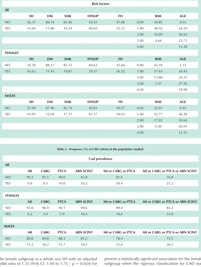
The percentages of individuals that presented RF for CAD in the group as a whole and in the subgroups of males and females are shown in table 1. Table 1 also shows the patient groups stratified by age and BMI categories. The percentages of patients that presented CAD criteria are shown in table 2.
The comparison of the male and female subgroups showed statistically significant differences in relation to SH (p = 0.038), smoking (p = 0.015) and FH (p < 0.001), of which the female subgroup presented a greater frequency of SH and FH and the males presented a greater frequency of smoking. No significant difference was found between the male and female subgroups in regard to the percentage of patients with DM and DYSLIP (p > 0.1). Additionally, a comparison of the two subgroups in relation to age groups and BMI categories also revealed statistically significant differences (p < 0.001).
The comparison of the criteria used to classify the patients with CAD (CABG, MI, PTCA, and ALT SCINT) confirmed that the male subgroup presented a greater frequency of these events (p < 0.001) when evaluated alone or when grouped according to the rigorous (CABG and/or MI and/or PTCA) or complaisant criteria (CABG and/or MI and/or PTCA and/or ALT SCINT).
Table 1 - Frequency (%) of risk factors for CAD in the population studied, and distribution of the individuals according to age groups and BMI
Risk factors All
SH DM SMK DYSLIP FH BMI AGE
NO 56.31 88.14 83.46 59.35 47.88 0.00 30.85 8.55 YES 43.69 11.86 16.54 40.65 52.12 1.00 48.42 24.10
2.00 16.09 30.24 3.00 4.64 22.73
4.00 14.38
FEMALES
SH DM SMK DYSLIP FH BMI AGE
NO 54.38 88.57 85.13 60.63 43.68 0.00 43.10 5.13 YES 45.62 11.43 14.87 39.37 56.32 1.00 37.63 18.45
2.00 13.80 29.25 3.00 5.47 27.26
4.00 19.90
MALES
SH DM SMK DYSLIP FH BMI AGE
NO 57.09 87.96 82.79 58.83 49.57 0.00 25.91 9.93 YES 42.91 12.04 17.21 41.17 50.43 1.00 52.77 26.38
2.00 17.02 30.64 3.00 4.30 20.91
4.00 12.15
Table 2 - Frequency (%) of CAD criteria in the population studied
Cad prevalence All
MI CABG PTCA ABN SCINT MI or CABG or PTCA MI or CABG or PTCA or ABN SCINT
NO 90.2 91.5 90.0 83.8 81.6 76.8
YES 9.8 8.5 10.0 16.2 18.4 23.2
FEMALES
MI CABG PTCA ABN SCINT MI or CABG or PTCA MI or CABG or PTCA or ABN SCINT
NO 93.8 96.0 94.1 89.6 89.4 85.2
YES 6.2 4.0 5.9 10.4 10.6 14.8
MALES
MI CABG PTCA ABN SCINT MI or CABG or PTCA MI or CABG or PTCA or ABN SCINT
NO 88.8 89.8 88.3 81.5 78.4 73.5
YES 11.2 10.2 11.7 18.5 21.6 26.5
the female subgroup as a whole was SH with an adjusted odds ratio of 1.35 (95% CI: 1.04 to 1.75 – p = 0.024) for the complaisant classification. Nevertheless, SH did not
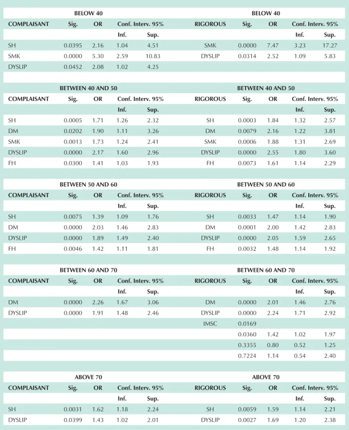
For the male subgroup various modifiable RF are associated with the presence of CAD using both the complaisant and rigorous classifications. However, the factors that presented the greatest association were DM and DYSLIP (Table 5).
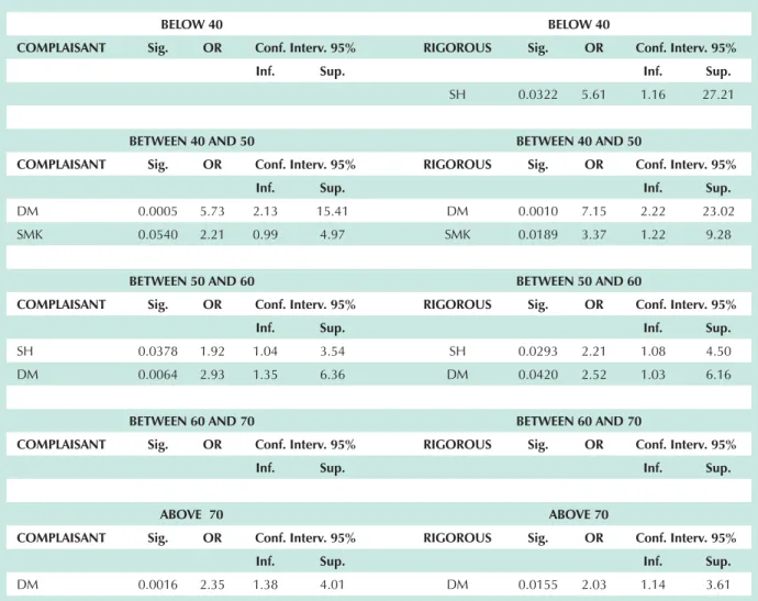
When classified by age groups, some RF that did not initially present a significant association with CAD or presented associations with marginal statistical significance revealed associations that were statistically significant or of greater significance (Tables 6 and 7). In this category the most significant RF were smoking in males under 40 years with an adjusted odds ratio of 5.30 (95% CI: 2.59 to 10.83 – p < 0.0001) for the complaisant classification and 7.47 (95% CI: 3.23 to 17.27 – p < 0.0001) for the rigorous classification; and the DM in females between the ages of 40 and 50 with an adjusted odds ratio of 5.73 (95% CI: 2.13 to 15.41 – p = 0.0005) for the complaisant classification and 7.15 (95% CI: 2.22 to 23.02 – p = 0.0010) for the rigorous
classification. An association between smoking and the presence of CAD for females between the ages of 40 and 50 was also observed, that is not observed in the analysis of the female subgroup as a whole, with an adjusted odds ratio of 2.21 (95% CI: 0.99 to 4.97 – p = 0.0540) for the complaisant classification and 3.37 (95% CI: 1.22 to 9.28 – p = 0.0189) for the rigorous classification. SH that only presented a significant association for the female subgroup with the rigorous criteria for CAD, presented a statistically significant association in various categories using both criteria when age stratification was used.
Discussion
The results of this analysis demonstrated that the main RF associated with a greater presence of CAD in the population studied were aging and male gender. When the modifiable risk factors are analyzed the strongest
Table 3 - Odds ratio (OR) of risk factors for CAD using the complaisant and rigorous criteria, for the entire group of patients
All COMPLAISANT Sig. OR Conf. Interv. 95%
Inf. Sup.
AGE 0 0.0000
1 0.0002 1.92 1.37 2.69
2 0.0000 2.90 2.09 4.02
3 0.0000 5.72 4.12 7.95
4 0.0000 10.45 7.45 14.67
SEX (M) 0.0000 2.84 2.46 3.30
SH 0.0000 1.34 1.19 1.51
DM 0.0000 1.97 1.67 2.31
SMK 0.0023 1.27 1.09 1.49
DYSLIP 0.0000 1.66 1.47 1.87
FH 0.0000 1.30 1.15 1.46
RIGOROUS Sig. OR Conf. Interv. 95%
Inf. Sup.
AGE 0 0.0000
1 0.0004 2.03 1.37 3.00
2 0.0000 3.26 2.23 4.77
3 0.0000 5.87 4.01 8.59
4 0.0000 10.29 6.97 15.19
SEX (M) 0.0000 3.13 2.66 3.70
SH 0.0000 1.36 1.20 1.55
DM 0.0000 1.78 1.50 2.11
SMK 0.0232 1.22 1.03 1.44
DYSLIP 0.0000 1.90 1.67 2.16
association with CAD was observed for DM and DYSLIP. However, SH, smoking and FH also presented statically significant associations with CAD. These results agree with other studies in medical literature3,5,16, despite differences
among the studies in relation to the importance of each risk factor. For example, Avezum et al5 in a study involving
patients in the metropolitan region of São Paulo, where many of the patients evaluated in our study live, observed that the risk factor with the greatest association with MIMI was smoking, while ours was DM. This difference could be the result of the type of criterion used to classify the CAD patient. While in the study of Avezum et al5 MI was
the only positivity criterion used, our study also included procedures (CABG and PTCA) and test results (ALT SCINT) to define the CAD. Additionally, our type of study was also different. Avezum et al used a case control study while ours was cross sectional. In a cross sectional study the significant event could have occurred in the past, causing a later modification in the modifiable RFs. For example, a patient that smoked and suffered a myocardial infarction could have quit smoking as a result of this event. Therefore, currently the patient could be diagnosed with CAD due to the history of myocardial infarction but having quit smoking some time ago would not be classified as a smoker. Despite the difference between the degree of association of CAD with smoking in comparison to the Avezum et al’ study5, an evaluation by age group reveals that, for some
categories, CAD and smoking association is even higher than in other studies5,16. There are various reasons that can
explain this greater association with smoking for some age
groups. For example, the greater association for females between the ages of 40 and 50 could be related to a synergy between smoking and taking the birth control pill. Catelli17
relates that smoking is associated with a greater risk for cardiovascular disease for both genders, however this risk is higher in females over 35 who use birth control pills. In turn, the significant association between smoking and CAD in young males, also described in other previous studies 18-20, could be the result of coronary endothelial alterations
and atherogenesis due to smoking, in an age group where other risk factors are less prevalent.
As mentioned above, the main risk factors associated with CAD in our study were DM and DYSLIP. Like smoking, the association between CAD and these factors also appears to be related to specific age groups. For example, DM which is the main risk factor associated with an increased frequency of CAD in females, seems to be more significant for females between the ages of 40 and 50.
SH, which did not present an expressive association with CAD for the female group as a whole, presented expressive association for some categories when analyzed by age group.
An important fact to note is that since many associations were tested, some of the statistically significant associations presented in the analysis could be the result of chance (type 1 error). One association that could be a result of this type of error was the one between the BMI category 1 and the increased presence of CAD in the males between the ages of 60 and 70 (Table 7), with an adjusted odds ratio of
Table 4 - Risk factors for CAD in the female group
Females
COMPLAISANT Sig. OR Conf. Interv. 95% Inf. Sup.
AGE 0 0.0000
1 0.8433 0.92 0.42 2.02
2 0.6818 0.85 0.40 1.81
3 0.0547 2.04 0.99 4.21
4 0.0003 3.81 1.85 7.86
SH 0.0239 1.35 1.04 1.75
DM 0.0000 2.11 1.52 2.93
RIGOROUS Sig. OR Conf. Interv. 95%
Inf. Sup.
AGE 0 0.0000
1 0.3955 0.67 0.27 1.67
2 0.7394 0.87 0.37 2.01
3 0.0841 2.04 0.91 4.56
4 0.0022 3.51 1.57 7.86
Table 5 - Risk factors for CAD in the male group
Males
COMPLAISANT Sig. OR Conf. Interv. 95% Inf. Sup.
AGE 0 0.0000
1 0.0001 2.14 1.48 3.11
2 0.0000 3.53 2.46 5.08
3 0.0000 6.88 4.77 9.94
4 0.0000 12.29 8.39 18.01
SH 0.0000 1.35 1.18 1.54
DM 0.0000 1.92 1.59 2.31
SMK 0.0011 1.33 1.12 1.59
DYSLIP 0.0000 1.85 1.61 2.11
FH 0.0000 1.34 1.17 1.53
RIGOROUS Sig. OR Conf. Interv. 95%
Inf. Sup.
AGE 0 0.0000
1 0.0001 2.42 1.56 3.74
2 0.0000 4.09 2.67 6.26
3 0.0000 7.21 4.69 11.07
4 0.0000 12.48 8.03 19.40
SH 0.0000 1.38 1.20 1.60
DM 0.0000 1.73 1.42 2.09
SMK 0.0155 1.26 1.04 1.51
DYSLIP 0.0000 2.09 1.81 2.42
FH 0.0000 1.41 1.22 1.62
1.42 (95% CI: 1.02 to 1.97 – p = 0.0360) for the rigorous classification. Besides the fact that BMI was not associated with an increased presence of CAD for any other group, the same occurred even in this same group of males between 60 and 70 years when the complaisant criteria was used. This association was only observed in the case of BMI category 1 but not for the higher BMI categories (categories 2 and 3). Nevertheless, for most of the statistically significant associations observed, the level of statistical significance was much lower than 0.05, therefore suggesting that results are actually a result of differences between the groups and not chance (type 1 error). It was also noted that some of the female age categories did not reveal associations between any of the RFs and the presence of CAD (age group categories 0 and 3). This could be due, in part, to the small number of patients in some groups and that the statistical test used was not powerful enough to reveal the associations in these small groups (type II error).
Considering the methods used in the present study, it is important to define the rigorous and complaisant criteria used to classify the CAD diagnosis. We understand that
neither on its own would prove to be adequate, since the rigorous criterion only included patients with an established diagnosis of CAD and did not include patients with a diagnosis suggested by the MPS, while the complaisant criterion would include those with diagnoses suggested by the MPS; on the other hand it could also consider some patients with false-positive MPS results. Based on this, we opted to separately analyze the two criteria and weigh the strong and weak points of each one in the analysis of the results obtained.
Note also that even though we analyzed the majority of the classic CAD RF, other known and important factors were not considered in this analysis due to lack of information. For example, we did not evaluate the patients stress levels21,
physical activity levels22, alcohol consumption23, diet24,
plasma homocysteine levels25, C-reative protein levels26,
abdominal circumference27, etc. In addition, some RFs
in-depth analysis considering the number of cigarettes smoked per day could have been more appropriate; for dyslipidemia, an analysis of the various cholesterol fractions; and for DM, a stratification by blood glucose levels. Likewise, BMI is currently considered a limited index to evaluate body morphology and has been substituted in various studies by abdominal fat measurements27-29.
Nevertheless, since this more detailed information was not available in our database, the analysis was restricted to the RFs obtained during the taking of the medical history.
Despite these limitations, the present study reaffirms the information in medical literature16,30, even though with
differing degrees of association for a population undergoing myocardial scintigraphy, and naturally reflects the limitations
of the database and methods used, as mentioned earlier. It also reconfirms the information on the evaluation of CAD RF not just related to gender but also to age groups, since some RFs, appear to have a more expressive association with CAD, depending on the gender31-33 and age group34,35.
In conclusion, the risk factors age and gender demonstrated the greatest association with CAD in the population studied and this association is more expressive for older and male patients. The modifiable RFs, in respect to primary prevention of CAD, that had the greatest association with coronary events were DM and DYSLIP for males and DM for females. In relation to specific age groups the most notable factors were smoking for young males (<40 years), and DM and smoking for females between the ages of 40 and 50.
Table 6 - Risk factors for CAD in the female patients according to age groups
BELOW 40 BELOW 40
COMPLAISANT Sig. OR Conf. Interv. 95% RIGOROUS Sig. OR Conf. Interv. 95%
Inf. Sup. Inf. Sup.
SH 0.0322 5.61 1.16 27.21
BETWEEN 40 AND 50 BETWEEN 40 AND 50
COMPLAISANT Sig. OR Conf. Interv. 95% RIGOROUS Sig. OR Conf. Interv. 95%
Inf. Sup. Inf. Sup.
DM 0.0005 5.73 2.13 15.41 DM 0.0010 7.15 2.22 23.02 SMK 0.0540 2.21 0.99 4.97 SMK 0.0189 3.37 1.22 9.28
BETWEEN 50 AND 60 BETWEEN 50 AND 60
COMPLAISANT Sig. OR Conf. Interv. 95% RIGOROUS Sig. OR Conf. Interv. 95%
Inf. Sup. Inf. Sup.
SH 0.0378 1.92 1.04 3.54 SH 0.0293 2.21 1.08 4.50 DM 0.0064 2.93 1.35 6.36 DM 0.0420 2.52 1.03 6.16
BETWEEN 60 AND 70 BETWEEN 60 AND 70
COMPLAISANT Sig. OR Conf. Interv. 95% RIGOROUS Sig. OR Conf. Interv. 95%
Inf. Sup. Inf. Sup.
ABOVE 70 ABOVE 70
COMPLAISANT Sig. OR Conf. Interv. 95% RIGOROUS Sig. OR Conf. Interv. 95%
Inf. Sup. Inf. Sup.
References
1. Yusuf S, Reddy S, Ounpuu S, Anand S. Global burden of cardiovascular diseases: part II: variations in cardiovascular disease by specific ethnic groups
and geographic regions and prevention strategies. Circulation. 2001; 104: 2855-64.
Table 7 - Risk factors CAD in the male patients according to age groups
BELOW 40 BELOW 40
COMPLAISANT Sig. OR Conf. Interv. 95% RIGOROUS Sig. OR Conf. Interv. 95%
Inf. Sup. Inf. Sup.
SH 0.0395 2.16 1.04 4.51 SMK 0.0000 7.47 3.23 17.27 SMK 0.0000 5.30 2.59 10.83 DYSLIP 0.0314 2.52 1.09 5.83 DYSLIP 0.0452 2.08 1.02 4.25
BETWEEN 40 AND 50 BETWEEN 40 AND 50
COMPLAISANT Sig. OR Conf. Interv. 95% RIGOROUS Sig. OR Conf. Interv. 95%
Inf. Sup. Inf. Sup.
SH 0.0005 1.71 1.26 2.32 SH 0.0003 1.84 1.32 2.57 DM 0.0202 1.90 1.11 3.26 DM 0.0079 2.16 1.22 3.81 SMK 0.0013 1.73 1.24 2.41 SMK 0.0006 1.88 1.31 2.69 DYSLIP 0.0000 2.17 1.60 2.96 DYSLIP 0.0000 2.55 1.80 3.60 FH 0.0300 1.41 1.03 1.93 FH 0.0073 1.61 1.14 2.29
BETWEEN 50 AND 60 BETWEEN 50 AND 60
COMPLAISANT Sig. OR Conf. Interv. 95% RIGOROUS Sig. OR Conf. Interv. 95%
Inf. Sup. Inf. Sup.
SH 0.0075 1.39 1.09 1.76 SH 0.0033 1.47 1.14 1.90 DM 0.0000 2.03 1.46 2.83 DM 0.0001 2.00 1.42 2.83 DYSLIP 0.0000 1.89 1.49 2.40 DYSLIP 0.0000 2.05 1.59 2.65 FH 0.0046 1.42 1.11 1.81 FH 0.0032 1.48 1.14 1.92
BETWEEN 60 AND 70 BETWEEN 60 AND 70
COMPLAISANT Sig. OR Conf. Interv. 95% RIGOROUS Sig. OR Conf. Interv. 95%
Inf. Sup. Inf. Sup.
DM 0.0000 2.26 1.67 3.06 DM 0.0000 2.01 1.46 2.76 DYSLIP 0.0000 1.91 1.48 2.46 DYSLIP 0.0000 2.24 1.71 2.92
IMSC 0.0169
0.0360 1.42 1.02 1.97 0.3355 0.80 0.52 1.25 0.7224 1.14 0.54 2.40
ABOVE 70 ABOVE 70
COMPLAISANT Sig. OR Conf. Interv. 95% RIGOROUS Sig. OR Conf. Interv. 95%
Inf. Sup. Inf. Sup.
2. Yusuf S, Reddy S, Ounpuu S, Anand S. Global burden of cardiovascular diseases: part I: general considerations, the epidemiologic transition, risk factors, and impact of urbanization. Circulation. 2001; 104: 2746-53.
3. Piegas LS, Avezum A, Pereira JC, Neto JM, Hoepfner C, Farran JA, et al. Risk factors for myocardial infarction in Brazil. Am Heart J. 2003; 146: 331-8.
4. Izar MC, Fonseca FA, Ihara SS, Kasinski N, Sang WH, Lopes IE, et al. Risk factors, biochemical markers, and genetic polymorphisms in early coronary artery disease. Arq Bras Cardiol. 2003; 80: 379-95.
5. Avezum A, Piegas LS, Pereira JC. Fatores de risco associados com infarto agudo do miocárdio na região metropolitana de São Paulo: uma região desenvolvida em um país em desenvolvimento. DEP - 20050415. Arq Bras Cardiol. 2005; 84: 206-13.
6. Huxley R, Barzi F, Woodward M. Excess risk of fatal coronary heart disease associated with diabetes in men and women: meta-analysis of 37 prospective cohort studies. BMJ. 2006; 332: 73-8.
7. Kanaya AM, Grady D, Barrett-Connor E. Explaining the sex difference in coronary heart disease mortality among patients with type 2 diabetes mellitus: a meta-analysis. Arch Intern Med. 2002; 162: 1737-45.
8. Lee WL, Cheung AM, Cape D, Zinman B. Impact of diabetes on coronary artery disease in women and men: a meta-analysis of prospective studies. Diabetes Care. 2000; 23: 962-8.
9. Orchard TJ. The impact of gender and general risk factors on the occurrence of atherosclerotic vascular disease in non-insulin-dependent diabetes mellitus. Ann Med. 1996; 28: 323-3.
10. Prediction of mortality from coronary heart disease among diverse populations: is there a common predictive function? Heart. 2002; 88: 222-8.
11. Toft J, Hesse B, Rabol A. The occurrence of false-positive technetium-99m sestamibi bull’s eye defects in different reference databases. A study of an age- and gender-stratified healthy population. Eur J Nucl Med. 1997; 24: 179-83.
12. Bouvier F, Bevegard S, Nejat M, Jensen-Urstad M. Myocardial sestamibi uptake in healthy subjects is related to age, gender and habitus. Clin Physiol. 1999; 19: 76-83.
13. Corbett JR, Kritzman JN, Ficaro EP. Attenuation correction for single photon emission computed tomography myocardial perfusion imaging. Curr Cardiol Rep. 2004; 6: 32-40.
14. Smanio PE, Watson DD, Segalla DL, Vinson EL, Smith WH, Beller GA. Value of gating of technetium-99m sestamibi single-photon emission computed tomographic imaging. J Am Coll Cardiol. 1997; 30: 1687-92.
15. Paul AK, Hasegawa S, Yoshioka J, Tsujimura E, Yamaguchi H, Tokita N, et al. Exercise-induced stunning continues for at least one hour: evaluation with quantitative gated single-photon emission tomography. Eur J Nucl Med. 1999; 26: 410-5.
16. Yusuf S, Hawken S, Ounpuu S, Dans T, Avezum A, Lanas F et al. Effect of potentially modifiable risk factors associated with myocardial infarction in 52 countries (the INTERHEART study): case-control study. Lancet. 2004; 364: 937-52.
17. Castelli WP. Cardiovascular disease: pathogenesis, epidemiology, and risk among users of oral contraceptives who smoke. Am J Obstet Gynecol. 1999; 180: S349-S356.
18. Dai J, Gao R, Chen J, Yao K, Yang Y, Qiao S, et al. The clinical features of myocardial infarction in patients younger than 35 years and over 45 years of age. Zhonghua Nei Ke Za Zhi. 1999; 38: 104-6.
19. Morillas PJ, Cabades A, Bertomeu V, Echanove I, Colomina F, Cebrian J, et al. Infarto agudo de miocardio en pacientes menores de 45 anos. Rev Esp
Cardiol. 2002; 55: 1124-31.
20. Zieske AW, McMahan CA, McGill HC Jr, Homma S, Takei H, Malcom GT, et al. Smoking is associated with advanced coronary atherosclerosis in youth. Atherosclerosis. 2005; 180: 87-92.
21. Blumenthal JA, Babyak M, Wei J, O’Connor C, Waugh R, Eisenstein E, et al. Usefulness of psychosocial treatment of mental stress-induced myocardial ischemia in men. Am J Cardiol. 2002; 89: 164-8.
22. Wessel TR, Arant CB, Olson MB, Johnson BD, Reis SE, Sharaf BL, et al. Relationship of physical fitness vs body mass index with coronary artery disease and cardiovascular events in women. JAMA. 2004; 292: 1179-87.
23. daLuz PL, Coimbra SR. Alcohol and atherosclerosis. An Acad Bras Cienc. 2001; 73: 51-5.
24. Singh RB, Dubnov G, Niaz MA, Ghosh S, Singh R, Rastogi SS, et al. Effect of an Indo-Mediterranean diet on progression of coronary artery disease in high risk patients (Indo-Mediterranean Diet Heart Study): a randomized single-blind trial. Lancet. 2002; 360: 1455-61.
25. Rasouli ML, Nasir K, Blumenthal RS, Park R, Aziz DC, Budoff MJ. Plasma homocysteine predicts progression of atherosclerosis. Atherosclerosis. 2005; 181: 159-65.
26. Boekholdt SM, Hack CE, Sandhu MS, Luben R, Bingham SA, Wareham NJ, et al. C-reactive protein levels and coronary artery disease incidence and mortality in apparently healthy men and women: The EPIC-Norfolk prospective population study 1993-2003. Atherosclerosis. 2006; 187 (2): 415-22.
27. Smith DA, Ness EM, Herbert R, Schechter CB, Phillips RA, Diamond JA, et al. Abdominal diameter index: a more powerful anthropometric measure for prevalent coronary heart disease risk in adult males. Diabetes Obes Metab. 2005; 7: 370-80.
28. Onat A, Avci GS, Barlan MM, Uyarel H, Uzunlar B, Sansoy V. Measures of abdominal obesity assessed for visceral adiposity and relation to coronary risk. Int J Obes Relat Metab Disord. 2004; 28: 1018-25.
29. Hoefle G, Saely CH, Aczel S, Benzer W, Marte T, Langer P, et al. Impact of total and central obesity on vascular mortality in patients undergoing coronary angiography. Int J Obes. (Lond) 2005; 29: 785-91.
30. Grundy SM, Pasternak R, Greenland P, Smith S Jr, Fuster V. Assessment of cardiovascular risk by use of multiple-risk-factor assessment equations: a statement for healthcare professionals from the American Heart Association and the American College of Cardiology. Circulation. 1999; 100: 1481-92.
31. Dabelea D, Kinney G, Snell-Bergeon JK, Hokanson JE, Eckel RH, Ehrlich J, et al. Effect of type 1 diabetes on the gender difference in coronary artery calcification: a role for insulin resistance? The Coronary Artery Calcification in Type 1 Diabetes (CACTI) Study. Diabetes. 2003; 52: 2833-9.
32. Juutilainen A, Kortelainen S, Lehto S, Ronnemaa T, Pyorala K, Laakso M. Gender difference in the impact of type 2 diabetes on coronary heart disease risk. Diabetes Care. 2004; 27: 2898-904.
33. Dotevall A, Hasdai D, Wallentin L, Battler A, Rosengren A. Diabetes mellitus: clinical presentation and outcome in men and women with acute coronary syndromes. Data from the Euro Heart Survey ACS. Diabet Med. 2005; 22: 1542-50.
34. Weitzman M, Cook S, Auinger P, Florin TA, Daniels S, Nguyen M, et al. Tobacco smoke exposure is associated with the metabolic syndrome in adolescents. Circulation. 2005; 112: 862-9.