Giancarlo C. Polesello, M.D., Ph.D., André Eugênio Omine Fernandes, M.D.,
Liszt Palmeira de Oliveira, M.D., Ph.D., João Paulo Tavares Linhares, M.D., and
Marcelo C. Queiroz, M.D., M.Sc.
Purpose: The main objective of this study was to investigate medial hip portals and evaluate their relation with anatomic structures in a cadaveric model.Methods: Placement of 3 medial arthroscopic portals was simulated in 10 fresh human paired cadaveric hip specimens by placing Steinmann pins into the joint underfluoroscopic control. Two portals were made at the groin, 1 anterior and 1 posterior to the adductor longus muscle, and the third portal was placed posterior to the adductor longus muscle, 5 cm distal to the groin. The specimens were then dissected, and the relation of the portals to the following structures was recorded: pectineus, adductor longus, gracilis, adductor brevis, adductor magnus, iliopsoas tendon, obturator nerve, femoral nerve, femoral artery, femoral vein, and profunda femoris artery.Results: Regarding the anteromedial portal, the closest neurovascular structure was the profunda femoris artery, which was 10.42.7 mm (range, 6 to 14 mm) distal to the portal. Regarding the posteromedial portal, the nearest neurovascular structure was the obturator nerve, which was 6.03.6 mm (range, 2 to 13 mm) posterior to the portal. Regarding the distal posteromedial portal, the nearest neurovascular structures were the obturator nerve, which was 4.63.0 mm (range, 1 to 9 mm) distal to the portal, and the profunda femoris artery, which was 10.5 3.9 mm (range, 6 to 17 mm) distal to the portal. Conclusions: The use of the medial portals did not cause any damage to the neurovascular structures evaluated. Despite this, the portals are in close relation to the obturator nerve and profunda femoris, and care should be taken.Clinical Relevance:This study investigated 3 medial hip portals in a cadaveric model and also defined safety parameters for this approach. Medial hip portals may be useful to directly approach medial hip pathologies.
A
rthroscopy has become an important tool for diagnosis and treatment of hip pathologies,1 and many portals have been described, mainly for the central, peripheral, and peritrochanteric compart-ments.2-4 Despite this, the medial peripheral compart-ment must be directly approached at times, and this can be difficult with the traditional portals previously described.Medial hip pathology may require a medial approach to the hip joint; however, access to the medial compartment
of the hip is controversial mainly because of possible injury to the obturator nerve and medial femoral circumflex artery.5Ludloff6and Ferguson7have depic-ted an open access method to the medial aspect of the hip through the anteromedial and posteromedial approaches for congenital hip dislocation in children, and the main surgical complications were femoral head osteonecrosis and joint stiffness.5 Chung et al.8 have described an arthroscopic medial portal to perform a lavage for septic arthritis in children. This portal was posterior to the adductor longus muscle with the hip positioned inflexion, abduction, and external rotation. Several technique modifications of this portal have been previously described for congenital hip dislocation treatment in children.9-11 Teloken et al.12 described a similar inferomedial portal for the extraction of afirearm projectile; a portal was positioned 3 cm poste-rior to the adductor longus tendon with the hip in extension and 30of abduction.
Although many different medial portals have been described, a detailed anatomic study has not been per-formed and the risks of these portals must be appraised. The main objective of this study was to investigate medial hip portals and evaluate their relation with From the Hip Group, Department of Orthopedic Surgery and
Traumatol-ogy, Faculdade de Ciências Médicas da Santa Casa de São Paulo (G.C.P., A.E.O.F., J.P.T.L., M.C.Q.), São Paulo, Brazil; and Arthroscopy Laboratory, Faculdade de Ciências Médicas da Universidade Estadual do Rio de Janeiro (L.P.d.O.), Rio de Janeiro, Brazil.
The authors report that they have no conflicts of interest in the authorship and publication of this article.
Received January 16, 2013; accepted September 6, 2013.
Address correspondence to Giancarlo C. Polesello, M.D., Ph.D., Hip Group, Department of Orthopedic Surgery and Traumatology, Faculdade de Ciências Médicas da Santa Casa de São Paulo, Rua Dr. Cesário Motta Júnior, 112, 01221-020, São Paulo, Brazil. E-mail:[email protected]
Ó2014 by the Arthroscopy Association of North America
0749-8063/1354/$36.00
http://dx.doi.org/10.1016/j.arthro.2013.09.004
anatomic structures in a cadaveric model. We hypoth-esized that the medial portals are safe and will not cause any injury to the main anatomic structures.
Methods
Ten fresh human paired hips were used for this study (mean age, 32 9 years; 2 male and 3 female cadavers). The ethics committee of our institution has approved this study.
Anteroposterior pelvic radiographs were performed to exclude any hip deformities, osteoarthritis, heterotopic ossification, congenital malformations, and skeletally immature cadavers.
The specimens were placed supine, and the hip was positioned in 70 of flexion, 70 of abduction, and
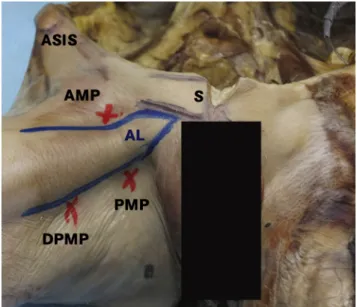
maximum external rotation6-9,13(Figs 1 and 2A). The anterior superior iliac spine, pubic symphysis, and adductor longus were marked with a surgical pen (Fig
1). The specimens were not immobilized. During portal placement, an assistant surgeon secured the specimens. For simulation of the portals, 3 Steinmann pins (4 mm200 mm) were positioned byfluoroscopy toward the transition of the head and femoral neck.3,4 Two portals were positioned in the inguinal skin crease, with 1 at the anterior edge of the adductor longus tendon, 10 mm distal from its origin, denomi-nated the anteromedial portal (AMP), and 1 at the posterior edge, 10 mm distal from its origin, denomi-nated the posteromedial portal (PMP). A third portal was placed at the posterior border of the adductor longus, 5 cm distal to the inguinal crease, denominated the distal posteromedial portal (DPMP). The approxi-mate insertion angles acquired by the Steinmann pins (Fig 2B) were measured with a goniometer in the axial (anterior and posterior) and coronal (superior and Fig 1. Right hip positioned inflexion, abduction, and external
rotation. (AL, adductor longus muscle; ASIS, anterior superior iliac spine; S, pubic symphysis.)
Fig 2. (A) Left hip positioned at 70 of
flexion, 70 of abduction, and maximum
external rotation, showing relation between portals (represented by Steinmann pins) and adductor longus muscle (AL). It should be noted that this picture was taken perpendicular to the adductor longus to better exemplify the anatomic structures. (D, distal; FH, femoral head; IL, inguinal ligament; L, lateral; M, medial; P, proximal; S, pubic symphysis; SA, sartorius muscle.) (B) Left hip radiograph showing Steinmann pins positioned in AMP, PMP, and DPMP toward transition of head and femoral neck.
inferior) planes. These measurements were performed once by thefirst author.4
Dissection of the medial aspect of the hip was per-formed by initially making a skinflap and then identi-fying the saphenous magna vein, obturator nerve, femoral bundle, and profunda femoris artery. After these structures were identified, the smallest distance between them and the Steinmann pins was measured with an electronic caliper (model 799A; Starrett, Athol, MA) (Fig 3).
Descriptive statistics were used for data analysis.
Results
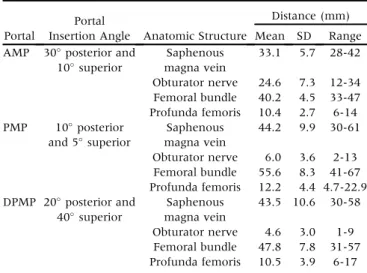
The main results are shown inTable 1.
Anteromedial Portal
The AMP was positioned at the superior border of the adductor longus muscle toward the medial articular surface of the hip (Fig 4B). The portal penetrated the pectineus muscle belly and then the joint capsule. In its path, the closest neurovascular structure was the pro-funda femoris bundle, which was, on average, 10.4
2.7 mm (range, 6 to 14 mm) lateral to the portal. No neurovascular structures were damaged.
Posteromedial Portal
The PMP was positioned at the posterior border of the adductor longus muscle toward the medial articular surface of the hip (Fig 4). The portal passed between the adductor longus and gracilis muscles superficially, and in some cases it penetrated the lateral border of the adductor longus muscle belly and then the joint capsule. In its path, the nearest neurovascular structure was the obturator nerve, which was, on average, 6.0
3.6 mm (range, 2 to 13 mm) posterior to the portal. No neurovascular structures were damaged.
Distal Posteromedial Portal
The DPMP was positioned at the posterior border of the adductor longus muscle, 5 cm distal to the inguinal crease toward the medial articular surface of the hip (Fig 4). The portal penetrated the medial posterior portion of the adductor longus and then the joint capsule. In its path, the nearest neurovascular struc-tures were, more superficially, the obturator nerve, which was, on average, 4.6 3.0 mm (range, 1 to 9 mm) distal to the portal, and the profunda femoris bundle, which was 10.53.9 mm (range, 6 to 17 mm) distal to the portal. No neurovascular structures were damaged.
Discussion
This study has described 3 medial portals for hip arthroscopy, and we have shown that no injury to the anatomic structures occurred. Nonetheless, the obtu-rator nerve and the profunda femoris bundle were put at risk.
The obturator nerve divides at the obturator foramen into an anterior branch, which passes anterior to the adductor muscle, innervating the adductor longus, adductor brevis, and gracilis muscles, and also sends sensory branches to the skin and fascia at the medial and proximal thigh. The posterior branch pierces and Table 1.Distances Between Portals and Anatomic Structures
Portal
Portal
Insertion Angle Anatomic Structure
Distance (mm)
Mean SD Range
AMP 30posterior and
10superior
Saphenous magna vein
33.1 5.7 28-42
Obturator nerve 24.6 7.3 12-34 Femoral bundle 40.2 4.5 33-47 Profunda femoris 10.4 2.7 6-14 PMP 10posterior
and 5superior
Saphenous magna vein
44.2 9.9 30-61
Obturator nerve 6.0 3.6 2-13 Femoral bundle 55.6 8.3 41-67 Profunda femoris 12.2 4.4 4.7-22.9 DPMP 20posterior and
40superior
Saphenous magna vein
43.5 10.6 30-58
Obturator nerve 4.6 3.0 1-9 Femoral bundle 47.8 7.8 31-57 Profunda femoris 10.5 3.9 6-17
Fig 4.(A) Patient supine with left hip positioned at 70 of flexion, 70 of abduction, and maximum
external rotation. The arthroscope is positioned in the AMP, and the shaver is positioned in the PMP. (D, distal; L, lateral; P, proximal; S, pubic symphysis.) (B) Patient supine with left hip positioned at 70 of flexion, 70 of abduction,
innervates the obturator externus muscle, and then it runs between the adductor magnus and brevis muscles, where it divides into a motor branch to the adductor magnus muscle and a sensory branch to the knee joint.14,15The 2 portals posterior to the adductor longus can endanger the anterior branch of the obturator nerve. To minimize this risk, the portals must be posi-tioned at the posterior border of the adductor longus, with the surgeon seeking to avoid the gracilis and the adductor brevis muscles.
The profunda femoris artery originates most fre-quently from the posterolateral aspect of the femoral artery and emits the medial and lateral circumflex femoral artery branches and 4 perforator arteries.16The AMP and PMP are in greater proximity to the profunda femoris artery, but no injuries occurred. The mean distance from the origin of the profunda femoris artery to the midpoint of the inguinal ligament is 35 to 50 mm16-18; therefore the AMP must be placed close to the inguinal crease so that it does not cause any injury in its path. Portals should be directed toward the femoral head-neck junction because the profunda femoris and medial circumflex femoral arteries are usually located distally to this. The greater the hip flexion and abduction, the more distal the deep femoral branch moves as it runs along the inferior edge of the iliopsoas muscle, minimizing the risk of injury during the procedure.
The distance between the emergences of the cir-cumflex arteries in relation to the profunda femoris artery is important because these structures can be injured during insertion of the portals. On average, the emergence of the lateral circumflex femoral artery ranges between 21 and 30 mm from the origin of the profunda femoris artery, and the emergence of the medial circumflex femoral artery ranges between 0 and 10 mm from the origin of the profunda femoris ar-tery.19There are anatomic variations of the circumflex arteries; they may emerge directly from the femoral artery or more proximally, increasing the risk of injury with these portals,19,20and it is more common that the medial circumflex femoral artery originates directly from the femoral artery and not from the lateral circumflex femoral artery.
The approximate location of the PMP has been described for use in joint lavage,13 septic arthritis drainage,8 and removal of foreign bodies,12 as well as for the treatment of congenital hip dislocation.9-11 Examples of cases in which it is necessary to approach the medial aspect of the femoral neck are osteoid osteoma, bone cysts, Brodie abscesses, and medial-region cam impingement. Because of this, we believed that the use of these portals was needed in cases in which a conventional arthroscopic approach would be very difficult. In addition, the medial portals allow a direct approach to the medial aspect of the femoral
neck, whereas this can only be performed in a tangen-tial manner with the other portals. It is important to note that there were no injuries caused by the portals in any of the specimens.
We have shown the possible use of 3 medial portals, but we believe that 2 portals (AMP and PMP) would be sufficient for an adequate approach to the medial hip joint. Although we had no injuries to the neurovascular structures, the posterior portals (PMP and DPMP) can cause injury to the obturator nerve. This can also be a complication of an open approach, so we believe that these procedures should only be performed by expert hip surgeons.
Limitations
This study has limitations. First, there was a small number of specimens, but this is similar to previous studies.3,4 The use of Steinmann pins that have a smaller diameter than the cannulas could alter the distance between the structures studied, but the cannulas have a blunt end, so a direct lesion to the structures is probably uncommon; however, some cannulas have sharp, not blunt, trocars that may directly injure structures. Nonetheless, this method has been used previously.3,4 We also noted that there is variability in the adductor longus muscle size between specimens, and this may lead to small changes in portal positioning. Dissection of specimens can lead to loss of anatomic relations between structures; however, this limitation was addressed by a careful and standardized dissection without any muscle removal. Only adult cadavers were used, so the findings from this study cannot be extrapolated to the pediatric population.
Conclusions
The use of the medial portals did not cause any damage to the neurovascular structures evaluated. Despite this, the portals are in close relation to the obturator nerve and profunda femoris, and care should be taken.
Acknowledgment
The authors thank Sheila Ingham, M.D., Ph.D. (Federal University of São Paulo, São Paulo, Brazil), for valuable advice, review of the manuscript, and help with the English version.
References
1. Banerjee P, McLean CR. Femoroacetabular impingement: A review of diagnosis and management.Curr Rev Muscu-loskelet Med2011;4:23-32.
3. Byrd TJW, Pappas JN, Pedley JM. Hip arthroscopy: An anatomic study of portal placement and relationship to the extra-articular structures. Arthroscopy 1995;11: 418-423.
4. Robertson WJ, Kelly BT. The safe zone for hip arthros-copy: A cadaveric assessment of central, peripheral, and lateral compartment portal placement. Arthroscopy 2008;24:1019-1026.
5. Hoppenfeld S, deBoer P. The hip and acetabulum. In: Surgical exposures in orthopaedics: The anatomic approach. Ed 3. Philadelphia: Lippincott Williams & Wil-kins, 2003;455-462.
6. Ludloff BK. The open reduction of the congenital hip dislocation by an anterior incision. Am J Orthop Surg 1913;10:438-454.
7. Ferguson AB Jr. Primary open reduction of congenital dislocation of the hip using a median adductor approach.
J Bone Joint Surg Am1973;55:671-689.
8. Chung WK, Slater GL, Bates EH. Treatment of septic arthritis of the hip by arthroscopic lavage.J Pediatr Orthop 1993;13:444-446.
9. Eberhardt O, Fernandez FF, Wirth T. Arthroscopic reduction of the dislocated hip in infants.J Bone Joint Surg Br2012;94:842-847.
10. Bulut O, Ozturk H, Tezeren G, Bulut S. Arthroscopic-assisted surgical treatment for developmental dislocation of the hip.Arthroscopy2005;21:574-579.
11. Gross R. Arthroscopy in hip disorders in children.Orthop Rev1977;6:43-49.
12. Teloken MA, Schmietd I, Tomlinson DP. Hip arthroscopy: A unique inferomedial approach to bullet removal. Arthroscopy2002;18:E21.
13. Strife JL, Towbin R, Crawford A. Hip arthrography in infants and children: The inferomedial approach. Radi-ology1984;152:536.
14. Tipton JS. Obturator neuropathy. Curr Rev Musculoskelet Med2008;1:234-237.
15. Anagnostopoulou S, Kostopanagiotou G, Paraskeuopoulos T, Chantzi C, Lolis E, Saranteas T. Anatomic variations of the obturator nerve in the inguinal region: Implications in conventional and ultrasound regional anesthesia techniques. Reg Anesth Pain Med2009;34:33-39.
16. Bannister LH, Berry MM, Collins P. Cardiovascular system. In: Gray’s anatomy. Ed 38. New York: Churchill Livingstone, Medical Division of Longmann Group UK Ltd, 1995;1566-1568.
17. Thitilertdecha S, Rungruang T, Voraphattropas C. The origin of profunda femoris artery in Thais. Siriraj Med J 2012;64(suppl 1):S34-S36.
18. Dixit DP, Mehta LA, Kothari ML. Variations in the origin and course of profunda femoris.J Anat Soc India2001;50:6-7. 19. Dixit D, Kubavat DM, Rathod SP, Pateld MM, Singel TC.
A study of variations in the origin of profunda femoris artery and its circumflex branches. Int J Biol Med Res 2011;2:1084-1089.