BILATERAL LAMELLAR KERATOPLASTY IN DESCEMETOCELE
TREATMENT IN DOG WITH BOTULISM BY USE OF EQUINE
RENAL CAPSULE AND CONJUNCTIVAL PEDICLE GRAFT
EMPREGO DE CERATOPLASTIA LAMELAR BILATERAL NO TRATAMENTO DE DESCEMETOCELE EM CÃO COM BOTULISMO, UTILIZANDO-SE CÁPSULA
RENAL EQÜINA E ENXERTO CONJUNTIVAL PEDICULADO
José Luiz Laus1 Paula Diniz Galera2 Ruben Pablo Schocken-Iturrino1
Marluce de Macedo Cavassani3 Alexandre Lima de Andrade4
CASE REPORT
1Professor DVM PhD, Veterinary College, UNESP, Jaboticabal, SP. Brasil, Rodovia Carlos Tonanni, km5, 14870-000, Fax-(016)
3224275. E.mail-jllaus@fcav.unesp.br. Author for correspondence.
2
Professor DVM - Veterinary College - UNIC, Cuiabá, MT. Brasil.
3Professor DVM - Veterinary College - UNESP, Araçatuba, SP. Brasil.
SUMMARY
A 3-year-old, male mixed breed dog with botulism and bilateral descemetocele was submitted to lamellar kerato-plasty with equine renal capsule preserved in glycerin in the right eye and conjunctival pedicle graft in the left eye. The evolution was satisfactory in both eyes, but better in the eye receiving the equine renal capsule, because the corneal transparence was more evident in that eye. On the other hand, the surgical period was more quickly in the eye receiving the equine renal capsule becau-se the preparation of the conjunctival pedicle before the kerato-plasty was not necessary.
Key words: keratoplasty, cornea, descemetocele, botulism.
RESUMO
Um animal da espécie canina, macho, de 3 anos de idade, com botulismo e descemetocele bilateral foi submetido à ceratoplastia lamelar com cápsula renal eqüina preservada em glicerina no olho direito, e enxerto conjuntival pediculado no olho esquerdo. Ambos os olhos mostraram evolução satisfatória porém, o olho receptor da cápsula renal eqüina apresentou transparência corneana mais evidente.
Palavras-chave: ceratoplastia, córnea, descemetocele, botulis-mo.
INTRODUCTION
A neurotoxin produced by Clostridium botulinum, which is an anaerobic gram-positive rod, is responsible for a non-contagious disease known as botulism. The main symptom of the disease is a neuronal inferior motor disturbance, which may result into a picture of total flaccid paralysis. The alteration of the activity of cranial nerves may de-crease the palpebral and pupilary reflexes (SWANGO et al., 1992). The interference of the motor enervation of the eyelids may result into a corneal drying with a severe damage to the cornea (SLATTER et at., 1990).
convergence power is developed by the cornea (WARING, 1984; HELPER, 1989). Its nerves origi-nate from ciliar nerve, which is derived from the ophthalmic nerve (DYCE et al., 1990).
Among the diseases commonly found in the cornea, the ulcerative keratitis is the most im-portant one due to the risks it offers to the eye func-tion. The treatment for that disease is accomplished by clinical and surgical procedures. Among the surgical ones, the keratoplasties are the most fre-quently employed.
The first reports on these techniques are dated from XVIII century. However, more successful techniques were presented from the se-cond half of the present century on. Procedures with auto and homografts were conducted successfully by GUNDERSEN (1958), JENSEN (1963), DICE et al. (1973), THOFT (1977), THOFT (1982), STARTUP (1984), NASISSE (1985), BRIGHTMAN et al. (1989), PORTNOY et al. (1989), KERN (1990), SLATTER (1990), HACKER (1991), MISHRA & REDDY (1991).
Superficial keratectomies in dogs have been repaired with equine pericardium by BARROS
et al. (1990), with equine renal capsule by
ANDRADE (1994), with homologous peritoneum by GARCIA et al. (1996) and sardine scales by LAUS (1994). LAUS et al. (1996) and MORALES et al. (1996) studied the corneal and both conjuncti-val pedicle and non-pedicle grafts, and could obser-ve that the pedicle grafts were better, since they provided with an immediate blood supply, though being indicated in deep corneal ulcers. Non-pedicle conjunctival grafts and corneal grafts were incorpo-rated more quickly by the cornea; however, this technique would be bettered indicated for less deep ulcers.
CASE REPORT
A male dog, aging 3 years old, mixed bre-ed with a diagnosis of botulism, was assistbre-ed at the Ophthalmogical Section of Veterinary College of São Paulo State University, UNESP, Jaboticabal-SP/ Brazil. This animal presented, as a consequence of botulism, a flaccid paralysis of the eyelids, followed by a deep bilateral corneal ulcer, with descemetoce-le. This case, due the severity, demanded the car-rying out of a surgical treatment which under such a condition, suggested the treatment with lamellar keratoplasty.
The animal, following the routine preli-minary procedures for a surgery like that, was then taken into the surgical center, being so, submitted to inhalatory anesthesia, with Halothane®, in a closed
circuit. The surgical procedure was done with the use of a surgical microscope.
The conjunctival pedicle graft is well-known technique, as being highly efficient in cases like that, and it was then employed by taking as basis, what had been described by SLATTER (1990). Lamellar keratoplasty with equine renal capsule was admitted, as we have mentioned before, had already been experimentally tested by our team, according to original publishing by ANDRADE (1994). As both eyes presented an ulceration, we then decided that the choice for which eye would receive what graft, should be randomized. This way, the left eye received the conjunctival pedicle graft, and the right eye, the equine renal capsule preserved in glycerin. The post-operative treatment was done by the use of chloranphenicol ophthalmic cream employing Epitezan®, at intervals of 06 hours for 20 days, and also Atropine eyedrops at 1%, every 24 h for 4 days.
The comparison between both employed techniques was made by comparing the surgical period spent on each of them, and also the evolution of cicatrization of the corneas receiving the surgical procedures. For the study of cicatrization evolution, photophobia, ocular discharge, oedema, neovascula-rization, pigmentation and transparence a slit-lamp was employed, as well as the fluoresceine test which was used with the same objectives. The utilized method for that comparing evaluation, was based on graduation (Nihil: absent; discreet: +; moderate: ++; intense: +++).
Otherwise, in the eye receiving conjunctival pedicle graft there is small anterior synechiae. (figures. 1 and 2).
RESULTS AND DISCUSSION
The ulcerative keratitis are one the most important ocular diseases. The treatment for these diseases, frequently require the utilization of surgical
methods known as keratoplasties.
In the last few decades, innovating te-chniques have been proposed. Among them, we could highlight the employment of biological mem-branes. In our work, we tried to assess the results of a keratoplasty with the employment of an equine renal capsule preserved in glycerin, by comparing them with a traditional procedure, such as conjuncti-val pedicle graft. In relation to the pedicle graft, many have been the researchers who have presented the advanta-ges and merits of the technique (PFEIFFER et al., 1977; THOFT, 1982; HAKAN-SON, et al.,1988). These authors des-cribed that patients who had been treated with that technique, evoluted fairly well. In spite of that, they could observe that in the first fifteen days after the surgery, the patients presented blepharospasm, photophobia, ocular discharge and hyperemia.
MORALES et al.
(1996), described that those findings were common and did occur indistinctively in all pati-ents. The same author also re-ported that, thirty days after the surgery, the pedicle graft became very outlined and a granulation tissue could be observed in the grafted areas. As usual, these findings maintained themselves for up to fifty days, when then the cornea started to regain its transparence.
In relation to biologi-cal membranes, BARROS et al. (1990) showed good results with the employment of equine peri-cardium preserved in glycerin. The authors also have reported that, besides the found habitual phenomena, there was a deposit of pigments in the cornea, which had been operated. GARCIA et al. (1996) studied the homolo-gous peritoneum and described similar results as the ones once presented by BARROS et al. (1990). Besides those works, we could bring to light the utilizati-on of sardine scales by LAUS (1994), in which the authors
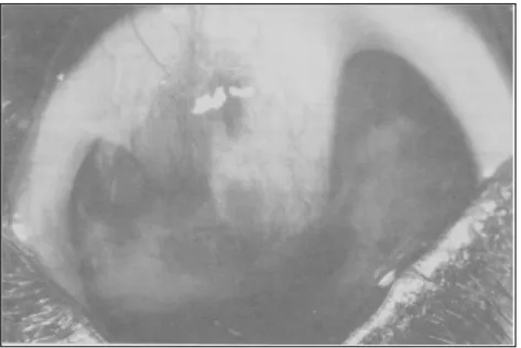
Figura 1 - The eye receiving the equine renal capsule 4 months after surgery. Notice that there are few vessels, there is no pigmentation and the graft can no longer be seen. The corneal transparence is not perfect, but it is better than the eye receiving the conjunc-tival pedicle graft.
showed the occurrence of new vessels and pigments, even after having a long period of post operative been elapsed.
Generally speaking, all the authors agree that lamellar keratoplasties result into a partial loss of transparence in the sites where the grafts are im-planted, due to the neo-vascular formation and pig-mentation.
About this present work, whose results are being discussed, it is relevant to state at first that, it is meant to be the very first work in which clini-cally, the equine renal capsule was tested, because until then, it had only been experimentally tested by ANDRADE (1994).
Since the ulcers were very similar in both eyes, we decided for utilizing this case to compare a traditional surgical procedure (conjunctival pedicle graft) with lamellar keratoplasty by employing an equine renal capsule preserved in glycerin, which up to that moment, had only been tested experimentally, showing fair results. It is worth mentioning that, this comparison was only possible, because the animal´s owner allowed such a procedure.
In relation to the comparison with con-junctival pedicle graft, the post operative assessment showed that in the first fifty days, blepharospasm, ocular discharge, hyperemia and oedema were evi-dent for both techniques. From thirty days after the post operative, the differences between both techni-ques began to appear. It could be observed that the equine renal capsule was incorporated by the cornea with the reduction of oedema and vascularization. In the eye receiving conjunctival pedicle graft, a vita-lity of the graft could be observed, which had been expected since it was a pedicle graft, according to MORALES et al. (1996).
At fifty days, the equine renal capsule could already be found totally incorporated by the cornea and the oedema was now very small. These remarks are in agreement with the ones by ANDRADE (1994). In that period, the conjunctival pedicle graft could be found still vitalized, according to MORALES et al. (1996).
Forty months after the surgery the cornea receiving renal capsule, showed a transparence de-gree fairly well whereas the other cornea still main-tained the conjunctival graft well vitalized. It is worth mentioning that, due the severity of ulcerati-on, we decided toward not cutting the pedicle,
Finally, by the comparative analysis one can assume that both methods were efficient. Howe-ver, advantages can be granted to renal capsule. It is said for this statement, some situations or conditions in which the conjunctiva cannot be utilized as for
example, in the bacterial infections of this structure, in cases in which it had been used but there was a dehiscence, and also when the ulcer occurs in equine species animals, because namely in this species the conjunctiva does not represent a fair material for the accomplishment of pedicle graft. Still about the advantages of renal capsule, we could mention a greater transparence of cornea, and the shorter time used for the accomplishment of keratoplasty. In relation to this time mentioned, it is easy to unders-tand why it is shorter, because by using renal cap-sule, we do not spend any time at all on the prepara-tion of conjunctival pedicle before the keratoplasty.
AKNOWLEDGEMENTS
Research supported by FAPESP - Proc 98/03153-0, Fort Dodge and Ethicon.
REFERENCES
ANDRADE, A.L. Emprego experimental da cápsula renal xenógena, conservada em glicerina, no reparo de ceratectomias superficiais em cães (Canis familiaris,
LINNAEUS, 1758): Avaliação clínica e morfológica. Jaboticabal, 1996. 74p. Tese (Mestrado em Veterinária) -Faculdade de Ciências Agrárias e Veterinárias, Universidade Estadual Paulista, 1996.
BARROS, P.S.M. Reparação cirúrgica da córnea de cães usando pericárdio de eqüino conservado em glicerina. In: CONGRESSO BRASILEIRO DA ANCLIVEPA, 13, 1990, Gramado. Resumos... Gramado: ANCLIVEPA, 1990, p. 11. BRIGHTMAN, A.H., MCLAUGHLIN, S.A., BROGDON, J.D.
Autogenous lamellar corneal grafting in dogs. J Am Vet Med Assoc, Schaumburg, v. 195, n. 4, p. 469-475, 1989.
DICE, P.F., SEVERIN, G.A., LUMB, W.V. Experimental autogenous and homologous corneal transplantation in the dog. J Am Anim Hosp Assoc, Mishawaka, v. 9, p. 245-269, 1973.
ZYCE, K.M., SACK, W.O., WENSING, C.J.G. Tratado de Anatomia Veterinária. Rio de Janeiro: Guanabara Koogan, 1990. Os órgãos dos sentidos, p. 225-235.
GARCIA, J.A., BARROS, P.S.M., LAUS, J.L., et al.Implante de peritônio homólogo conservado após ceratectomia lamelar em cães. Braz J Vet Res and Anim Sci., São Paulo, v. 33, supl., p. 290-294, 1996.
GUNDERSEN, T. Conjuntival flaps in the treatment of corneal disease with reference to a new technique of application. Arch Ophthalmol, Chicago, v. 60, p. 880-888, 1958.
HACKER, D.V. Frozen corneal grafts in dogs and cats: a report on 19 cases. J Am Anim Hosp Assoc, Mishawaka, v. 27, p. 387-398, 1991
Ciência Rural, v. 29, n. 2, 1999. HELPER, L.C. Magrane’s canine ophthalmology. 4. ed.
Philadelphia: Lea & Febiger, 1989. Diseases and surgery of the cornea and sclera, p. 102-149.
JENSEN, E.C. Experimental corneal transplantation in the dog. J Am Vet Med Assoc, Chicago, v. 142, p. 11-22, 1963.
KERN, T.J. Ulcerative keratitis. Vet Clin North Am Small Anim Pract, Philadelphia, v. 20, n. 3, p. 643-666, 1990.
LAUS, J.L. Emprego da escama de sardinha (Sardinella Brasiliensis - STEIDACHNER, 1859), conservada em
glicerina, como sucedâneo de córneas no reparo de ceratectomias superficiais: Estudo experimental em cães. (Canis familiaris - LINNAEUS, 1758). Jaboticabal, 1994.
71p. Tese (Livre Docência em Medicina Veterinária) -Faculdade de Ciências Agrárias e Veterinárias, Universidade Estadual Paulista, 1994.
LAUS, J.L., SOUZA, M.S.B.; MORALES, A., et al. Comparação entre ceratoplastias lamelares por enxertos autógenos, livres, de córnea e pediculados de conjuntiva. Estudo experimental no cão (Canis familiaris - LINNAEUS, 1758). Braz J Vet Res Anim Sci, São Paulo, v. 33, n. 1, p. 41-46, 1996.
MISHRA, C.G., REDDY, T.V. Lamellar homogenous corneal transplantation in mules. Indian Vet J, Madras, v. 68, p. 367-369, 1991.
MORALES, A.; LAUS, J.L.; SOUZA, M.S.B., et al. Comparação entre enxertos autógenos livres e pediculados de conjuntiva no reparo de ceratectomias superficiais. Estudo experimental no cão (Canis familiaris - LINNAEUS, 1758). Braz J Vet Res Anim Sci, São Paulo, v. 33, n. 1, p. 28-31, 1996.
NASISSE, M.P. Canine ulcerative keratitis. Comp Cont Educ
for Pract Vet, Baton Rouge, v. 7, n. 9, p. 686-701, 1985.
PFEIFFER, R.L., GELLAT, K.N., GWIN, R.M. Tarsoconjunctival pedicle grafts for deep corneal ulceration in the dog and cat. J Am Anim Hosp Assoc, Lakewood, v. 13, p. 387-391, 1977.
PORTNOY, S.L., INSLER, M.S., KAUFMAN, H.E. Surgical management of corneal ulceration and perforation. Surv Ophthalmol, Boston, v. 34, n. 1, p. 47-58, 1989.
SAMUELSON, D.A. Ophthalmic embryology and anatomy. In: GELLAT, K.N. Veterinary ophthalmology. 2.ed. Philadelphia: Lea & Febiger, 1991, p. 3-123.
SLATTER, D. Fundamentals of veterinary ophthalmology. 2.ed. Philadelphia: Saunders, 1990. Cornea and sclera, p. 257-303.
STARTUP, F.C. Corneal ulceration in the dog. J Small Anim Pract, London, v. 25, p. 737-752, 1984.
SWANGO, L.J., BANKEMPER, K.W., KONG, L.I. Infecções bacterianas, riquetsiais, protozoais, e outras. In: ETTINGER, S.J. Tratado de medicina interna veterinária. 3. ed. São Paulo. Manole, 1992, p. 286-287
THOFT, R.A. Conjunctival transplantation. Arch Ophthalmol, Chicago, v. 95, p. 1425-1427, 1977.
THOFT, R.A., Indications for conjunctival transplantation. Ophthalmology, Philadelphia, v. 89, n. 4, p. 335-339, 1982.