Resistance in
Caenorhabditis elegans
by Direct and
Indirect Mechanisms
Larissa de Freitas Bonomo1, David Nunes Silva1, Patrı´cia Ferreira Boasquivis1, Franciny Aparecida Paiva1, Joyce Ferreira da Costa Guerra1, Talita Alves Faria Martins3, A´ lvaro Gustavo de Jesus Torres3,
Igor Thadeu Borges Raposo de Paula1, Washington Luiz Caneschi1, Philippe Jacolot4, Nicolas Grossin5, Frederic J. Tessier4, Eric Boulanger5, Marcelo Eusta´quio Silva1,6, Maria Lu´cia Pedrosa1,2,
Riva de Paula Oliveira1,3*
1Nu´cleo de Pesquisa em Cieˆncias Biolo´gicas, Universidade Federal de Ouro Preto, Ouro Preto, Brazil,2Departamento de Cieˆncias Biolo´gicas, Universidade Federal de Ouro Preto, Ouro Preto, Brazil,3Departamento de Biodiversidade, Evoluc¸a˜o e Meio Ambiente, Universidade Federal de Ouro Preto, Ouro Preto, Brazil,4Institut polytechnique LaSalle Beauvais, Beauvais, France,5Faculte´ de Me´decine – Poˆle Recherche, Universite´ de Lille 2, Lille, France,6Departamento de Alimentos, Universidade Federal de Ouro Preto, Ouro Preto, Brazil
Abstract
Ac¸aı´ (Euterpe oleracea Mart.) has recently emerged as a promising source of natural antioxidants. Despite its claimed
pharmacological and nutraceutical value, studies regarding the effects of ac¸aı´in vivoare limited. In this study, we use the Caenorhabditis elegansmodel to evaluate thein vivoantioxidant properties of ac¸aı´ on an organismal level and to examine its mechanism of action. Supplementation with ac¸aı´ aqueous extract (AAE) increased both oxidative and osmotic stress resistance independently of any effect on reproduction and development. AAE suppressed bacterial growth, but this antimicrobial property did not influence stress resistance. AAE-increased stress resistance was correlated with reduced ROS
production, the prevention of sulfhydryl (SH) level reduction andgcs-1activation under oxidative stress conditions. Our
mechanistic studies indicated that AAE promotes oxidative stress resistance by acting through DAF-16 and the osmotic stress response pathway OSR-1/UNC-43/SEK-1. Finally, AAE increased polyglutamine protein aggregation and decreased proteasome activity. Our findings suggest that natural compounds available in AAE can improve the antioxidant status of a whole organism under certain conditions by direct and indirect mechanisms.
Citation:Bonomo LdF, Silva DN, Boasquivis PF, Paiva FA, Guerra JFdC, et al. (2014) Ac¸aı´ (Euterpe oleraceaMart.) Modulates Oxidative Stress Resistance in Caenorhabditis elegansby Direct and Indirect Mechanisms. PLoS ONE 9(3): e89933. doi:10.1371/journal.pone.0089933
Editor:Aamir Nazir, CSIR-Central Drug Research Institute, India
ReceivedOctober 4, 2013;AcceptedJanuary 24, 2014;PublishedMarch 3, 2014
Copyright:ß2014 Bonomo et al. This is an open-access article distributed under the terms of the Creative Commons Attribution License, which permits
unrestricted use, distribution, and reproduction in any medium, provided the original author and source are credited.
Funding:This work received financial support from the Conselho Nacional de Desenvolvimento Cientı´fico e Tecnolo´gico (CNPq Process 473015/2008 0), Fundac¸a˜o de Amparo a` Pesquisa de Minas Gerais (FAPEMIG Process CBBAPQ- 01153-08 and CBB-PPM-00491-10) and Universidade Federal de Ouro Preto (UFOP). Research fellowships were sponsored by CAPES (Bonomo, L.F.; Boasquivis, P.F.; de Paula, I.T.B.R; Guerra, J.F.C.), CNPq (Oliveira, R.P., Pedrosa, M.L., Silva, M.E.), FAPEMIG (Paiva, F.A.; Silva, D.N.) and UFOP (Caneschi, W.L.). The funders had no role in study design, data collection and analysis, decision to publish, or preparation of the manuscript.
Competing Interests:The authors have declared that no competing interests exist. * E-mail: rivaoliveira@nupeb.ufop.br
Introduction
Ac¸aı´ (Euterpe oleraceaMart.) is an exotic fruit originally native to Central and South America that grows in the floodplains of the Amazon region [1]. Traditionally, ac¸aı´ is used as a medicinal plant and as a staple food in many parts of Brazil. In recent years, ac¸aı´ pulp has gained international attention as a functional food due to its nutritional benefits and therapeutic promise. Composition analysis shows that ac¸aı´ pulp contains approximately 13% protein, 48% lipids, 1.5% total sugar and several other nutrients such as lignans, dietary fiber and polyphenols [2]. The main polyphenols found in ac¸aı´ are anthocyanins, proanthocyanidins (specifically cyanidin 3-O-glucoside and cyanidin 3-O-rutinoside) and other flavonoids [2,3]. The overall pharmacological properties of ac¸aı´ are related to its antiproliferative, anti-inflammatory, antioxidant and cardioprotective effects [4].
Ac¸aı´ pulp and its polyphenolic fractions show high antioxidant activity based on various in vitro assays, predominantly against DPPH, superoxide and hydroxyl radicals and hypochlorous acid [5–11].In vitro cell-based assays have also demonstrated that the pulp can reduce ROS production in human erythrocytes and polymorphonuclear (PMN) cells exposed to oxidative stress [8,9]. Brain tissue cells pretreated with ac¸aı´ decreased H2O2-induced
damage to lipids and proteins and reduced the activities of the antioxidant enzymes superoxide dismutase (SOD) and catalase (CAT) to basal levels [12].
significant increase in serum antioxidant capacity and a reduction of lipid peroxidation [8,14]. In another study, ac¸aı´ puree improved select markers of metabolic disease risk in overweight adults [15]. The high superoxide and peroxyl radical scavenging capacity of ac¸aı´ pulp suggests that ac¸aı´ has anti-aging properties. InDrosophila melanogaster, dietary ac¸aı´ supplementation increased the lifespan of female flies fed a high fat diet and also under oxidative stress induced bysod1RNAi [16]. In another fruit fly,Anastrepha ludens, ac¸aı´ supplementation promoted survival in flies on diets with high fat and high sugar while decreasing lifetime reproductive output [17].
In addition to flies, genetic and pharmacological modifications of stress resistance and lifespan mechanisms have been well elucidated in the nematode Caenorhabditis elegans. A number of genes and pathways have been identified to modulate lifespan inC. elegans. These highly conserved pathways include the insulin/IGF-1 receptor-like signaling pathway, which depends on the transcription factor DAF-16/FOXO. Reduced insulin/IGF-1 signaling activates DAF-16 nuclear localization, which in turn induces the expression of genes that increase lifespan and promote resistance to various stresses [18]. SKN-1/Nrf is another transcription factor regulated by the insulin/IGF-1 signaling pathway [19]. SKN-1 contributes to the increased stress tolerance and longevity resulting from reduced insulin/IGF-1 signaling independently of DAF-16 [19]. Using this model, several studies have demonstrated the beneficial effects of natural products in promoting stress resistance and longevity, including extracts from Ginkgo biloba[20], blueberry [21],Cinnamomum cassiabark [22] and cranberry [23].
In the present study, we use theC. elegansmodel to evaluate the in vivoantioxidant properties of ac¸aı´ on an organismal level and to unveil its mechanism of action. Our results indicate that ac¸aı´ aqueous extract (AAE) increases both oxidative and osmotic stress resistance independently of any effect on reproduction and development. AAE also suppressed bacterial growth, but this antimicrobial property did not influence stress resistance. AAE-increased stress resistance was correlated with reduced ROS production, the prevention of SH level reduction and gcs-1 activation under oxidative stress conditions. AAE promotes oxidative stress resistance by acting through DAF-16 and the osmotic stress response pathway OSR-1/UNC-43/SEK-1. Our findings suggest that AAE modulates oxidative stress responsesin vivoby both direct and indirect mechanisms.
Materials and Methods
Chemicals and Reagents
M199, Fungizone, penicillin, streptomycin and L-glutamine were purchased from Gibco by Life Technologies (Saint Aubin, France). Methanol, HPLC-grade acetonitrile, HPLC-grade water, formic acid, DPPH (2,2-diphenyl-1-picryl-hydrazyl), Trolox (6-hydroxy-2,5,7,8-tetramethylchroman-2-carboxylic acid) and cya-nidin 3-O-rutinoside and kanamycin were purchased from Sigma-Aldrich (St. Louis, MO, USA). Cyanidin 3-O-glucoside was purchased from Sigma-Aldrich (Saint Quentin Fallavier, France). Carboxy-H2DCFDA was purchased from Invitrogen (Eugene,
Oregon, USA). Lyophilized ac¸aı´ fruit (Euterpe oleraceaMart.) was obtained from Liote´cnica Alimentos LTDA (Embu, SP, Brazil).
Caenorhabditis elegansStrains and Maintenance
The following strains were used in this study: Bristol N2
(wild-type; WT); LD1171, Is003 (gcs-1::GFP); CL2166,
dvls19[pAF15(gst-4::GFP::NLS)]; CF1553, muls84[pAD76( sod-3::GFP)]; VP198, kbIs5 [gpdh-1p::GFP + rol-6(su1006)]; TJ 356,
zIs356[pGP30(DAF-16::GFP)+pRF4(rol-6)]; BA17, fem-1(hc17); EU1, skn-1(zu67) IV/nT1; CF1038, daf-16(mu86); AU3, nsy-1(ag3); KU4, sek-1(km4); VC8, jnk-1(gk7); AM1, osr-1(rm1); and MT2605,unc-43(n498n1186). AllC. elegansstrains were maintained at 20uC on solid nematode growth medium (NGM) seeded withE. coli (OP50) as a food source according to Brenner [24]. The synchronization of worm cultures was achieved by hypochlorite treatment of gravid hermaphrodites.
Human Umbilical Vein Endothelial Cells (HUVECs) Culture HUVECs isolated previously by Boulanger et al. [25] were collected from the veins of umbilical cords obtained from Jeanne de Flandres Maternity (Lille University Hospital, France). Cells were cultured in M199 (2.5mg/mL Fungizone, 100 U/mL penicillin, 100mg/mL streptomycin, 2 mM L-glutamine and 150 mM Hepes) supplemented with 20% FBS. HUVECs were maintained in a humidified atmosphere containing 5% CO2 at
37uC.
Ac¸aı´ Aqueous Extract Preparation and Treatment For all the experimental procedures, lyophilized ac¸aı´ fruit was diluted with S basal solution and filter sterilized to obtain an aqueous extract (AAE). Control solution (S basal) or 100 mg/mL AAE was then mixed with anE. coli(OP50) pellet at OD 1 and seeded onto NGM plates. In experiments conducted with dead bacteria, NGM plates seeded withE. coliOP50, with or without 100 mg/mL AAE, were treated with 10 mM Kanamycin (KAN).
Anthocyanin Quantification
Total anthocyanin was quantified by the pH differential method as described by Giustiet al. [26]. Diluted samples were added to 0.025 M chloride buffer (pH 1.0) and 4.0 M sodium acetate buffer (pH 4.5). After incubation in the dark for 30 min at room temperature, absorbances were determined simultaneously as absorption maxima for the visible light spectrum and at 700 nm (SP-220, Biospectro, PR, Brazil). The total anthocyanin content was expressed in mg of cyanidin-3-glucoside equivalent per 100 g of ac¸aı´ powder. A molar absorptivity of 26,900 M21cm21and a molecular mass of 449.2 g/mol were used for cyanidin-3-glucoside.
The quantification of cyanidin glucoside and cyanidin 3-O-rutinoside was performed in duplicate using a modified method from Gordonet al. [27]. Briefly, 0.25 g of sample was diluted in 3 mL of methanol/water/acetic acid (50:49.5:0.5, v/v/v). After shaking for 5 min, the sample was sonicated for 20 sec and centrifuged at 8,000 rpm for 10 min at 10uC. The supernatant was collected, and the pellet was re-extracted twice. The three supernatants were pooled, transferred into a 10 mL volumetric flask and diluted with methanol/water/acetic acid mix. After filtration through a 0.45mm filter, each sample was analyzed by LC-MS/MS. Mass spectrometry analyses were carried out on a Thermo Scientific TSQ Quantum Discovery MAX triple-stage quadrupole mass spectrometer (Thermo Fisher Scientific, Courta-boeuf, France) with an electrospray ionization (ESI) probe coupled to an Accela HPLC system (Thermo Fisher Scientific, Courta-boeuf, France). The analytical separation was performed on a Symmetry Shield RP18 column, 15062.1 mm, 3.5mm (Waters,
Monitoring (SRM) mode. The specific transitions m/z 449.0R m/z 287.1 and m/z 595.0 R m/z 287.1 were used for the detection and quantification of cyanidin 3-O-glucoside and cyanidin 3-O-rutinoside, respectively. As no stable isotopically labeled internal standard for anthocyanins is commercially available, the quantification was performed using the standard addition method and expressed as mg/100 g of dry matter.
DPPH Radical Scavenging Assay
The DPPH Radical Scavenging Activity of AAE was deter-mined as described by Brand-Willianset al. [28]. In short, 100mL of ac¸aı´ extract (1, 10 and 100 mg/mL) was added to 3.9 mL of 60mM DPPH dissolved in 80% methanol. The mixture was homogenized and kept in the dark for 30 min at room temperature, after which the absorbance at 515 nm was deter-mined (SP-220, Biospectro, PR, Brazil). A calibration curve was prepared using 2,5,7,8-tetramethylchroman-2-carboxylic acid (Trolox) in the concentration range 200–800mM. The percentage of inhibition was determined according to the following equation: % Scavenging activity = (1 – Abs Sample 515/Abs Control 515) 6100. The DPPH radical scavenging assay was conducted twice.
Body Length and Brood Size Assays
To measure body length, first-larval-stage animals (L1) were treated with control solution (S basal) or 100 mg/mL AAE until the third larval stage (L3) and then transferred to NGM plates with E. coli OP50 until the next day. Images were captured (Axio Imager Z2, Zeiss, NY, USA) of one-day-old animals, and body length was measured along the animal axis using NIH Image J software.
To determine total progeny production, ten fourth-larval-stage (L4) worms, previously treated with control solution (S Basal) or 100 mg/mL AAE since L1, were placed onto the E. coli OP50 lawn on individual NGM plates in the presence or absence of 100 mg/mL AAE. During the egg-laying period, nematodes were transferred onto new plates every 24 h for five days until the end of the reproductive period. The F1 progeny from each individual worm was counted after approximately two days. The total progeny numbers for each plate were calculated and divided by the number of animals [29]. Both experiments were conducted three times.
Longevity
The longevity assay was performed with the adult sterile strain fem-1(hc17)as the wild-type strain to avoid progeny overgrowth in lifespan. Therefore, eggs were shifted to 25uC, the nonpermissive temperature forfem-1(hc17)fertility. Lifespan was scored every day after hermaphrodites completed the final larval molt, from the first day of adulthood (defined as t = 0) until death. We analyzed approximately 90 hermaphrodites treated with control solution (S basal) or 100 mg/mL AAE and divided into three NGM plates of 30 animals each. Animals were scored as dead if they displayed no spontaneous movement or failed to respond when prodded. Dead worms that displayed internally hatched progeny, an extruded gonad or desiccation caused by crawling off the agar were excluded from the data [21]. The longevity assay was conducted three times.
Stress Resistance Assays
To evaluate oxidative stress resistance, N2 wild-type animals andskn-1(zu67),daf-16(mu86),nsy-1(ag3),sek-1(km4),jnk-1(gk7), osr-1(rm1)and unc-43(n498n1186)mutants were treated with control solution (S basal) or 100 mg/mL AAE from L1 until L4 and then
with 7.5 mMtert-butyl hydrogen peroxide (t-BOOH) in M9. To perform the oxidative stress resistance assay in dead bacteria, NGM plates seeded withE. coliOP50, with or without 100 mg/ mL AAE, were treated with 10 mM KAN. Survival was measured at 3, 6, 9 and 12 h. We analyzed five wells, each with approximately ten worms, for each experimental group. Worms were prodded with a platinum wire and scored as dead if they displayed no pharyngeal pumping or movement [30].
Thermotolerance assays were performed with hermaphrodites on adult day 5, after the majority of egg-laying had ceased. Animals treated at 20uC with control solution (S basal) or 100 mg/ mL AAE from L1 until adult day 5 were transferred onto 3-cm NGM agar plates supplemented as indicated above and then incubated at 35uC for 12 h. Survival was monitored at 6, 9 and 12 h and scored as the number of animals responsive to gentle touch as a fraction of the original number of animals on the plate. Animals that had died from desiccation on the sides of the plate were excluded [21]. To quantify the percent of motile worms under acute osmotic stress, animals were treated with control solution (S basal) or 100 mg/mL AAE for 68 h from L1 and then transferred to new plates containing 500 mM NaCl. The percentage of worms that moved outside a 7-mm circle was monitored at 15, 30 and 60 min. All experiments measuring oxidative stress resistance were conducted at least twice. The heat stress and motility under acute osmotic stress assays were each performed three times.
Bacterial Growth Curve
E. coliOP50 growth was evaluated over 4 h in the presence of 100 mg/mL AAE. All OD readings at 600 nm were normalized to the OD of the control group at time zero. Bacterial growth was measured in three individual experiments.
In vivoMeasurement of ROS
ROS production was measured inC. elegansand HUVECs using the fluorescent probe 29, 79-dichlorofluorescein diacetate (H2DCFDA). ROS production in C. elegans was performed as
described by Shiet al. [31]. Synchronized L1 animals were treated with control solution (S basal) or 100 mg/mL AAE for 48 h. L4 worms were incubated in 1 mL PBS containing 1 mM H2O2for
2 h. Subsequently, the worms were washed twice and incubated in 0.5 mL PBS containing 50mM H2DCFDA for 1 h. Thirty animals
in experimental triplicates of each group were then transferred into the wells of a 96-well microtiter plate containing 200mL PBS. The fluorescence quantification was carried out on a multilabel microplate Reader VICTOR X3 (Perkin Elmer, Massachusetts, USA) using excitation at 485 nm and emission at 535 nm.
ROS production in HUVECs was performed as described by Montiel-Da´valoset al. [32]. HUVECs were grown in 12-well plates to a density of 16105 cells per well. Confluent cells were then pretreated with or without 2.5 mg/mL AAE for 16 h, followed by incubation for 30 min with 10mM H2DCFDA at 37uC in the
dark. After this, the cells were washed with PBS and then treated with or without 0.25 mM H2O2for 1 h. Cells were washed once
and harvested in PBS. Fluorescence was detected with a flow cytometer using excitation at 488 nm and emission at 525 nm. In bothC. elegansand HUVECs, the fluorescence of the control group was used to normalize the values from all other groups. The experiments were repeated at least four times.
Quantification of Total Sulfhydryl (SH) Levels
t-BOOH for 1 h. The animals were then washed with M9 buffer and sonicated in 2-mL microcentrifuge tubes. The resulting homogenate was centrifuged, and the supernatant was collected, discarding cellular debris and intact worms. The total protein content was determined according to the method described by Lowryet al. [33] using bovine serum albumin (BSA) as a standard. The total and free serum sulfhydryl groups were estimated using Ellman’s reagent according to Sedlak and Lindsay [34]. The protein-bound sulfhydryl groups were determined as the difference between the total and free sulfhydryl groups. The experiment was repeated twice.
Reporter Gene Expression
Transgenic worms containing reporter genes were treated with control solution (S basal) or 100 mg/mL AAE for 48 h starting at L1, followed by the presence or absence of oxidative stress. The stress condition was 7.5 mM t-BOOH for 1 h forgcs-1::GFPand gst-4::GFPgene expression analysis and 10 mM t-BOOH for 1 h for sod-3::GFP. To analyze the subcellular localization of DAF-16::GFP, synchronized L1 transgenic worms were treated with control solution (S basal) or 100 mg/mL AAE for 48 h with or without subsequent 7.5 mM t-BOOH treatment for 1 h. Twenty worms from each group were mounted onto microscope slides coated with 1% agarose, anaesthetized with 0.5 mM sodium azide and capped with coverslips. Photographs were taken on a fluorescence microscope (Axio Imager Z2, Zeiss, NY, USA), and GFP fluorescence signals were measured using NIH Image J software. ForDAF-16::GFP, expression patterns were classified as cytosolic, intermediate or nuclear. The experiment was conducted three times.
Analysis of gene expression by qPCR
Synchronized L1 larvae were grown in plates containingE. coli OP50 bacteria resuspended in basal solution or AAE 100 mg/mL until L4 stage. Total RNA from worms was isolated using BRAZOL (LCG Biotecnologia, Sa˜o Paulo, Brazil) according to manufacturer’s instructions and cDNA was synthesized using High-Capacity cDNA Reverse Transcription Kits (Applied Biosystems). The qPCR was performed on a Applied Biosystems 7500 Real-Time PCR System (Applied Biosystems, Carlsbad, CA, USA) using a Power SYBR Green PCR master mix (Applied Biosystems). qPCR levels were normalized to the expression of ama-1, which encodes the large subunit of RNA polymerase II. The fold change was normalized to that observed in untreatedC. eleganssamples. Primers sequences forama-1,daf-16,gst-7,ctl-1, sod-3 and osr-1 are listed in Table S1. The gene expression was analyzed in three individual experiments.
Polyglutamine (PolyQ) Aggregation Quantification Transgenic worms carrying the reporter gene vha-6::Q44::YFP were treated with control solution (S basal) or 100 mg/mL AAE since L1. Photographs of one-, four-, eight- and twelve-day-old animals were taken with a fluorescence microscope (Axio Imager Z2, Zeiss, NY, USA), and the numbers of aggregates were counted. The experiment was repeated three times.
Proteasome Activity Quantification
In vitro 26S proteasome activity assays were performed as described by Kisselev and Goldberg [35]. Approximately 5,000 N2 wild-type animals were treated with control solution (S basal) or 100 mg/mL AAE for 48 h at L1. L4 worms were then harvested and sonicated. The lysates were centrifuged at 20,0006g for 20 min at 4uC. Protein extract was quantified using the QuantiPro BCA Assay Kit (Sigma Aldrich, St. Louis, MO, USA). For measuring the chymotrypsin-like activity of the proteasome, succinyl-Leu-Leu-Val-Tyr-4-methyl-coumaryl-7-amide (SLLVY-MCA) (Sigma-Aldrich, St. Louis, MO, USA) was used both in the presence or absence of 20mM MG-132, a proteasome inhibitor. Enzyme kinetics were monitored in a temperature-controlled microplate reader VICTOR X3 (Perkin Elmer, Massachusetts, USA) every 15 min for 1 h at 37uC; the excitation and emission wavelengths were 380 and 460 nm, respectively. Proteasome activity was calculated as the difference between the total activity and the activity remaining in the presence of 20mM MG-132. The proteasome activity quantification was conducted three times.
Data Analysis
Statistical analysis was performed using GraphPad Prism version 5.00 for Windows (San Diego, CA). The results were plotted as the mean6SEM (standard error of the mean) of at least two individual experiments. Data were subjected to the Kolmo-gorov-Smirnov test for normality. For data with a normal distribution, Student’sttest was used to compare pairs of groups, whereas a one-way ANOVA followed by Tukey’s post-test was used to compare three or more groups. Nonparametric data were analyzed using the Mann-Whitney test when comparing two groups and the Kruskal-Wallis test followed by Dunn’s post-test for comparing three or more groups. All survival curves were analyzed by the Log-rank (Mantel-Cox) test. The statistical significance was determined as p,0.05.
Table 1.Determination of anthocyanins present in ac¸aı´ aqueous extract (AAE).
Compounds Concentration (mg/100 g)
Total monomeric anthocyaninsa 31.0
62.4 Cyanidin 3-O glucosideb 8.860.9 Cyanidin 3-O rutinosideb 8.7
60.6
The results are expressed as the mean6SEM (standard error of the mean). aTotal monomeric anthocyanins measured by the pH differential method. bCyanidin 3-O glucoside and Cyanidin 3-O rutinoside determined by LC-MS/MS analysis.
doi:10.1371/journal.pone.0089933.t001
Table 2.In vitroantioxidant activity of ac¸aı´ aqueous extract (AAE) measured by DPPH assay.
% inhibition (Mean±SEM)
AAE (mg/
mL) AAE AAE+OP50
AAE+OP50+
KAN AAE+KAN
1 5.0760.75a 4.38
60.98a 20.71
60.28b 0.10
60.16b 10 53. 3065.61a 49.53
64.07a 18.55
62,18b 25.68
62.22b 100 79.6163.33a 77.30
63.49a 73.46
64.54a 78.35
61.78a
DPPH, 2,2-diphenyl-1-picrylhydrazyl; Trolox,
6-hydroxy-2,5,7,8-tetramethylchroman-2-carboxylic acid; OP50,E. colistrain; KAN, Kanamycin. Different subscript letters indicate significant differences by one-way ANOVA followed by Tukey’s post-test.
Results
AAE phytochemical composition and antioxidant capacityin vitroandin vivo
We first characterized the composition of anthocyanins and the in vitro antioxidant capacity of our extract. AAE presents 31.062.4 mg/100 g of the total anthocyanins measured by the pH differential method. Cyanidin 3-O glucoside and cyanidin 3-O rutinoside are the two major anthocyanins in ac¸aı´ pulp; AAE presents 8.860.9 mg/100 g of cyanidin 3-O glucoside and 8.760.9 mg/100 g of cyanidin 3-O rutinoside, as determined by LC-MS/MS analysis (Table 1). Ac¸aı´’s polyphenolic compounds have already been associated with itsin vitroantioxidant capacity
[3,36,37]. We next determined the antioxidant capacity of 1, 10 and 100 mg/mL AAE by the DPPH radical scavenging activity method. We observed that AAE displays increasing in vitro antioxidant capacity in a dose-dependent manner (Table 2). At 100 mg/mL AAE, the highest concentration tested, the DPPH inhibition was 79.61%, which is equivalent to 800mM Trolox, the reference standard, which shows 80.90% inhibition. Moreover, the percentage of DPPH inhibition for any concentration tested was not significantly altered when the extracts were mixed withE. coli OP50. Thus, we used AAE at 100 mg/mL with liveE. coliOP50 for the subsequent experiments.
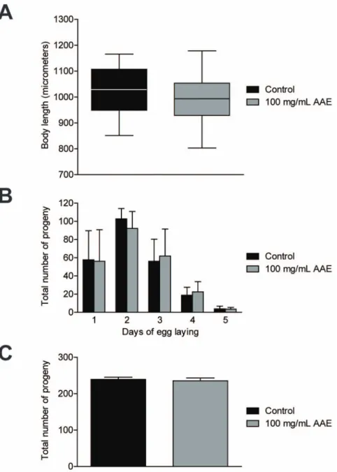
AAE does not alterC. elegansdevelopment and progeny As toxic compounds usually delayC. elegans development and progeny [38], we characterized the effect of AAE treatment on these two biological parameters. Body length was determined in one-day-old adults treated with or without 100 mg/mL AAE from L1 until L3 and then transferred to NGM plates withE. coliOP50. We did not observe a significant difference in body length between control (1020.0611.6mm) and AAE-treated animals (994.3610.5mm) (Figure 1A). To measure the total number of progeny, animals were treated with control solution (S basal) or 100 mg/mL AAE from L1 until the end of their reproductive period. There was no significant difference in the total number of progeny between control (239.466.1) and AAE-treated animals (235.667.5) (Figure 1B). In addition, there was no difference in the egg-laying profile between the two groups (Figure 1C). These results suggest that 100 mg/mL AAE is not toxic toC. elegans, as it does not interfere with development and progeny (Figure 1A, 1B and 1C).
AAE increases resistance to oxidative and hyperosmotic stress conditions
As AAE showsin vitroantioxidant activity and is not toxic toC. elegans, we next evaluated whether AAE treatment has a protective effect under normal and oxidative, heat and hyperosmotic stress conditions. To monitor longevity under normal conditions, fem-1(hc17)mutants grown at 25uC were treated with control solution (S basal) or 100 mg/mL AAE beginning at L1. AAE treatment had no effect on theC. elegansaging profile (p = 0.2997) (Figure 2A). Oxidative stress resistance assays were performed in wild-type animals treated with control solution (S basal) or 100 mg/mL AAE from L1 until L4 and then with 7.5 mM t-BOOH in M9. AAE showed increased oxidative stress resistance when compared to the control (p = 0.0002) (Figure 2B). The effect of AAE on heat tolerance was analyzed at 35uC in five-day-old wild-type animals with or without 100 mg/mL AAE treatment from L1. AAE did not protectC. elegans against heat stress when compared to the control group (p = 0.7388) (Figure 2C). Finally, we analyzed motility under acute osmotic stress at 500 mM NaCl. For the Figure 2. Effect of ac¸aı´ aqueous extract (AAE) onC. elegansgrown under normal and stress conditions. A)fem-1(hc17)mutants were treated at 25uC with control solution (S basal) or 100 mg/mL AAE beginning at L1. Surviving and dead animals were counted daily until all nematodes had died. Log-rank (Mantel-Cox) analysis showed no significant difference between the curves.B) Animals were treated with control solution (S basal) or 100 mg/mL AAE from L1 until L4 and then submitted to 7.5 mM t-BOOH in M9. The survival was measured at 6, 9 and 12 h. The survival curves show that AAE treatment increasedC. elegansoxidative stress resistance. ***p,0.001 by the Log-rank (Mantel-Cox) test.C) Animals were treated with control solution (S basal) or 100 mg/mL AAE beginning at L1. After five days at 20uC, the animals were incubated at 35uC and survival was monitored at 6, 9 and 12 h. There was no significant difference between curves by the Log-rank (Mantel-Cox) test.D) Animals were treated with control solution (S basal) or 100 mg/mL AAE for 68 h beginning at L1 and then transferred to new plates containing 500 mM NaCl. The percentage of worms that moved outside a 7-mm circle was monitored at 15, 30 and 60 min. *p,0.05 by a two-tailed Student’st-test.
motility assay, L1 wild-type animals were treated with 100 mg/ mL AAE for 68 h and then transferred to NGM plates with 500 mM NaCl. AAE treatment significantly increased motility under the osmotic stress condition (Figure 1D). After 1 h, 48.31% of the animals treated with 100 mg/mL AAE showed motility compared to 27.70% from the control group (p,0.05). We conclude that 100 mg/mL AAE protects against oxidative and hyperosmotic stress conditions but not under normal and heat stress conditions.
The protective effect of AAE against oxidative stress is independent of its antimicrobial effect
As bacteria play a role inC. elegansmortality and stress resistance [39,40], we investigated whether the increased oxidative stress promoted by AAE could be a secondary response to a possible AAE antimicrobial property. First, we evaluated E. coli OP50 growth, measuring the OD over 4 h in the presence or absence of 100 mg/mL AAE. AAE treatment decreased bacteria growth at all times analyzed (Figure 3A). Because we observed that 100 mg/ mL AAE exerted an antimicrobial effect, we repeated the oxidative stress resistance assay in animals treated with AAE on dead bacteria. We observed that wild-type animals treated with 100 mg/mL AAE presented increased survival on 7.5 mM
t-BOOH compared to controls (p,0.0001) (Figure 3B). These results show that AAE treatment does not increase C. elegans oxidative stress resistance through an antimicrobial property.
AAE improves redox status inC. elegans and HUVECs under oxidative stress conditions
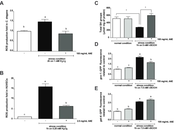
Potential antioxidants can act directly to neutralize reactive oxygen species (ROS) and thus prevent the cellular response to increased oxidative stress to combat and remove ROS. To evaluate the capacity of AAE to neutralize ROSin vivo, we assessed ROS production in two experimental models, C. elegans and HUVECs, using the H2DCFDA fluorescent probe. We did not
detect an effect of AAE treatment on basal ROS levels in eitherC. elegansor HUVECs (data not shown). We observed a significant increase in fluorescence intensity in worms (Figure 4A) and cells (Figure 4B) treated with their respective stress conditions. However, AAE treatment significantly reduced intracellular ROS accumulation inC. elegansunder stress conditions (p,0.01) (Figure 4A). In HUVECs, the stress condition increased ROS production by 16-fold relative to the control, and AAE diminished this increase by up to 6-fold (p,0.001) (Figure 4B).
As AAE exhibited an in vivo antioxidant capacity, we next evaluated whether AAE would improve redox status in wild-type C. elegans. We assessed the levels of total sulfhydryl (SH) groups in animals treated with control solution (S basal) or 100 mg/mL AAE from L1 until L4. The treatment ofC. eleganswith 100 mg/ mL of AAE for 48 h did not increase the levels of total protein sulfhydryl groups (515.97632.73 vs. 512.2652.75mmol/mg of protein). The stress condition significantly decreased the levels of SH groups (515.97632.73 vs. 279.7365.51mmol/mg of protein) (p,0.05). However, when the worms were first exposed to AAE and then submitted to the stress condition with t-BOOH, this pretreatment was able to prevent the reduction of protein sulfhydryl group levels induced by t-BOOH (601.18638.25 vs. 279.7365.51mmol/mg of protein) (p,0.05) (Figure 4C).
To correlate thein vivoantioxidant effect of AAE treatment with a molecular response inC. elegans, we analyzed the gene expression of c-glutamyl cysteine synthetase-1 (gcs-1) and glutathione-s-transferase-4 (gst-4). GCS-1 is the limiting enzyme of glutathione (GSH) synthesis and is expressed at high levels in the intestine in response to oxidative stress but at low levels under normal conditions in an SKN-1-dependent manner [41]. gst-4::GFP is expressed primarily in the muscles and the hypodermis under normal conditions, and is increased in response to a variety of oxidative stress treatments [42,43]. Transgenic worms containing reporter genes were treated with control solution (S basal) or 100 mg/mL AAE for 48 h beginning at L1 and either in the presence or absence of oxidative stress. The fluorescence signals of gcs-1::GFP and gst-4::GFP animals treated with 100 mg/mL of AAE for 48 h were not significantly increased compared to the control group (Figure 4D–E). Exposure to 7.5 mM t-BOOH for 1 h increased gcs-1::GFP and gst-4::GFP expression, and AAE significantly prevented the upregulation ofgcs-1::GFP(Figure 4D) but not gst-4::GFP (Figure 4E). Taken together, these results suggest that AAE functions as a direct antioxidant through the removal of ROS, preventing the decrease in SH groups mediated by the stress condition.
Increased oxidative stress resistance induced by AAE treatment depends on DAF-16 and OSR-1/UNC-43/SEK-1
Certain flavonoids protect against oxidative stress either directly, by radical scavenging, or indirectly, by inducting antioxidant enzymes and thus increasing the stress resistance of Figure 3. Effect of ac¸aı´ aqueous extract (AAE) on the bacterial
growth and oxidative stress resistance of wild-typeC. elegans
grown on dead bacteria. A)E. coliOP50 growth was evaluated over 4 h in the presence of 100 mg/mL AAE. The OD of the control group at time zero was used to normalize all other OD readings. * Treatment of 100 mg/mL AAE decreased bacteria growth at all times analyzed with p,0.05, determined by a two-tailed Student’st-test.B) Animals were treated with or without 100 mg/mL AAE, mixed with eitherE. coliOP50 orE. coliOP50 treated with 10 mM Kanamycin (KAN), from L1 until L4 and then submitted to 7.5 mM t-BOOH in M9. The survival was measured at 6, 9 and 12 h. The survival curves show that AAE treatment increased C. elegans oxidative stress resistance independent of its antibacterial effect. ***p,0.001 related to the respective control by the Log-rank (Mantel-Cox) test.
the organism. It has been shown inC. elegansthat phytochemicals and plant extracts activate several stress response pathways [21,23,44]. To verify whether, in addition to its direct role, AAE protection under oxidative stress could also require these pathways, we performed the oxidative stress resistance assay in wild-type and mutant animals treated with 100 mg/mL AAE or control solution (S basal).
DAF-16/FOXO, the downstream target of insulin-like signaling in C. elegans, is a transcription factor required for both lifespan regulation and stress resistance [45,46]. The nuclear translocation of DAF-16 is positively regulated by c-Jun N-terminal kinase (JNK-1) in parallel with insulin-like signaling [46]. We therefore investigated whether oxidative stress resistance induced by AAE treatment depends on JNK-1/DAF-16 signaling. AAE treatment did not increasedaf-16(mu86)mean survival (p = 0.1782), whereas it increased the mean survival ofjnk-1(gk7)(p,0.0001) (Figure 5A;
Table 3). These results suggest that AAE may increase oxidative stress resistance via DAF-16 independently of JNK-1.
SKN-1/Nrf is a transcription factor that promotes the expression of antioxidant or detoxification enzymes, thus increas-ing stress resistance and longevity in C. elegans [19,30]. Under oxidative stress, SKN-1 functions depend on p38 MAPK signaling through the NSY-1/SEK-1/PMK-1 pathway [47]. We measured the survival of nsy-1(ag3), sek-1(km4) and skn-1(zu67) mutants treated with AAE. AAE treatment prolonged the mean survival of nsy-1(ag3) (p = 0.0005) and skn-1(zu6) (p = 0.0002) but not sek-1(km4)(p = 0.7483) (Figure 5B; Table 3). These results suggest that the AAE antioxidant effect does not depend on SKN-1, and may act via the p38 pathway, but only through SEK-1.
In C. elegans, survival under hyperosmotic stress requires the OSR-1/UNC-43/SEK-1 stress response pathway [48]. We also tested whether the protective effect of AAE treatment under Figure 4. Effect of ac¸aı´ aqueous extract (AAE) on redox status in wild-typeC. elegansand HUVECs. A)C. eleganswas treated with control solution (S basal) or 100 mg/mL AAE for 48 h and then submitted to the presence or absence of 1 mM H2O2for 2 h. The results are expressed as H2DCFDA fluorescence relative to the untreated control.B) HUVECs were treated with or without 2.5 mg/mL AAE for 16 h and then incubated in 0.25 mM H2O2for 1 h. The fluorescence was measured by flow cytometry. The results are expressed as H2DCFDA fluorescence relative to the untreated control. Different letters indicate significant differences by one-way ANOVA followed by Tukey’s post-test.C) To measure the levels of total SH groups, animals were treated with control solution (S basal) or 100 mg/mL AAE from L1 until L4 and then incubated with or without 5 mM t-BOOH for 1 h. *p values were determined by a two-tailed Student’st-test, and groups were significantly different when p,0.05 inC. elegans. Transgenic worms containing reporter genes were treated with control solution (S basal) or 100 mg/mL AAE for 48 h beginning at L1 and then with or without the oxidative stress condition. After a 1-h hour recovery period, photographs were taken on a fluorescence microscope. For (D)gcs-1::GFP and (E)gst-4::GFPanimals, GFP fluorescence signals were measured using NIH Image J software. Different letters correspond to significant differences by the Kruskal-Wallis test followed by Dunn’s post-test.
oxidative stress conditions depends on OSR-1 and UNC-43. AAE treatment did not extend the mean survival ofunc-43(n498n1186) (p = 0.7320) (Figure 5C, Table 3). In osr-1(rm1) mutants, AAE treatment reduced the mean survival under oxidative stress (p = 0.0009) (Figure 5C, Table 3). These findings suggest that AAE modulates oxidative stress resistance through OSR-1 and two of its downstream effectors: UNC-43 and SEK-1.
AAE treatment inducesctl-1andgst-7expression in a DAF-16-dependent manner
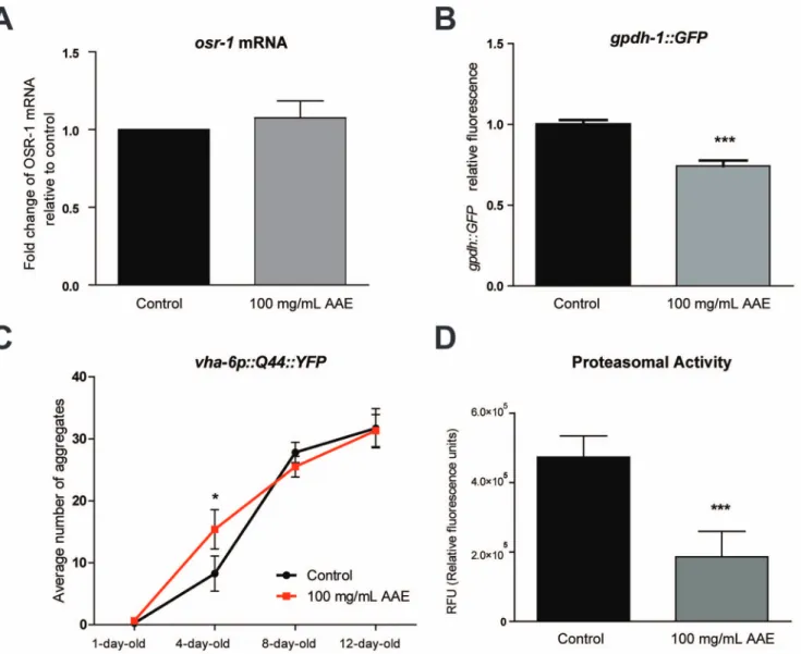
As AAE treatment increased oxidative stress resistance inC. elegansthrough DAF-16, we investigated whether AAE treatment
activates DAF-16 nuclear translocation under normal and stress conditions. The results showed that under normal growth conditions, AAE treatment did not induce DAF-16::GFP nuclear translocation (Figure 6A–B). Moreover, the AAE treatment did not alterdaf-16 mRNA levels in wild-type animals (Figure 6C). The stress condition used in this study (7.5 mM t-BOOH for 1 h) also did not increase the fraction of animals showing DAF-16::GFP nuclear localization (Figure 6B). However, under stress conditions, AAE treatment significantly reduced DAF-16::GFP nuclear localization (Figure 6B). One possible explanation for this finding is that AAE modifies DAF-16 to increase its activity but not its concentration. To further test this hypothesis, we measured the Figure 5. Contribution of genetic background to oxidative stress resistance induced by ac¸aı´ aqueous extract (AAE) treatment. Animals were treated with 100 mg/mL AAE or control solution (S basal) from L1 until L4 and then submitted to 7.5 mM t-BOOH in M9. Survival was measured at 3, 6, 9 and 12 h.A) Survival curves for transcription factordaf-16 (mu86)and JNK MAPK pathwayjnk-(gk7)mutants.B) Survival curves for skn-1(zu67)and p38 MAPK pathway,nsy-1(ag3),sek-1(km4)mutants.C) Survival curves for osmotic stress resistance pathway,unc-43(n498n1186)and osr-1(rm1)mutants. *p,0.05 and ***p,0.001 by the Log-rank (Mantel-Cox) test (see Table 3 for more details).
expression of the DAF-16 stress target genes after AAE treatment in wild-type animals anddaf-16mutants (Figure 6D). The results showed that the transcripts levels of ctl-1 and gst-7 were upregulated in wild-type animals treated with AAE in a manner that was dependent upon daf-16 (Figure 6D). However, AAE treatment did not induce neithersod-3mRNA (Figure 6D) nor sod-3::GFP expression (Figure S1). Taken together, these results suggest that AAE treatment increases oxidative stress resistance by activating antioxidant genes via DAF-16.
AAE increases polyglutamine protein aggregation and decreases proteasome activity
OSR-1 is a secreted protein that couples with SEK-1/MAPKK through UNC-43/CaMKII to promote resistance to osmotic stress [48]. A loss of OSR-1 function constitutively activates gpdh-1 expression and glycerol accumulation [49]. Glycerol replaces inorganic ions in the cytoplasm and functions as a chemical chaperone that aids in the refolding of misfolded proteins [50].
It is possible that AAE increases both oxidative and osmotic stress resistance by directly blocking OSR-1 activity and inducing gpdh-1 expression. To test this hypothesis, we measured osr-1 mRNA levels andgpdh-1::GFPexpression in animals treated or not with 100 mg/mL AAE for 48 h. The AAE treatment did not modifyosr-1gene expression under normal conditions (Figure 7A) and reduced gpdh-1::GFP expression (Figure 7B). Nevertheless, AAE treatment could interfere in OSR-1 activity which is coupled with the pathway UNC-43/SEK-1 in order to promote oxidative stress resistance.
Alternatively, AAE may function as a mild stressor, increasing hypertonic stress and/or protein homeostasis. High ionic strength is a well-known disruptor of protein secondary structure that can increase protein aggregation [51]. To test the hypothesis that AAE could increase hypertonic strength, we quantified aging-induced polyglutamine protein aggregation in vha-6::Q44::YFP transgenic worms. Q44 animals exhibit a diffuse fluorescence throughout the intestine until they reach adulthood, when focal fluorescent aggregates gradually increase. At one-day-old, Q44::YFP aggre-gation was not different between the control and 100 mg/mL AAE-treated groups (Figure 7C), whereas four-day-old Q44::YPF adult animals treated with 100 mg/mL AAE had approximately twice as many aggregates as control animals (p,0.05) (Figure 7C). Although the average number of aggregates increased with age, the AAE treatment had no effect on Q44::YFP aggregation in adult worms at eight- and twelve-day-old (Figure 7C). This result
suggests that AAE treatment accelerates the aggregation formation at the young adult stage. To evaluate whether AAE treatment impairs protein homeostasis, we measured proteasome activity in animals treated with control solution (S basal) from L1 until L4. Proteasome chymotrypsin-like activity was monitored by SLLVY-MCA digestion in L4 worm extracts containing equal amounts of total protein. AAE decreased proteasome degradation activity by 2.5-fold relative to the controls (p,0.05) (Figure 7D). These results suggest that AAE may increase oxidative and osmotic stress resistance via increased ionic strength and/or impairment of protein homeostasis.
Discussion
Ac¸aı´ (Euterpe oleraceaMart.) has been highly praised in recent few years for having a wide range of health-promoting and therapeutic benefits due to its nutritional value and high levels of polyphenolic compounds, especially anthocyanin and proanthocyanidin. Al-though a great number of studies have reported that ac¸aı´ treatment in vitro provides antiproliferative, anti-inflammatory, antioxidant and cardioprotective effects [4],in vivostudies are still lacking. Here, we employed anin vivoapproach to investigate the antioxidant properties of ac¸aı´ and its underlying mode of action by using the model organismC. elegans. Our results reveal that ac¸aı´ protects against oxidative stress through both direct and indirect mechanisms. AAE increases resistance to oxidative and osmotic stress independently of any effect on reproduction, development and bacterial growth. AAE treatment directly reduces ROS production and prevents SH level reduction andgcs-1activation under oxidative stress conditions. Oxidative stress resistance is also indirectly mediated by AAE through DAF-16 and the OSR-1/ UNC-43/SEK-1 osmotic stress pathway.
Many dietary polyphenols have antioxidant activity, and this activity is generally attributed to their ability to directly neutralize reactive oxygen and nitrogen species. The findings presented here support a direct mechanism of action for AAE through the reduction of ROS production induced by hydrogen peroxide (H2O2) in C. elegans and HUVECs. Many authors have already
demonstrated that dietary supplementation with polyphenols and phytochemicals significantly decreases ROS production under stress conditions in C. elegans. For example, the flavonoids epicatechin, quercetin, rutin and Ginkgo biloba extract EGb761 reduce the ROS accumulation induced by thermal stress [52–54]. Similarly, treatment with myricetin, a flavonoid commonly used as Table 3.Effect of ac¸aı´ aqueous extract (AAE) on the oxidative stress resistance of wild-type and mutantC. elegans.
Median survival (h) na
Control Treated Pvs. Control (log-rank)b Control Treated
N2 7.4 8.0 ,0.0001 665 (12) 669 (12)
osr-1(rm-1) 5.4 4.7 0.0009 167 (3) 166 (3)
unc-43(n498n1186) 6.6 6.7 0.7320 186 (3) 183 (3)
daf-16(mu86) 5.7 6.0 0.1782 185 (3) 160 (3)
jnk-1(gk7) 7.0 7.5 ,0.0001 165 (3) 162 (3)
nsy-1(ag3) 8.9 9.5 0.0005 63 (2) 80 (2)
sek-1(km4) 7.3 7.6 0.7483 109 (2) 108 (2)
skn-1(zu67) 6.7 7.3 0.0002 163 (3) 157 (3)
aTotal number of hermaphrodites analyzed. The number in parentheses indicates the number of independent trials. bComparisons were performed using the Log-rank (Mantel-Cox) test.
a natural chemopreventive, also reduces ROS accumulation in wild-type animals exposed to H2O2[55]. Conversely, Guhaet al.
[23] demonstrated that cranberry extract (CBE) supplementation was not effective in reducing ROS levels in worms exposed to paraquat or in protecting worms exposed to oxidative stress.
Many antioxidants can also increase oxidative stress resistance by inducing the transcription of cytoprotective proteins. Flavo-noids and phytochemicals have been shown to increase the
expression of protective genesin C. eleganssuch asgst-4[42],gcs-1 [56] andsod-3[31]. In our work, AAE treatment did not alter the expression ofgst-4,gcs-1 orsod-3under normal conditions, but it prevented gcs-1 activation under oxidative stress conditions. Moreover, AAE treatment prevented SH level reduction under oxidative stress conditions. Similarly, GSH levels in epicatechin-treated worms under thermal stress were restored to normal levels [52]. These findings support a direct mode of action for AAE. Figure 6. Effect of ac¸aı´ aqueous extract (AAE) onDAF-16::GFPnuclear localization and the expression of its stress-inducible targets. Transgenic worms containing reporter genes were treated with control solution (S basal) or 100 mg/mL AAE for 48 h from L1 and then with or without the oxidative stress condition.A, B)DAF-16::GFP, worms were classified as cytosolic, intermediate or nuclear according to their subcellular distribution of DAF-16.C) mRNA level ofdaf-16in wild-type animals treated or not with 100 mg/mL AAE for 48 h.D) mRNA levels of DAF-16 stress-inducible genessod-3,ctl-1 andgst-7in wild-type anddaf-16animals treated or not with 100 mg/mL AAE.#p = 0.07, *p,0.05, **p,0.01 by a two-tailed Student’st-test.
doi:10.1371/journal.pone.0089933.g006
Figure 7. Effect of ac¸aı´ aqueous extract (AAE) on protein homeostasis. A) mRNA level ofosr-1in wild-type animals treated or not with 100 mg/mL AAE for 48 h.B) Transgenic worms carrying the reporter genegpdh-1::GFPwere treated or not with 100 mg/mL AAE for 48 h and then photographs were taken on a fluorescent microscope. GFP fluorescence signals were measured using NIH Image J software. ***p,0.001 by a two-tailed Student’st-test.C) Transgenic worms carrying the reporter genevha-6::Q44::YFPwere treated with control solution (S basal) or 100 mg/mL AAE starting at L1. Photographs of one-, four-, eight- and twelve-day-old animals were taken on a fluorescence microscope, and the numbers of aggregates were counted. *p,0.05 by a one-tailed Student’st-test.D) Animals were treated with 100 mg/mL AAE or control solution (S basal) from L1 until L4. Proteasome chymotrypsin-like activity was monitored by SLLVY-MCA digestion in L4 worm extracts containing equal amounts of total protein. ***p,0.001 by a two-tailed Student’st-test.
These results can be partially explained by the high levels of polyphenols present in ac¸aı´. In this scenario, the radical-scavenging properties of AAE’s polyphenols, demonstrated both in vitroandin vivo, directly neutralize ROS produced under stress condition. The depletion of sulfhydryl groups, the most abundant and important nonenzymatic defense molecules, especially GSH, reflects intracellular oxidation. Thus, a reduction of the cellular stress environment promoted by AAE is able to avert the oxidation of sulfhydryl groups and the activation of gcs-1, the limiting enzyme of glutathione (GSH) synthesis (Figure 8).
We have demonstrated that AAE treatment also increases acute osmotic stress resistance inC. elegans. This activity can be explained not only by the radical-scavenging properties of AAE but also by an indirect activity through the activation of protective signaling pathways. Our genetic analysis indicates that stress resistance mediated by AAE is dependent on the DAF-16 and OSR-1/ UNC-43/SEK-1 osmotic stress pathways.
The DAF-16/FOXO transcription factor is regulated by the insulin signaling pathway and is considered a key regulator of many important biological processes, including lifespan, metabo-lism and stress responses [57]. The nuclear translocation of DAF-16 and sod-3expression were reported for different extracts and polyphenols such as cranberry extract [23] and Monascus -fermented dioscorea [31], quercetin [58], myricetin and kaemp-ferol [59]. Our results showed that AAE treatment does not increase oxidative stress resistance indaf-16(mu86)mutant animals, suggesting that AAE treatment protects against oxidative stress in a DAF-16-dependent manner. Nevertheless, AAE treatment does not increase DAF-16 nuclear localization or sod-3 expression under normal conditions. One possible explanation for the requirement of DAF-16 on AAE-induced oxidative stress resis-tance without nuclear localization is that AAE might modify DAF-16 to increase its activity but not its concentration. Furthermore, DAF-16 activation by AAE in the nucleus might upregulate specific subsets of genes other thansod-3. In fact, AAE treatment increased the gene expression of ctl-1 and gst-7 in a DAF-16-dependent manner.
OSR-1 is a master regulator of C. elegans survival in hyperosmotic environments [48]; OSR-1 couples with SEK-1/ MAPKK through UNC-43/CaMKII to promote resistance to chronic osmotic stress. The requirement of UNC-43 has been reported for the extended lifespan induced by blueberry [21], cranberry [23] and quercetin [44]. However, the prolongevity effect induced by these phytochemicals depends differently on OSR-1 and SEK-1. For example, SEK-1, but not OSR-1, is required for quercetin-induced longevity [44], whereas OSR-1, but not SEK-1, is required for cranberry-induced longevity [23]. Our genetic study indicates that OSR-1 is required for AAE-increased oxidative stress resistance as well as UNC-43 and SEK-1. Wilson et al. [21] found that blueberry polyphenol-induced longevity is dependent on the OSR-1/UNC-43/SEK-1 pathway. These results suggest that the OSR-1/UNC-43/SEK-1 pathway is a key target for anthocyanins, the predominant polyphenols present in ac¸aı´ and blueberry.
Notably, prolongevity and thermotolerance are common features observed inC. elegansexposed to phytochemical interven-tions [21,23,44,54]. The ability of these phytochemicals to increase resistance to heat stress may be due to their similar polyphenol content. Despite this similarity, neither of these effects were observed in the animals treated with AAE. Although blueberry and ac¸aı´ extracts have similar polyphenol content, other bioactive metabolites are found in ac¸aı´ extract. These compounds include peonidin rutinoside and peonidin glucoside, cyanidin arabinosylarabinoside, cyanidin arabinoside and cyanidin 3-acetyl hexose [2,60]. As a number of experiments have shown that different phytochemicals may interact and synergize to exert their biological functions, it is not unexpected that AAE treatment does not increase thermotolerance inC. elegans.
Guhaet al. [23] observed that CBE treatment decreases osmotic stress resistance in C. elegans. The authors explained this susceptibility through a depression of CaMKII/p38 MAPK signaling by CBE, based on the observation thatosr-1 mRNA is upregulated by CBE treatment.osr-1 upregulation by flavonoids was also demonstrated by Xue et al. [61]. In our study, we Figure 8. Hypothetical model of the mode of action of ac¸aı´ aqueous extract (AAE) on C. elegans.Text marked within a rectangle represents modulators or data observed experimentally in this manuscript. AAE modulates oxidative stress resistance by direct and indirect mechanisms. AAE removes ROS directly and preventsgcs-1activation and SH level reduction which in turn increases oxidative stress resistance. AAE also promotes oxidative stress resistance indirectly through DAF-16/OSR-1/UNC-43/SEK-1. In addition, AAE increases osmotic stress resistance, possibly as a result of impaired protein homeostasis and/or increased ionic strength.
demonstrated that AAE treatment increases acute osmotic stress resistance in C. elegans. However, the AAE treatment did not modify osr-1 gene expression under normal conditions and reduced gpdh-1::GFP expression. These results did not support our hypothesis that the mechanism of AAE-increased osmotic stress resistance requires neitherosr-1downregulation nor glycerol synthesis.
As stress resistance mediated by AAE treatment was dependent on OSR-1, we hypothesize that ac¸aı´ might act directly by blocking OSR-1 protein activity or may function as a mild stressor (Figure 8). OSR-1 inhibition by AAE in turn activates UNC-43 and SEK-1 and consequently promotes oxidative stress resistance. If AAE treatment generates a hypertonic environment, we could expect increased protein aggregation and proteasomal activity. In agreement with our hypothesis, we observed that AAE treatment increased the number of Q44::YFP protein aggregates. However, proteasomal activity was significantly reduced, which might also contribute to the increased polyglutamine aggregation.
Our study highlights thein vivoantioxidant and stress resistance properties of AAE, supporting previous in vitro experiments on cultured cells and short-term rodent studies. This work also helps to reveal the underlying molecular mechanisms by which AAE modulates stress responses. We propose that AAE modulates oxidative stress resistance by direct and indirect mechanisms. AAE removes ROS directly and preventsgcs-1activation and SH level reduction, which in turn protects against oxidative stress. AAE also promotes oxidative stress resistance indirectly through DAF-16 and OSR-1/UNC-43/SEK-1. In addition, AAE increases osmotic stress resistance, possibly as a result of increased ionic strength and/or protein homeostasis impairment (Figure 8). Regardless of the specific mechanism involved, our findings indicate that the natural compounds available in AAE can improve the antioxidant status of a whole organism under certain conditions.
Supporting Information
Figure S1 Effect of ac¸aı´ aqueous extract (AAE) on sod-3::GFP expression. Transgenic worms carrying the reporter gene sod-3::GFP were treated with control solution (S basal) or 100 mg/mL AAE for 48 h from L1 and then with or without the oxidative stress condition. After a 1-h recovery period, photo-graphs were taken on a fluorescence microscope. GFP fluores-cence signals were measured using NIH Image J software. Different letters correspond to significant differences by the Kruskal-Wallis test followed by Dunn’s post-test. AAE treatment alone nor the exposure to 10 mM t-BOOH for 1 h significantly upregulatedsod-3::GFPexpression.
(TIF)
Table S1 List of primers for qPCR.
(DOCX)
Acknowledgments
We thank Ce´line Leridon for helping with anthocyanin quantification. We also thank theCaenorhabditisGenetics Centre (CGC), which is funded by the NIH National Center for Research Resources (NCRR), and Dr. T. Keith Blackwell for supplying theC. elegansstrains.
Author Contributions
Conceived and designed the experiments: RPO EB MLP. Performed the experiments: LFB DNS PFB FAP JFCG TAFM AGJT ITBRP WLC PJ. Analyzed the data: LFB DNS PFB FAP JFCG TAFM AGJT ITBRP WLC PJ. Contributed reagents/materials/analysis tools: NG FT EB MES MLP. Wrote the paper: LFB RPO.
References
1. Mun˜iz-Miret NVR, Hiraoka M, Montagnini F, Mendelsohn RO (1996) The economic value of managing the ac¸aı´ palm (Euterpe oleracea Mart.) in the floodplains of the Amazon estuary, Para´, Brazil. Forest Ecology and Management 87: 163–173.
2. Schauss AG, Wu X, Prior RL, Ou B, Patel D, et al. (2006) Phytochemical and nutrient composition of the freeze-dried amazonian palm berry,Euterpe oleraceae Mart. (acai). J Agric Food Chem 54: 8598–8603.
3. Kang J, Xie C, Li Z, Nagarajan S, Schauss AG, et al. (2011) Flavonoids from acai (Euterpe oleraceaMart.) pulp and their antioxidant and anti-inflammatory activities. Food Chemistry 128: 152–157.
4. Heinrich M, Dhanji T, Casselman I (2011) Ac¸ai (Euterpe oleraceaMart.) — A phytochemical and pharmacological assessment of the species’ health claims. Phytochemistry Letters 4: 10–21.
5. Girones-Vilaplana A, Valentao P, Moreno DA, Ferreres F, Garcia-Viguera C, et al. (2012) New beverages of lemon juice enriched with the exotic berries maqui, acai, and blackthorn: bioactive components andin vitrobiological properties. J Agric Food Chem 60: 6571–6580.
6. Hogan S, Chung H, Zhang L, Li J, Lee Y, et al. (2010) Antiproliferative and antioxidant properties of anthocyanin-rich extract from ac¸ai. Food Chemistry 118: 208–214.
7. Rufino MdSM, Pe´rez-Jime´nez J, Arranz S, Alves RE, de Brito ES, et al. (2011) Ac¸aı´ (Euterpe oleraceae) ‘BRS Para´’: A tropical fruit source of antioxidant dietary fiber and high antioxidant capacity oil. Food Research International 44: 2100–2106.
8. Jensen GS, Wu X, Patterson KM, Barnes J, Carter SG, et al. (2008) In vitro and in vivo antioxidant and anti-inflammatory capacities of an antioxidant-rich fruit and berry juice blend. Results of a pilot and randomized, double-blinded, placebo-controlled, crossover study. J Agric Food Chem 56: 8326–8333. 9. Honzel D CS, Redman KA, Schauss AG, Endres JR, Jensen GS (2008)
Comparison of chemical and cell-based antioxidant methods for evaluation of foods and natural products: generating multifaceted data by parallel testing using erythrocytes and polymorphonuclear cells. Journal of Agricultural and Food Chemistry 56: 8319–8325.
10. Schauss AG, Wu X, Prior RL, Ou B, Huang D, et al. (2006) Antioxidant capacity and other bioactivities of the freeze-dried Amazonian palm berry, Euterpe oleraceaeMart. (acai). J Agric Food Chem 54: 8604–8610.
11. Wu GS (2004) The functional interactions between the p53 and MAPK signaling pathways. Cancer Biol Ther 3: 156–161.
12. Spada PD, Dani C, Bortolini GV, Funchal C, Henriques JA, et al. (2009) Frozen fruit pulp ofEuterpe oleraceaeMart. (Acai) prevents hydrogen peroxide-induced damage in the cerebral cortex, cerebellum, and hippocampus of rats. J Med Food 12: 1084–1088.
13. Guerra JF, Magalhaes CL, Costa DC, Silva ME, Pedrosa ML (2011) Dietary acai modulates ROS production by neutrophils and gene expression of liver antioxidant enzymes in rats. J Clin Biochem Nutr 49: 188–194.
14. Jensen GS, Ager DM, Redman KA, Mitzner MA, Benson KF, et al. (2011) Pain reduction and improvement in range of motion after daily consumption of an acai (Euterpe oleraceaMart.) pulp-fortified polyphenolic-rich fruit and berry juice blend. J Med Food 14: 702–711.
15. Udani JK, Singh BB, Singh VJ, Barrett ML (2011) Effects of Acai (Euterpe oleracea Mart.) berry preparation on metabolic parameters in a healthy overweight population: a pilot study. Nutr J 10: 45.
16. Sun X, Seeberger J, Alberico T, Wang C, Wheeler CT, et al. (2010) Acai palm fruit (Euterpe oleraceaMart.) pulp improves survival of flies on a high fat diet. Exp Gerontol 45: 243–251.
17. Liedo P, Carey JR, Ingram DK, Zou S (2012) The interplay among dietary fat, sugar, protein and acai (Euterpe oleraceaMart.) pulp in modulating lifespan and reproduction in a Tephritid fruit fly. Exp Gerontol 47: 536–539.
18. Murphy CT (2006) The search for DAF-16/FOXO transcriptional targets: approaches and discoveries. Exp Gerontol 41: 910–921.
19. Tullet JM, Hertweck M, An JH, Baker J, Hwang JY, et al. (2008) Direct inhibition of the longevity-promoting factor SKN-1 by insulin-like signaling inC. elegans. Cell 132: 1025–1038.
20. Wu Z, Smith JV, Paramasivam V, Butko P, Khan I, et al. (2002)Ginkgo biloba extract EGb 761 increases stress resistance and extends life span ofCaenorhabditis elegans. Cell Mol Biol (Noisy-le-grand) 48: 725–731.
21. Wilson MA, Shukitt-Hale B, Kalt W, Ingram DK, Joseph JA, et al. (2006) Blueberry polyphenols increase lifespan and thermotolerance inCaenorhabditis elegans. Aging Cell 5: 59–68.
23. Guha S, Cao M, Kane RM, Savino AM, Zou S, et al. (2012) The longevity effect of cranberry extract inCaenorhabditis elegansis modulated by daf-16 and osr-1. Age (Dordr) 5: 1559–1574.
24. Brenner S (1974) The genetics ofCaenorhabditis elegans. Genetics 77: 71–94. 25. Boulanger E, Grossin N, Wautier MP, Taamma R, Wautier JL (2007)
Mesothelial RAGE activation by AGEs enhances VEGF release and potentiates capillary tube formation. Kidney Int 71: 126–133.
26. Giusti MMWR (2001) Anthocyanins. Characterization and measurement with UV-visible spectroscopy. In: Wrolstad RE AT, An H, et al., editor. Current Protocols in Food Analytical Chemistry.New York: Wiley. pp. Unit F1.2.1–13. 27. Gordon AGCA, Correˆa Cabral LM, Cordeiro de Freitas S, Araujo Dib Taxi CM, Marino Donangelo C, et al. (2012) Chemical characterization and evaluation of antioxidant properties of Ac¸aı´ fruits (Euterpe oleraceaeMart.) during ripening. Food Chemistry 133 256–263.
28. Brand-Willians WCM, Berset C (1995) Use of a Free Radical Method to Evaluate Antioxidant Activity. Lebensmittel-Wissenschaft & Technologie 28: 25–30.
29. Reisner K, Lehtonen M, Storvik M, Jantson T, Lakso M, et al. (2011) Trans fat diet causes decreased brood size and shortened lifespan inCaenorhabditis elegans delta-6-desaturase mutant fat-3. J Biochem Mol Toxicol 25: 269–279. 30. Oliveira RP, Porter Abate J, Dilks K, Landis J, Ashraf J, et al. (2009)
Condition-adapted stress and longevity gene regulation byCaenorhabditis elegansSKN-1/Nrf. Aging Cell 8: 524–541.
31. Shi YC, Yu CW, Liao VH, Pan TM (2012) Monascus-fermented dioscorea enhances oxidative stress resistance via DAF-16/FOXO inCaenorhabditis elegans. PLoS One 7: e39515.
32. Montiel-Davalos A, Ibarra-Sanchez Mde J, Ventura-Gallegos JL, Alfaro-Moreno E, Lopez-Marure R (2010) Oxidative stress and apoptosis are induced in human endothelial cells exposed to urban particulate matter. Toxicol In Vitro 24: 135–141.
33. Lowry OH, Rosebrough NJ, Farr AL, Randall RJ (1951) Protein measurement with the Folin phenol reagent. J Biol Chem 193: 265–275.
34. Sedlak J, Lindsay RH (1968) Estimation of total, protein-bound, and nonprotein sulfhydryl groups in tissue with Ellman’s reagent. Anal Biochem 25: 192–205. 35. Kisselev AF, Goldberg AL (2005) Monitoring activity and inhibition of 26S
proteasomes with fluorogenic peptide substrates. Methods Enzymol 398: 364– 378.
36. Song J, Zhao M, Liu X, Zhu Y, Hu X, et al. (2013) Protection of cyanidin-3-glucoside against oxidative stress induced by acrylamide in human MDA-MB-231 cells. Food Chem Toxicol 58: 306–310.
37. Noratto GD, Angel-Morales G, Talcott ST, Mertens-Talcott SU (2011) Polyphenolics from acai (Euterpe oleraceaMart.) and red muscadine grape (Vitis rotundifolia) protect human umbilical vascular Endothelial cells (HUVEC) from glucose- and lipopolysaccharide (LPS)-induced inflammation and target microRNA-126. J Agric Food Chem 59: 7999–8012.
38. Hasegawa K, Miwa S, Tsutsumiuchi K, Taniguchi H, Miwa J (2004) Extremely low dose of acrylamide decreases lifespan inCaenorhabditis elegans. Toxicol Lett 152: 183–189.
39. Garigan D, Hsu AL, Fraser AG, Kamath RS, Ahringer J, et al. (2002) Genetic analysis of tissue aging inCaenorhabditis elegans: a role for heat-shock factor and bacterial proliferation. Genetics 161: 1101–1112.
40. Gems D, Riddle DL (2000) Defining wild-type life span inCaenorhabditis elegans. J Gerontol A Biol Sci Med Sci 55: B215–219.
41. An JH, Blackwell TK (2003) SKN-1 linksC. elegansmesendodermal specification to a conserved oxidative stress response. Genes Dev 17: 1882–1893. 42. Hasegawa K, Miwa J (2010) Genetic and cellular characterization of
Caenorhabditis elegans mutants abnormal in the regulation of many phase II enzymes. PLoS One 5: e11194.
43. Hasegawa K, Miwa S, Isomura K, Tsutsumiuchi K, Taniguchi H, et al. (2008) Acrylamide-responsive genes in the nematodeCaenorhabditis elegans. Toxicol Sci 101: 215–225.
44. Pietsch K, Saul N, Menzel R, Sturzenbaum SR, Steinberg CE (2009) Quercetin mediated lifespan extension inCaenorhabditis elegansis modulated by age-1, daf-2, sek-1 and unc-43. Biogerontology 10: 565–578.
45. Murphy CT, McCarroll SA, Bargmann CI, Fraser A, Kamath RS, et al. (2003) Genes that act downstream of DAF-16 to influence the lifespan ofCaenorhabditis elegans. Nature 424: 277–283.
46. Oh SW, Mukhopadhyay A, Svrzikapa N, Jiang F, Davis RJ, et al. (2005) JNK regulates lifespan inCaenorhabditis elegansby modulating nuclear translocation of forkhead transcription factor/DAF-16. Proc Natl Acad Sci U S A 102: 4494– 4499.
47. Inoue H, Hisamoto N, An JH, Oliveira RP, Nishida E, et al. (2005) TheC. elegansp38 MAPK pathway regulates nuclear localization of the transcription factor SKN-1 in oxidative stress response. Genes Dev 19: 2278–2283. 48. Solomon A, Bandhakavi S, Jabbar S, Shah R, Beitel GJ, et al. (2004)
Caenorhabditis elegansOSR-1 regulates behavioral and physiological responses to hyperosmotic environments. Genetics 167: 161–170.
49. Wheeler JM, Thomas JH (2006) Identification of a novel gene family involved in osmotic stress response inCaenorhabditis elegans. Genetics 174: 1327–1336. 50. Lamitina T, Huang CG, Strange K (2006) Genome-wide RNAi screening
identifies protein damage as a regulator of osmoprotective gene expression. Proc Natl Acad Sci U S A 103: 12173–12178.
51. Choe KP, Strange K (2007) Molecular and genetic characterization of osmosensing and signal transduction in the nematode Caenorhabditis elegans. FEBS J 274: 5782–5789.
52. Gonzalez-Manzano S, Gonzalez-Paramas AM, Delgado L, Patianna S, Surco-Laos F, et al. (2012) Oxidative status of stressedCaenorhabditis eleganstreated with epicatechin. J Agric Food Chem 60: 8911–8916.
53. Kampkotter A, Nkwonkam CG, Zurawski RF, Timpel C, Chovolou Y, et al. (2007) Investigations of protective effects of the flavonoids quercetin and rutin on stress resistance in the model organismCaenorhabditis elegans. Toxicology 234: 113–123.
54. Kampkotter A, Pielarski T, Rohrig R, Timpel C, Chovolou Y, et al. (2007) The Ginkgo bilobaextract EGb761 reduces stress sensitivity, ROS accumulation and expression of catalase and glutathione S-transferase 4 inCaenorhabditis elegans. Pharmacol Res 55: 139–147.
55. Buchter C, Ackermann D, Havermann S, Honnen S, Chovolou Y, et al. (2013) Myricetin-Mediated Lifespan Extension inCaenorhabditis elegansIs Modulated by DAF-16. Int J Mol Sci 14: 11895–11914.
56. Ishikado A, Sono Y, Matsumoto M, Robida-Stubbs S, Okuno A, et al. (2012) Willow bark extract increases antioxidant enzymes and reduces oxidative stress through activation of Nrf2 in vascular endothelial cells andCaenorhabditis elegans. Free Radic Biol Med.
57. Mukhopadhyay A, Oh SW, Tissenbaum HA (2006) Worming pathways to and from DAF-16/FOXO. Exp Gerontol 41: 928–934.
58. Kampkotter A, Timpel C, Zurawski RF, Ruhl S, Chovolou Y, et al. (2008) Increase of stress resistance and lifespan ofCaenorhabditis elegansby quercetin. Comp Biochem Physiol B Biochem Mol Biol 149: 314–323.
59. Grunz G, Haas K, Soukup S, Klingenspor M, Kulling SE, et al. (2012) Structural features and bioavailability of four flavonoids and their implications for lifespan-extending and antioxidant actions in C. elegans. Mech Ageing Dev 133: 1–10.
60. Del Pozo-Insfran D, Brenes CH, Talcott ST (2004) Phytochemical composition and pigment stability of Acai (Euterpe oleraceaMart.). J Agric Food Chem 52: 1539–1545.