Selenocysteine modulates resistance to
environmen-tal stress and confers anti-aging effects in
C. elegans
Jun-Sung Kim, So-Hyeon Kim, Sang-Kyu Park*
Soonchunhyang University, College of Medical Sciences, Department of Medical Biotechnology, Asan, Chungnam, Republic of Korea.
OBJECTIVE:The free radical theory of aging suggests that cellular oxidative damage caused by free radicals is a leading cause of aging. In the present study, we examined the effects of a well-known anti-oxidant amino acid derivative, selenocysteine, in response to environmental stress and aging usingCaenorhabditis elegans as a model system.
METHOD:The response to oxidative stress induced by H2O2or ultraviolet irradiation was compared between
the untreated control and selenocysteine-treated groups. The effect of selenocysteine on lifespan and fertility was then determined. To examine the effect of selenocysteine on muscle aging, we monitored the change in motility with aging in both the untreated control and selenocysteine-treated groups.
RESULTS: Dietary supplementation with selenocysteine significantly increased resistance to oxidative stress. Survival after ultraviolet irradiation was also increased by supplementation with selenocysteine. Treatment with selenocysteine confers a longevity phenotype without an accompanying reduction in fertility, which is frequently observed in lifespan-extending interventions as a trade-off inC. elegans. In addition, the age-related decline in motility was significantly delayed by supplementation of selenocysteine.
CONCLUSION:These findings suggest that dietary supplementation of selenocysteine can modulate response to stressors and lead to lifespan extension, thus supporting the free radical theory of aging.
KEYWORDS: Selenocysteine; Stress Response; Lifespan; Aging;C. elegans.
Kim JS, Kim SH, Park SK. Selenocysteine modulates resistance to environmental stress and confers anti-aging effects in C. elegans. Clinics. 2017;72(8):491-498
Received for publication onJanuary 27, 2017;First review completed onApril 21, 2017;Accepted for publication onMay 30, 2017 *Corresponding author. E-mail: [email protected]
’ INTRODUCTION
Aging is one of the most complex biological pathways, with hundreds of theories attempting to explain the aging process. The leading theory is the free radical theory of aging, which suggests that the accumulation of oxidative damage to cellular macromolecules caused by free radicals is the major cause of normal aging (1). The mitochondrial decline theory of aging emphasizes the function of the mitochondria in aging. The theory is based on the fact that the mitochondrion is the most powerful free radical-producing organelle in the cell through its electron transport chain reaction (2). Other well-known theories of aging include the genomic instability theory, Hayflick limit theory, telomerase theory, and mem-brane theory (3, 4). Despite these various theories, there is no single theory of aging that can explain all phenomena obser-ved in the aging process. Therefore, people believe that the various theories of aging are closely inter-related (4).
Based on the free radical theory of aging, the effects of anti-oxidants that can ameliorate cellular oxidative damage on lifespan and age-related alterations have been studied. Dietary supplementation with resveratrol, an anti-oxidant abundant in red wine, increases lifespan and age-related phy-siological changes in many model organisms (5, 6). Resverat-rol also has a positive effect in ameliorating many age-related diseases, including cancer and Alzheimer’s disease (7, 8). Another well-known anti-oxidant, vitamin E, also extends lifespan and retards age-related transcriptional changes in the brain and muscles of mice (9). In Caenorhabditis elegans, ani-mals grown in media prepared with electrolyzed-reduced water have shown an extended lifespan compared with animals grown in media prepared with distilled water (10). Electrolyzed-reduced water has been shown to have anti-oxidant activity (11). A recent study demonstrated that N-acetyl-L-cysteine, a cysteine derivative exhibiting strong anti-oxidant activity, confers a longevity phenotype and increased resistance to environmental stressors (12). N-acetyl-L-cysteine induces the expression ofhsp-16.2andsod-3, which are positively correlated with an individual’s lifespan inC. elegans(12).
Selenocysteine is a cysteine derivative containing selenium (13). Selenium is known for its anti-cancer and anti-inflam-matory properties (14, 15). Selenocysteine is incorporated into various anti-oxidant enzymes, including glutathione perox-idase and thioredoxin reductase, and acts as the active site in DOI:10.6061/clinics/2017(08)07
Copyright&2017CLINICS–This is an Open Access article distributed under the terms of the Creative Commons License (http://creativecommons.org/licenses/by/ 4.0/) which permits unrestricted use, distribution, and reproduction in any medium or format, provided the original work is properly cited.
the cellular anti-oxidant defense system (16). Selenium defi-ciency is associated with many diseases, including cancer, cardiovascular disease, and osteoarthritis (17). In contrast, excess selenium generates reactive oxygen species (ROS) and triggers apoptotic cell death (17).In vitroanalysis reveals that selenocysteine has a lower pKa than cysteine, which can create an acidic environment (18). In aged rats, selenium reduces oxidative stress, apoptosis, and memory impair-ment (19). Synthetic oligopeptides containing selenocysteine decrease the production of reactive oxygen species and suppress apoptosis through the regulation of the Bcl-2/Bax ratio (20). In humans, mutations in selenocysteine synthase, an enzyme catalyzing the synthesis of selenocysteine, cause early-onset neurological damages, such as cerebellar atrophy (21). In contrast,Drosophilamutants deficient in the biosyn-thesis of selenocysteine exhibit normal viability, lifespan, and response to oxidative stress (22). These findings sug-gest that the effect of selenocysteine may be species-specific. Selenocysteine-containing thioredoxin reductase is required for molting, the removal of old cuticle from the epidermis ofC. elegans(23). Thioredoxin reductase naturally decreases with aging (23). Treatment with the selenium-containing xylofuranoside, a compound synthesized from D-xylose, reduces Mn-induced toxicity in C. elegans(24). Xylofurano-side also induces the up-regulation ofsod-3and the nuclear localization of DAF-16, a transcription factor involved in stress response and aging inC. elegans(24).
In the present study, we examined the effect of selenocys-teine in response to environmental stressors and aging. The change in resistance to oxidative stress induced by hydrogen peroxide by selenocysteine supplementation was monito-red using C. elegans as the model system. The survival of worms after ultraviolet (UV) irradiation was used to com-pare untreated and selenocysteine-treated worms. The effect of supplementation with selenocysteine on the organism’s lifespan and reproductive capabilities was measuredin vivo. We also investigated the effect of selenocysteine on the age-related decline of motility, one of the age-age-related physiolo-gical changes observed inC. elegans.
’ MATERIALS AND METHODS
Worm strains and culture
The N2 CGCb strain ofC. elegans, purchased from theC. elegansGenetics Center (CGC, Minneapolis, USA), was used as the wild-type control. Solid nematode growth media (NGM) plates containing 25 mM NaCl, 1.7% agar, 2.5 mg/mL peptone, 5 mg/mL cholesterol, 1 mM CaCl2, 1 mM MgSO4,
and 50 mM KH2PO4(pH6.0) were used as the growth media.
All experiments were conducted at 20o
C. Escherichia coli OP50 was added to each NGM plate as a source of food.
Survival under oxidative stress
Five L4/young adult worms were transferred to a fresh NGM plate and permitted to lay eggs for 5 h. After removing the five adult worms, the progeny were grown on NGM plates for 3 days. Age-synchronized worms were treated with different concentrations (0, 1, 2.5, or 5 mM) of selenocysteine (Sigma-Aldrich, St. Louis, USA) for 24 h. The worms were then exposed to 1 or 2 mM H2O2 in S-basal without
cho-lesterol (5.85 g sodium chloride, 1 g potassium phosphate dibasic, and 6 g potassium phosphate monobasic for 1 L sterilized distilled water). The survival of worms under oxidative-stress conditions was monitored after 6 h. Worms
not responding to any mechanical stimuli were considered dead. Three independent replicate experiments were per-formed. Statistical significance was measured using the standard two-tailed Student’s t-test. P-values less than 0.05 were considered significant.
Resistance to UV irradiation
Age-synchronized worms were cultured in NGM plates containing different concentrations of selenocysteine (0, 1, 2.5, and 5 mM) for 24 h and exposed to UV light (20 J/cm2/min) for 1 min using a 254 nm UV crosslinker (BLX-254, VILBER Lourmat Co., Torcy, France). After UV irradiation, the plates were transferred back to the 20o
C incubator. Living and dead worms were scored every day until all worms were dead. For statistical analysis, we employed the log-rank test (25).
Lifespan assay
Sixty age-synchronized 3-day-old worms were transferred to fresh NGM plates containing different concentrations of selenocysteine (0, 1, 2.5, or 5 mM). 5-Fluoro-2’ -deoxyruri-dine (12.5 mg/L) was added to prevent internal hatching. Thereafter, worms were transferred to fresh NGM plates containing different concentrations of selenocysteine and 12.5 mg/L of 5-fluoro-2’-deoxyruridine every other day until all worms were dead. The number of living and dead worms was scored every day. The log-rank test was used for sta-tistical analysis (25).
Fertility assay
Five L4/young adult worms were transferred to a fresh NGM plate containing different concentrations of selenocys-teine (0, 1, 2.5, or 5 mM) and permitted to lay eggs for 5 h. The eggs were maintained at 20o
C for 2 days. Ten 2-day-old worms were transferred to 10 fresh NGM plates individually containing different concentrations of selenocysteine every day until each worm laid no eggs. Eggs spawned on each day by an individual worm were incubated at 20o
C for 48 h, and the number of progeny produced was recorded. The average number of progeny produced by 10 individual worms treated with different concentrations of selenocys-teine was compared with that of the control by ANOVA.
Locomotion assay
The effects of selenocysteine on the age-related decline in motility was monitored using age-synchronized worms (n=100). Each worm’s response to mechanical stimuli was classified into three levels. Worms that moved spontaneously without mechanical stimuli were labeled‘‘phase 1’’. Worms that moved their whole body or solely their head after worm picker stimulation were labeled‘‘phase 2’’or‘‘phase 3’’, res-pectively. Each worm’s response to mechanical stimuli was recorded at days 5, 10, 15 and 20 after hatching.
’ RESULTS
Effect of selenocysteine on survival under oxidative stress induced by H2O2
control was 73.3±11.63% (mean±SEM), and that in the
selenocysteine-treated groups was greater than 98%; the values were not significantly different (0.05opo0.1). When the authors used a higher concentration of H2O2(2 mM) to
induce oxidative stress, supplementation with selenocysteine resulted in a significant difference in survival between the control and selenocysteine-treated groups. Only 6.7±3.33%
of worms survived in the untreated control groups, whereas 28.9±9.09 (p=0.083) and 44.4±5.88% (p=0.005) of worms
survived in the experimental groups pre-treated with 2.5 and 5 mM selenocysteine, respectively (Figure 1).
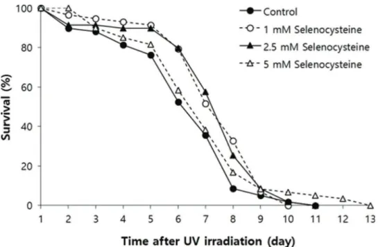
Effect of selenocysteine on survival after UV irradiation
Next, we examined the effect of selenocysteine on survival after UV irradiation. As shown in Figure 2, the mean survival time of the untreated control group was 6.39 days. In the selenocysteine-treated groups, the mean survival times were extended by up to 7.36 days with 1 or 2.5 mM selenocysteine (15.1% increase, po0.05). Unlike the effect on the response to oxidative stress, 5 mM selenocysteine failed to show a significant effect on the survival rate and time after UV irradiation. The mean survival time was 6.93 days, which was not statistically significantly different from that of the control (p=0.302).
Lifespan-modulating effect of selenocysteine in C. elegans
We tested the effect of selenocysteine on lifespan in C. elegans. In the first experiment, the mean lifespan was increased from 16.3 days in the untreated control group to 20.9 days (27.6% increase) with 1 mM selenocysteine, 20.6 days (25.9% increase) with 2.5 mM selenocysteine, and 22.2 days (35.9% increase) with 5 mM selenocysteine (Table 1). The maximum lifespan was also increased from 24 days to
27 days with 5 mM selenocysteine (Figure 3). Independent repeated experiments showed the same significant increase in lifespan with all concentrations of selenocysteine tested. The survival curve shown in Figure 3 was drawn using the average values of three independent lifespan assays.
Impact on reproduction by selenocysteine inC. elegans
Next, we examined the effect of selenocysteine on orga-nisms’ reproduction. As shown in Figure 4, the gravid period was shifted by selenocysteine supplementation. The untreated control worms produced progeny from day 2 to day 6 after hatching. However, the worms treated with sele-nocysteine produced small amounts of progeny on day 2 and maintained fertility until day 7 after hatching. Among the different concentrations of selenocysteine tested, 5 mM of selenocysteine caused a significant difference in the number of progeny produced compared with the untreated control. On day 2, 21.7±13.14 (mean±SEM) progeny were produced
by the untreated control, whereas only 0.1±0.13 progeny
were produced by the worms treated with 5 mM selenocys-teine. The numbers of progeny produced on day 3 were 128.1±5.82 and 108.8±4.62 in the untreated control and the
5 mM selenocysteine-treated groups, respectively (p=0.098). However, more progeny were produced at a later stage of the gravid period by the worms treated with 5 mM seleno-cysteine. The number of progeny produced was increa-sed from 23.3±6.17 in the untreated control to 51.5±6.49
in the worms treated with 5 mM selenocysteine on day 5 (p=0.031). The worms treated with 5 mM selenocysteine continued to produce 1.8±0.59 progeny, whereas no
pro-geny were produced by the untreated control on day 7 (p=0.049) (Figure 4). The total number of progeny produced during the gravid period was not significantly affected by the supplementation with selenocysteine.
Effect of selenocysteine on age-related decline in motility
To examine the effect of selenocysteine supplementation on muscle aging, we monitored changes in motility over time in worms untreated and treated with selenocysteine. The age-related decline in motility was retarded by supplementa-tion with selenocysteine (Figure 5). Ten days after hatching, more worms were classified as‘‘phase 1’’, including worms that could move spontaneously without any mechanical stimuli in the selenocysteine-treated group, compared with the untreated control. At 10 days of age, 50.5% of animals were classified as ‘‘phase 1’’ in the untreated control and 71.0% of the selenocysteine-treated worms classified as ‘‘phase 1’’. On day 15, only 19.2% of the worms were classi-fied as ‘‘phase 1’’in the control, whereas 71.0% of worms were still classified as‘‘phase 1’’in the selenocysteine-treated
group. In contrast, the number of worms that could move only their head after stimulation,‘‘phase 3’’, was less in the selenocysteine-treated group than in the untreated control. The percentage of worms classified as ‘‘phase 3’’ was decreased from 36.4% in the untreated control worms to 10.0% by the supplementation with selenocysteine. Thirty percent of the selenocysteine-treated worms were still able to move freely without any stimuli (phase 1), whereas no worm was classi-fied as‘‘phase 1’’in the untreated control on day 20 (Figure 5).
’ DISCUSSION
A positive correlation between increased resistance to environmental stressors and lifespan extension was observed with numerous genetic and nutritional interventions. One of the most conserved age-modulating cellular pathways
Figure 2 -Effects of selenocysteine on survival after UV irradiation. After 24 h of selenocysteine pre-treatment, animals were irradiated with 20 J/cm2/min of UV light for 1 min. The time-course % survival of age-synchronized worms (n=60) was then monitored every day until all worms were dead in the untreated and selenocysteine-treated groups. Mean survival time was significantly increased with 1 or 2.5 mM selenocysteine (po0.05).
Table 1-Effect of selenocysteine on lifespan inC. elegans.
Selenocysteine (mM) Mean lifespan (day) Maximum lifespan (day) p-value1 % effect2
1stexperiment 0 16.3 24
1 20.9 26 o0.001 27.6
2.5 20.6 25 o0.001 25.9
5 22.2 27 o0.001 35.9
2ndexperiment 0 17.0 28
1 23.9 29 o0.001 33.3
2.5 21.9 28 0.002 21.7
5 22.6 28 o0.001 25.8
3rdexperiment 0 20.1 24
1 21.5 27 0.003 7.0
2.5 22.2 29 o0.001 10.3
5 21.6 29 0.010 7.2
1p-values were calculated using the log-rank test by comparing the survival of the untreated control group (0 mM selenocysteine) with that of the experimental groups treated with different concentrations of selenocysteine.
among various organisms, from yeasts to humans, is insulin/ IGF-1-like signaling (26). Mutations in daf-2, a receptor for insulin/IGF-1-like ligand, and age-1, an intracellular adaptor molecule involved in insulin/IGF-1-like signaling, lead to an extended lifespan inC. elegans(27). Long-livingdaf-2orage-1 mutants also exhibit increased survival after exposure to environmental stressors, including oxidative stress, heat shock, and UV irradiation (28). Dietary restriction confers the longe-vity phenotype inC. elegans,Drosophila melanogaster, and mice (29, 30). Dietary-restricted animals show increased resistance to oxidative stress and reduced incidence of a number of
age-related diseases (31). Supplementation with anti-oxidants, such as resveratrol, N-acetyl-L-cysteine, and curcumin, causes an increase in both lifespan and resistance to stressors in C. elegans (6, 12, 32). Extracts from Acanthpanax sessiliflorus have both anti-oxidant and lifespan-extending properties in vivo(33). In mice, the effect of anti-oxidants on lifespan is still controversial. For example, vitamin E supplementation results in increased lifespan, whereas supplementation with coen-zyme Q10or lycopene fails to produce a longevity phenotype,
despite reducing the incidence of tumors (34, 35). In the present study, we observed increased survival under oxidative
Figure 3 -Lifespan-extending effect of selenocysteine. Age-synchronized young adult worms (n=60) were transferred to NGM plates containing differenct concentrations of selenocysteine, and the number of alive/dead worms was recorded every day. Worms that were lost or showed internal hatching during the assay were excluded. For all concentrations of selenocysteine tested, the mean lifespan was markedly increased compared with that of the untreated control (po0.05).
stress and UV irradiation via supplementation of selenocys-teine. These findings suggest that dietary supplementation with selenocysteine can increase resistance to environmental stresses in a dose-dependent manner inC. elegansand provide evidence for the in vivoanti-stress activity of selenocysteine. Having observed increased resistance to oxidative stress and UV irradiation by selenocysteine, the authors asked whether dietary supplementation with selenocysteine could affect the lifespan of C. elegans. A lifespan assay revealed that seleno-cysteine can in fact significantly extend both mean and maxi-mum lifespan inC. elegans. Our findings indicate that dietary supplementation with selenocysteine does confer a longevity phenotype inC. elegans, possibly by modulating the response to environmental stressors, supporting the free radical theory of aging. Future studies should focus on the effect of sele-nocysteine on ROS level and the activity of anti-oxidant enzymes, the identification of the underlying cellular mechan-isms involved, and the relationship with other known lifespan-extending genetic pathways.
The disposable soma theory of aging was first hypothe-sized by Thomas Kirkwood in 1977 and suggested that an organism should distribute limited cellular resources to reproductive ability and the maintenance of somatic cells (36). Previous studies have shown that numerous mutants with extended lifespan exhibit reduced fertility or a delayed gravid period in C. elegans, which suggests that reduced reproductive activity is a natural trade-off of lifespan exten-sion (37, 38). For example, long-living daf-2 mutants have exhibited reduced fertility (37). A number of lifespan-extend-ing dietary interventions also accompany reduced fertility and/or a delayed gravid period inC. elegans. Dietary supple-mentation with the anti-oxidant resveratrol increases lifespan and reduces fecundity (39). Complete knockout of germ cells also increases lifespan inC. elegans (40). In contrast, dietary supplementation with N-acetyl-L-cysteine confers a longevity
phenotype and enhances fertility (12). Here, we demonstrated that the total number of progeny produced during a gravid period was not affected by selenocysteine. However, a delay of the gravid period in worms treated with 5 mM selenocysteine was observed. These results indicate that the lifespan-extend-ing effects of selenocysteine accompany a delayed gravid period as a possible trade-off for producing the longevity phe-notype. Another study showed that polyphenols from blue-berries extended lifespan and caused a delay in the decline of pumping rate as a trade-off, suggesting that there can be alternative trade-offs for lifespan extension (41).
Muscle tissue demands high amounts of ATP produced by mitochondria for proper functioning and is susceptible to ROS produced by mitochondria as a byproduct during ATP generation. The loss of muscle mass and strength is defined as sarcopenia (42). Recent studies suggest that age-related degeneration of muscle is associated with age-related dysfun-ction of mitochondria, which generate less ATP and produce more free radicals (43, 44). Anti-oxidantSod-1-deficient mice show a premature aging phenotype and early-onset sarcope-nia (45). In contrast, over-expression of mitochondrial catalase, an anti-oxidant enzyme, reduces the accumulation of oxida-tive damage and delays age-related decline of muscle function in mice (46). In C. elegans, an individual’s motility declines with age. Therefore, the age-related decline of motility is one of the most widely used biomarkers for aging inC. elegans. Dietary supplementation with silymarin, a flavanone deriva-tive found in milk thistle (Silybum marianum), extends lifespan and increases locomotion rate inC. elegans(47). Silymarin also markedly protects amyloid beta-induced toxicity expressed in muscle (47). Phycoerythin, a strong anti-oxidant isolated from marine cyanobacteria, confers a longevity phenotype and enhances indicators of health, including pharyngeal pumping and locomotion rate (48). Our study showed that dietary supplementation with selenocysteine significantly delayed the
age-related decline of motility in C. elegans. This result sug-gests that selenocysteine has an anti-aging effect on muscle tissue. Future studies should determine the underlying mecha-nisms involved in the effect of selenocysteine.
Hormesis is defined as the beneficial response to the expo-sure of a low dose of harmful interventions. In C. elegans, increased resistance to stress and extended lifespan have been observed due to the hormesis effect of free radicals, heat stress, and dietary restriction (DR) (49). The effect of DR in particular has been reported in various model organisms (50). DR increases resistance to stressors and extends lifespan in yeasts, worms, flies, and mice (50). Supplementation with N-acetyl-L-cysteine increases resistance to oxidative stress at low doses and decreases resistance to oxidative stress at high doses, which suggests a possible hormesis effect of N-acetyl-L-cysteine (12). Because both beneficial and harmful effects of selenocysteine have been reported, our observations could be due to a hormesis effect. Further studies should focus on the identification of cellular pathways involved in seleno-cysteine’s effects to fully understand thein vivoactivity of selenocysteine.
The longevity phenotype observed in this study usingC. elegans cannot be directly carried over to higher organisms. Therefore, a study of the effect of selenocysteine in other model organisms should follow. In addition, the effect of selenocysteine on age-related disorders will be useful for practical applications of selenocysteine.
’ ACKNOWLEDGMENTS
This work was supported by the Soonchunhyang University Research Fund and the Basic Science Research Program through the National Research Foundation of Korea funded by the Ministry of Education (2015R1D1A 1A01057435).
’ AUTHOR CONTRIBUTIONS
Park SK conceived and designed the study and reviewed the manuscript. Kim JS performed and analyzed all experiments and wrote the manuscript. Kim SH performed repeated experiments for stress response and provided a critical review of the manuscript.
’ REFERENCES
1. Viña J, Borras C, Abdelaziz KM, Garcia-Valles R, Gomez-Cabrera MC. The free radical theory of aging revisited: the cell signaling disruption theory of aging. Antioxid Redox Signal. 2013;19(8):779-87, http://dx.doi.org/ 10.1089/ars.2012.5111.
2. Payne BA, Chinnery PF. Mitochondrial dysfunction in aging: Much pro-gress but many unresolved questions. Biochim Biophys Acta. 2015;1847 (11):1347-53, http://dx.doi.org/10.1016/j.bbabio.2015.05.022.
3. Bernadotte A, Mikhelson VM, Spivak IM. Markers of cellular senescence. Telomere shortening as a marker of cellular senescence. Aging (Albany NY). 2016;8(1):3-11, http://dx.doi.org/10.18632/aging.100871. 4. da Costa JP, Vitorino R, Silva GM, Vogel C, Duarte AC, Rocha-Santos T. A
synopsis on aging-Theories, mechanisms and future prospects. Ageing Res Rev. 2016;29:90-112, http://dx.doi.org/10.1016/j.arr.2016.06.005. 5. Howitz KT, Bitterman KJ, Cohen HY, Lamming DW, Lavu S, Wood JG,
et al. Small molecule activators of sirtuins extend Saccharomyces cerevi-siae lifespan. Nature. 2003;425(6954):191-6, http://dx.doi.org/10.1038/ nature01960.
6. Wood JG, Rogina B, Lavu S, Howitz K, Helfand SL, Tatar M, et al. Sirtuin activators mimic caloric restriction and delay ageing in metazoans. Nat-ure. 2004;430(7000):686-9, http://dx.doi.org/10.1038/nature02789. 7. Ferguson LR. Role of plant polyphenols in genomic stability. Mutat Res.
2001;475(1-2):89-111, http://dx.doi.org/10.1016/S0027-5107(01)00073-2. 8. Jang M, Cai L, Udeani GO, Slowing KV, Thomas CF, Beecher CW, et al.
Cancer chemopreventive activity of resveratrol, a natural product derived from grapes. Science. 1997;275(5297):218-20, http://dx.doi.org/10.1126/ science.275.5297.218.
9. Park SK, Page GP, Kim K, Allison DB, Meydani M, Weindruch R, et al. alpha- and gamma-Tocopherol prevent age-related transcriptional altera-tions in the heart and brain of mice. J Nutr. 2008;138(6):1010-8. 10. Park SK, Park SK. Electrolyzed-reduced water increases resistance to
oxidative stress, fertility, and lifespan via insulin/IGF-1-like signal in C. elegans. Biol Res. 2013;46(2):147-52, http://dx.doi.org/10.4067/S0716-97602013000200005.
11. Shirahata S, Kabayama S, Nakano M, Miura T, Kusumoto K, Gotoh M, et al. Electrolyzed-reduced water scavenges active oxygen species and protects DNA from oxidative damage. Biochem Biophys Res Commun. 1997;234(1):269-74, http://dx.doi.org/10.1006/bbrc.1997.6622.
12. Oh SI, Park JK, Park SK. Lifespan extension and increased resistance to environmental stressors by N-acetyl-L-cysteine in Caenorhabditis elegans. Clinics. 2015;70(5):380-6, http://dx.doi.org/10.6061/clinics/2015(05)13. 13. Areti S, Verma SK, Bellare J, Rao CP. Selenocysteine vs Cysteine: Tuning
the Derivatization on Benzenesulfonyl Moiety of a Triazole Linked Dansyl Connected Glycoconjugate for Selective Recognition of Selenocysteine and the Applicability of the Conjugate in Buffer, in Serum, on Silica Gel, and in HepG2 Cells. Anal Chem. 2016;88(14):7259-67, http://dx.doi.org/ 10.1021/acs.analchem.6b01518.
14. Rayman MP. The importance of selenium to human health. Lancet. 2000;356(9225):233-41, http://dx.doi.org/10.1016/S0140-6736(00)02490-9. 15. Schrauzer GN. Anticarcinogenic effects of selenium. Cell Mol Life Sci.
2000;57(13-14):1864-73, http://dx.doi.org/10.1007/PL00000668. 16. Li F, Lutz PB, Pepelyayeva Y, Arner ES, Bayse CA, Rozovsky S. Redox
active motifs in selenoproteins. Proc Natl Acad Sci U S A. 2014;111 (19):6976-81, http://dx.doi.org/10.1073/pnas.1319022111.
17. Lee KH, Jeong D. Bimodal actions of selenium essential for antioxidant and toxic pro-oxidant activities: the selenium paradox (Review). Mol Med Rep. 2012;5(2):299-304, http://dx.doi.org/10.3892/mmr.2011.651. 18. Byun BJ, Kang YK. Conformational preferences and pK(a) value of
sele-nocysteine residue. Biopolymers. 2011;95(5):345-53, http://dx.doi.org/ 10.1002/bip.21581.
19. Balaban H, Naziroglu M, Demirci K, Ovey IS. The Protective Role of Selenium on Scopolamine-Induced Memory Impairment, Oxidative Stress, and Apoptosis in Aged Rats: The Involvement of TRPM2 and TRPV1 Channels. Mol Neurobiol. 2017;54(4):2852-2868, http://dx.doi. org/10.1007/s12035-016-9835-0.
20. Jiang Q, Pan Y, Cheng Y, Li H, Li H. Protection of rat liver against hepatic ischemia-reperfusion injury by a novel selenocysteine-containing 7-mer peptide. Mol Med Rep. 2016;14(3):2007-15, http://dx.doi.org/10.1111/ jcmm.13129.
21. Puppala AK, French RL, Matthies D, Baxa U, Subramaniam S, Simonovic M. Structural basis for early-onset neurological disorders caused by mutations in human selenocysteine synthase. Sci Rep. 2016;6:32563, http://dx.doi.org/10.1038/srep32563.
22. Hirosawa-Takamori M, Chung HR, Jäckle H. Conserved selenoprotein synthesis is not critical for oxidative stress defence and the lifespan of Drosophila. EMBO Rep. 2004;5(3):317-22, http://dx.doi.org/10.1038/sj. embor.7400097.
23. Stenvall J, Fierro-Gonzalez JC, Swoboda P, Saamarthy K, Cheng Q, Cacho-Valadez B, et al. Selenoprotein TRXR-1 and GSR-1 are essential for removal of old cuticle during molting in Caenorhabditis elegans. Proc Natl Acad Sci U S A. 2011;108(3):1064-9, http://dx.doi.org/10.1073/pnas. 1006328108.
24. Wollenhaupt SG, Soares AT, Salgueiro WG, Noremberg S, Reis G, Viana C, et al. Seleno- and telluro-xylofuranosides attenuate Mn-induced toxi-city in C. elegans via the DAF-16/FOXO pathway. Food Chem Toxicol. 2014;64:192-9, http://dx.doi.org/10.1016/j.fct.2013.11.030.
25. Peto R, Peto J. Asymptotically efficient rank invariant test procedures. J R Statist Soc A. 1972;135(2):185-207, http://dx.doi.org/10.2307/2344317. 26. Longo VD, Finch CE. Evolutionary medicine: from dwarf model systems
to healthy centenarians? Science. 2003;299(5611):1342-6, http://dx.doi. org/10.1126/science.1077991.
27. Johnson TE, Henderson S, Murakami S, de Castro E, de Castro SH, Cypser J, et al. Longevity genes in the nematode Caenorhabditis elegans also mediate increased resistance to stress and prevent disease. J Inherit Metab Dis. 2002;25(3):197-206, http://dx.doi.org/10.1023/A:1015677828407. 28. Johnson TE, de Castro E, Hegi de Castro S, Cypser J, Henderson S,
Tedesco P. Relationship between increased longevity and stress resistance as assessed through gerontogene mutations in Caenorhabditis elegans. Exp Gerontol. 2001;36(10):1609-17, http://dx.doi.org/10.1016/S0531-5565(01)00144-9.
29. Sohal RS, Weindruch R. Oxidative stress, caloric restriction, and aging. Science. 1996;273(5271):59-63, http://dx.doi.org/10.1126/science.273. 5271.59.
30. Walker G, Houthoofd K, Vanfleteren JR, Gems D. Dietary restriction in C. elegans: from rate-of-living effects to nutrient sensing pathways. Mech Ageing Dev. 2005;126(9):929-37, http://dx.doi.org/10.1016/j.mad.2005. 03.014.
32. Liao VH, Yu CW, Chu YJ, Li WH, Hsieh YC, Wang TT. Curcumin-medi-ated lifespan extension in Caenorhabditis elegans. Mech Ageing Dev. 2011;132(10):480-7, http://dx.doi.org/10.1016/j.mad.2011.07.008. 33. Park JK, Kim CK, Gong SK, Yu AR, Lee MY, Park SK. Acanthopanax
sessiliflorus stem confers increased resistance to environmental stresses and lifespan extension in Caenorhabditis elegans. Nutr Res Pract. 2014;8 (5):526-32, http://dx.doi.org/10.4162/nrp.2014.8.5.526.
34. Lee CK, Pugh TD, Klopp RG, Edwards J, Allison DB, Weindruch R, et al. The impact of alpha-lipoic acid, coenzyme Q10 and caloric restric-tion on life span and gene expression patterns in mice. Free Radic Biol Med. 2004;36(8):1043-57, http://dx.doi.org/10.1016/j.freeradbiomed. 2004.01.015.
35. Navarro A, Gomez C, Sanchez-Pino MJ, Gonzalez H, Bandez MJ, Boveris AD, et al. Vitamin E at high doses improves survival, neurological per-formance, and brain mitochondrial function in aging male mice. Am J Physiol Regul Integr Comp Physiol. 2005;289(5):R1392-9, http://dx.doi. org/10.1152/ajpregu.00834.2004.
36. Ehrlich S. Effect of fertility and infertility on longevity. Fertil Steril. 2015;103(5):1129-35, http://dx.doi.org/10.1016/j.fertnstert.2015.03.021. 37. Hughes SE, Evason K, Xiong C, Kornfeld K. Genetic and pharmacological
factors that influence reproductive aging in nematodes. PLoS Genet. 2007;3(2):e25, http://dx.doi.org/10.1371/journal.pgen.0030025. 38. Larsen PL. Aging and resistance to oxidative damage in Caenorhabditis
elegans. Proc Natl Acad Sci U S A. 1993;90(19):8905-9, http://dx.doi.org/ 10.1073/pnas.90.19.8905.
39. Gruber J, Tang SY, Halliwell B. Evidence for a trade-off between survival and fitness caused by resveratrol treatment of Caenorhabditis elegans. Ann N Y Acad Sci. 2007;1100:530-42, http://dx.doi.org/10.1196/annals. 1395.059.
40. Hsin H, Kenyon C. Signals from the reproductive system regulate the lifespan of C. elegans. Nature. 1999;399(6734):362-6, http://dx.doi.org/ 10.1038/20694.
41. Joseph JA, Shukitt-Hale B, Denisova NA, Bielinski D, Martin A, McEwen JJ, et al. Reversals of age-related declines in neuronal signal transduction,
cognitive, and motor behavioral deficits with blueberry, spinach, or strawberry dietary supplementation. J Neurosci. 1999;19(18):8114-21. 42. Del Campo A, Jaimovich E, Tevy MF. Mitochondria in the Aging Muscles
of Flies and Mice: New Perspectives for Old Characters. Oxid Med Cell Longev. 2016;2016:9057593, http://dx.doi.org/10.1155/2016/9057593. 43. Marzetti E, Calvani R, Bernabei R, Leeuwenburgh C. Apoptosis in skeletal
myocytes: a potential target for interventions against sarcopenia and physical frailty - a mini-review. Gerontology. 2012;58(2):99-106, http://dx. doi.org/10.1159/000330064.
44. Suliman HB, Piantadosi CA. Mitochondrial Quality Control as a Ther-apeutic Target. Pharmacol Rev. 2016;68(1):20-48, http://dx.doi.org/10.1124/ pr.115.011502.
45. Muller FL, Song W, Liu Y, Chaudhuri A, Pieke-Dahl S, Strong R, et al. Absence of CuZn superoxide dismutase leads to elevated oxidative stress and acceleration of age-dependent skeletal muscle atrophy. Free Radic Biol Med. 2006;40(11):1993-2004, http://dx.doi.org/10.1016/j.freeradbiomed. 2006.01.036.
46. Umanskaya A, Santulli G, Xie W, Andersson DC, Reiken SR, Marks AR. Genetically enhancing mitochondrial antioxidant activity improves mus-cle function in aging. Proc Natl Acad Sci U S A. 2014;111(42):15250-5, http://dx.doi.org/10.1073/pnas.1412754111.
47. Kumar J, Park KC, Awasthi A, Prasad B. Silymarin extends lifespan and reduces proteotoxicity in C. elegans Alzheimer0s model. CNS Neurol Disord Drug Targets. 2015;14(2):295-302, http://dx.doi.org/10.2174/ 1871527314666150116110212.
48. Sonani RR, Singh NK, Awasthi A, Prasad B, Kumar J, Madamwar D. Phycoerythrin extends life span and health span of Caenorhabditis ele-gans. Age (Dordr). 2014;36(5):9717, http://dx.doi.org/10.1007/s11357-014-9717-1.
49. Rattan SI. Hormesis in aging. Ageing Res Rev. 2008;7(1):63-78, http://dx. doi.org/10.1016/j.arr.2007.03.002.