DIFFERENT METHODS AND THERMAL INACTIVATION
OF EXOGENOUS PME IN MANGO JUICE
Determinação da atividade da pectina metilesterase por diferentes métodos e inativação térmica da PME exógena no suco de manga
Samantha Lemke Gonzalez1, Regina Cristina Aparecida Lima2,
Eliana Beleski Borba Carneiro2, Mareci Mendes de Almeida2,Neiva Deliberali Rosso3
ABSTRACT
Pectin methylesterase (PME) hydrolyzes methyl ester groups in pectin chains to form carboxylic groups, releasing methanol and H3O+. The aim of this study was to determine PME activity in samples of pectinases by UV-VIS spectroscopy, to measure the
acid and methanol produced in the reaction of pectin with pectinase and to verify the thermal inactivation of exogenous PME in mango juice. The activity of PME in samples of pectinase was determined by potentiometry, UV-VIS spectroscopy, and by the action of alcohol oxidase. The reaction showed greater activity at pH 4.0 to 4.5 and at a temperature of 45° C. PME activity determined by UV-VIS spectroscopy with bromophenol blue indicator showed a good correlation with the activity determined by potentiometry and with alcohol oxidase. The results showed that bromophenol blue indicators can be used to determine PME activity in samples of pectinases where the optimum pH is located in the acidic range. The thermal inactivation of exogenous PME in mango juice occurred at 75° C for 20 min of exposure.
Index terms: Pectin metilesterase, activity, thermal inactivation, mango juice.
RESUMO
A PME hidrolisa os grupos metil éster na cadeia da pectina, formando grupos carboxílicos, liberando metanol e H3O+.
Objetivou-se, com o presente estudo, determinar a atividade da PME em amostras de pectinases por espectroscopia Uv-vis para quantificar o ácido e o metanol produzido na reação da pectina com as pectinases e verificar a inativação térmica da PME exógena no suco de manga. A atividade da PME nas três amostras de pectinases foi determinada por potenciometria, espectroscopia Uv-Vis, e pela ação da álcool oxidase. A reação mostrou uma maior atividade em H de 4,0 a 4,5 e a temperatura de 45º C. A atividade da PME, determinada por UV-Vis com o indicador azul de bromofenol apresentou uma boa correlação com a atividade determinada por potenciometria e com a álcool oxidase. Os resultados mostraram que o indicador azul de bromofenol pode ser utilizado para determinar a atividade da PME em amostras de pectinases em que o pH ótimo situa-se na faixa ácida. A inativação térmica da PME no suco de manga ocorreu na temperatura de 75º C, por 20 min de exposição.
Termos para indexação: Pectina metilesterase, atividade, inativação térmica, suco de manga.
(Received in november 30, 2010 and approved in march 22, 2011)
2Universidade Federal de Santa Catarina/UFSC – Cidade Universitária Trindade – Florianópolis, SC 2Universidade Estadual de Ponta Grossa/UEPG – Ponta Grossa, PR
3Universidade Estadual de Ponta Grossa/UEPG – Avenida Carlos Cavalcanti – n. 4748 – 84030-900 – Ponta Grossa, PR – [email protected]
INTRODUCTION
Pectin methylesterase (PME), EC 3.1.1.11, is an enzyme that acts mainly in the hydrolysis of methyl ester groups in pectin chains to form carboxylate groups, releasing methanol and H3O+ (JAYANI et al., 2005).
According to Zocca et al. (2007) reaction of pectin with PME in the presence of an appropriate indicator at the optimum pH for the enzyme activity can be monitored by UV-VIS. A study showed that PME activity can be determined with an indicator dye known as bromocresol green (CECI; LOZANO, 1998).
Methanol is toxic to human beings, being an undesirable component in the production of alcoholic
drinks and juices in general. Wood e Siddiqui (1971) used UV-VIS spectroscopy to determine methanol levels in KMnO4. Klavons e Bennett (1986) showed that an alcohol oxidase can be used for methanol oxidation and subsequent reaction with 2,4-pentanedione. Mangos e Haas (1997) determined PME activity in reactions with pectin and the methanol released was oxidized by alcohol oxidase.
Riahi e Ramaswamy (2003) evaluated the kinetics of the inactivation of exogenous PME in apple juice. The study showed that an increase in pressure over time has a significant effect on PME inactivation. Plaza et al. (2008)
observed that the PME was thermostable at 50° C at pH 5.0 and polyols showed a higher protective effect for the enzyme than sugars. Slavov et al. (2009) investigated the behavior of high methoxy pectin in the presence of a fungal PME and of a PME from orange. In the gel, the degree of methylation decreased slowly with orange PME and rapidly with Aspergillus PME.
Mango fruit can be processed to produce nectar, juice and drinks. According to Ollé et al. (2000) and Kashyap et al. (2001) exogenous pectinolytic enzymes promote a decrease in viscosity, an increase in concentration and clarification of the mango juice nectar. The literature presents few studies using indicators and no studies appear to be available regarding the use of bromophenol blue indicator for the determination of PME activity. Some studies have employed alcohol oxidase and acetyl acetone to quantify the methanol. The aim of this study was to determine PME activity in samples of pectinases by UV-VIS spectroscopy to measure the acid and methanol produced in the reaction of pectin with PME, and to verify the thermal inactivation of exogenous PME in mango juice.
MATERIALS AND METHODS
Materials
The enzyme preparations Panzym Univers, Pectinex 100L Plus and Panzym Clears (Novozymes) consisted of a mixture of pectinolytic enzymes, predominantly PME and polygalacturonase, produced by submerged fermentation of Aspergillus sp. High methoxyl apple pectin (Herbstreith;
Fox) was employed, the water used was deionized and other reagents were analytical grade (Merck, Sigma and Reagen).
Determination of PME activity by titration
PME activity was determined by titration, using an experimental system of double-walled glass cells coupled with a thermostatized bath (Microchemistry) and a pH meter Micronal (B474 model) equipped with a combined electrode, calibrated daily with pH 4.0 and 7.0 buffers. The experiments were performed with the controlled addition of 0.05 mol L-1
NaOH free CO2. The experimental solution consisted of 0.150 g apple pectin, 5.00 mg mL-1, 0.100 mol L-1 NaCl and
the volume was filled to 30 mL with deionized water. After solubilization of the components, 50 μL of pectinolytic enzyme was added. The temperature and pH were kept constant. According to Fachin et al. (2002) PME activity is proportional to the initial rate of NaOH consumption over time (VNaOH /ttime). In this study, the unit used to express the PME activity was mmol L-1 of carboxylic acid produced
per second. The influence of pH and temperature on the activity of pectinase Panzym Univers was verified. The effect of pH on enzyme activity was determined for the values of 3.0, 3.5, 4.0, 4.25, 4.5, 5.0 and 5.5, keeping the reaction temperature constant at 45 ± 0.1° C. The effect of temperature on the reaction was measured at 25, 35, 45, 55, 65 and 75° C, keeping the pH value constant at 4.0. All tests were performed in duplicate.
Determination of PME activity by UV-VIS
Acid Quantification
The pH of the experimental solution was adjusted to 4.5 and then 150 mL of a solution of bromophenol blue indicator (Reagen) was added while the temperature was kept constant at 25° C. Aliquots of 3 mL of reaction solution were transferred to a quartz cuvette with a 1.0 cm optical path and 5 µL of pectinase was added. The solution was homogenized by vortex, and the kinetics of the reaction were monitored at 592 nm every 30 s for 30 min with a MultiSpec-1501 Shimadzu spectrophotometer. The same conditions were applied to the procedure with the bromocresol green indicator (Reagen). All experiments were conducted in duplicate. The assay was calibrated with a standard curve of D-galacturonic acid for both indicators. The standard curve was built from the D-galacturonic acid concentration in relation to the absorbance, thus with the exact acid concentration at different times, the PME activity was determined.
Quantification of methanol with alcohol oxidase
PME activity can be determined by monitoring the release of methanol produced during pectin hydrolysis by the action of the enzyme. In this method, methanol is oxidized to formaldehyde by the action of alcohol oxidase enzymes. It reacts with acetyl acetone or Purpald resulting in compounds that absorb in the visible region. According to Jacobsen e Dickinson (1974), the product of Purpald reactions with formaldehyde is 6-mercapto-s-triazol-(4,3-b)-s-tetrazine with maximum absorbance at 550 nm, while the reaction product of acetyl acetone with formaldehyde is 3.5-diacetyl-1,4-dihydro-2,6-dimethylpyridine which absorbs at 412 nm (NASH, 1953)
Reagent Purpald and Acetyl Acetone
then wrapped in an ice bath to stop PME reactions in the substrate. The methanol released in the reaction was measured according to the method described by Anthon e Barrett (2004). All experiments were performed in duplicate. A standard curve was constructed with known volumes of methanol, 99.8% (0-100μL) and the absorbance was plotted according to the methanol concentration. From the standard curve the concentration of methanol produced at each time interval was calculated. The PME activity was then calculated from a graph showing the methanol concentration in relation to time.
Determination of exogenous PME residual activity in mango juice
Mangoes of the Tommy Atkins cultivar were acquired in the local market of the city of Ponta Grossa, PR, Brazil. They were selected regarding color and texture, the size ranged between 9.5-11.7cm long and 8.6 – 10.3 cm wide, and the weight varied from 394-614g. Juice was extracted with a Britania Turbo Juicer and was then centrifuged to separate off suspended solids, with an Eppendorf Centrifuge 5810R centrifuge at 4º C and 3220 g for 15 minutes. The juice was packaged in 30 mL bottles and stored in a freezer at -22º C. 10 mL of preheated juice and 50 μL Pectinex 100L Plus were added to a test tube. The vial was then shaken and sealed and subjected to temperatures of 45-75º C with a thermostat (Microquimica) for 10, 20 and 30 min while shaking. After this period the pH of the juice-PME was recorded and the same value was set for the system containing the substrate. Then the juice-PME was added to the reaction and the titration procedure was employed to determine the exogenous PME residual activity in mango juice.
RESULTS AND DISCUSSION
Determination of optimum pH and temperature for Panzym Univers
The optimum pH for the PME in the pectinase sample, that is, the pH interval where the maximum PME activity occurred, was between 4.0 and 4.5. The same range was determined for samples of Pectinex 100L Plus and Panzym Clears (GONZALEZ; ROSSO, 2011). According to Duvetter et al. (2005)for samples of commercial pectinases obtained from Aspergillus aculeatus and Aspergillus niger the PME optimum pH was also 4.0 to
4.5. The optimum temperature was 45º C, which is consistent with the temperature verified by Nikolic e Mojovic (2007) for PME in a sample of pectinase produced by Aspergillus niger.
Determination of PME activity by UV-VIS
Acid Quantification
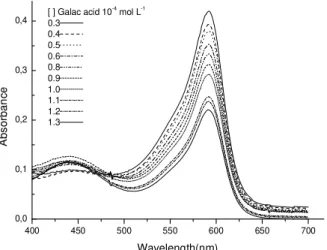
The PME activity in the pectinase samples was determined in the presence of bromophenol blue indicator. In this regard, a study was carried out on the stability of this indicator in the pH range of 5.0 to 3.7. It was observed that with increasing pH there was an increase in absorbance, with the maximum at 592 nm. It was also observed that the absorption band does not suffer displacement. Since the bromophenol blue indicator showed good stability in the pH range studied and the color change is located mainly between 3.0 and 4.6 pH, (ATKINS; JONES, 2006) values near the optimum for PME activity (DUVETTER et al., 2005), it was considered as appropriate for use in determining the PME activity.
In the reaction of PME with pectin at 25° C and pH 4.5, there was a decrease in pH due to the formation of H+.
As H+ is combined with the indicator, there is an increase
in the concentration of protonated species and a decrease in the concentration of deprotonated species, promoting a proportional decrease in the absorption band at 592 nm (Figure 1). A study by Hagerman e Austin(1986) determined the PME activity using the bromothymol blue indicator. The color change of this indicator occurs at pH 6.0–7.6, and the maximum absorbance at 620 nm. The bromothymol blue indicator is suitable for determination of acid produced in reactions where the optimum pH of the enzyme activity is within the range of the color change of the indicator. It should also be considered that, in addition to enzymatic hydrolysis at pH values above 7.0, pectin hydrolysis can occur. According to Fayyaz et al. (1995) a reaction conducted at pH 8.0 to determine the PME activity is not safe due to the occurrence of pectin autohydrolysis under alkaline conditions. Considering these factors, bromothymol blue can not be used to determine the PME activity in samples of commercial pectinase in which the optimum pH lies between 4.0 and 4.5.
A 592, pH 4.5 = 0.44903 - 1713.311 x [H+]
ΔA = Absorbance variation in 592 nm at pH 4.5; [H+] = H+
concentration in mol L-1
(1)
400 450 500 550 600 650 700
0,0 0,1 0,2 0,3
0,4 [ ] Galac acid 10
-4
mol L-1
0.3 0.4 0.5 0.6 0.8 0.9 1.0 1.1 1.2 1.3
A
b
s
o
rb
a
n
c
e
Wavelength(nm)
Figure 1 – Behavior of bromophenol blue at different concentrations (0.3-1.3 x 10⁻⁴ molL-1) of D–galacturonic
acid at 25o C.
The PME activity of three pectinase samples (Pectinex 100L Plus, Panzym Univers and Panzym Clears) was determined at pH 4.5 and 25° C. To determine the PME activity the standard curve of D-galacturonic acid was used and the concentration of [H+] was calculated from Equation 1.
Thus, from the variation in the acid concentration over the time interval the PME activity was determined. Data from one experiment is shown in Table 1, which shows the variation in the absorbance at 592 nm for the respective reaction times. The absorbance variation was converted into acid concentration variation, Equation 1, for each time interval.
Table 1 – Absorbance (A) and variation of A (A) at 592 nm versus time, and the H+ concentration variation ([H+])
corresponding to each time interval.
Time (s) A at 592 nm A [H+] (molL-1)
0 0.809847
90 0.760955 4.889 x 10⁻² 2.335 x 10⁻⁴
180 0.724774 3.618 x 10⁻² 2.409 x 10⁻⁴
270 0.706426 1.835 x 10⁻² 2.513 x 10⁻⁴
A linear response was observed for the plot Δ[H+] in mol L-1 versus time in seconds, Equation 1,
presenting o correlation coefficient of 0.99537. The PME activity of pectinase samples shown in Table 2 was calculated from the straight line equation. The slope is equal to 0.9908 x 10⁻⁷ mol L-1s-1 and most of the enzyme
activity that can be expressed as 0.9908 x10⁻⁴ mmol L-1s-1
of galacturonic acid.
The PME activity in three samples was also determined with the bromocresol green indicator at pH 4.5 and 25° C. It should be noted that the color change of this indicator occurs in the pH range of 3.8 to 5.4. From the standard curve of this indicator with the D-galacturonic acid and Equation 2, the PME activity for each sample of pectinase was calculated. The curve showed a linear response and the correlation coefficient was 0.9948. ∆A 617, pH 4.5 = 0.1245 + 393.363 x [H+]
A = Absorbance variation in 617 nm at pH 4.5; [H+] = H+
concentration in mol L-1.
Table 2 presents the values of PME for the three pectinases samples with both indicators and their respective standard deviations. It can be observed that the PME activity values for the three samples were lower for the bromocresol green indicator in relation to those obtained with the bromophenol blue indicator. These differences can be attributed to the lower sensitivity of bromocresol green, the range of the color change of this indicator and its stability in relation to the pH at which the pectin reaction in the presence of the enzyme was conducted (VILARIŇO et al., 1993). The pH of the reaction was 4.5, close to the optimum pH of each sample, and within the pH range at which the indicator changed color, that is, from 3.8 to 5.4. For both indicators the color stability was observed for 20 min in the pectin solution at pH 4.5 and 25° C and with constant ionic strength. It was found that the color of the bromocresol green indicator in the reaction with pectin did not remain constant, the absorbance of the solution varied even in the absence of PME. On the other hand, bromophenol blue changes color in the pH range of 3.0 to 4.6, the region closest to pH 4.5 at which the reaction was conducted. The color of this indicator remained constant throughout the period of the reaction. Therefore, the data obtained for bromophenol blue is considered to be more reliable.
Table 2 – PME activity determined by UV-VIS with the two indicators at pH 4.5 and 25° C.
PME Activity (mmol.L-1s-1)
Enzyme/ Indicators bromophenol blue bromocresol green
Pectinex 100L Plus 0.950 x 10⁻⁴ 0.01 1.347 x 10-50.02
Panzym Univers 1.015 x 10⁻⁴ 0.01 2.730 x 10-50.03
Panzym Clears 1.343 x 10⁻⁴ 0.01 3.538 x 10-50.02
Table 3 shows the PME activities, for the three samples of pectinases obtained from the procedures by UV-VIS and titrationat 25° C and pH 4.5. In the UV-VIS spectroscopy study, the bromophenol blue indicator showed good stability in solution with pectin for a reaction of 20 min. The PME activity determined by this procedure shows good correlation when compared with the values obtained from the NaOH volume variation versus the time variation, ΔVNaOH/ ΔDtime, and pH variation versus the time variation ΔpH/Δtime (GONZALEZ; ROSSO, 2011). PME activity and methanol quantification
Purplad
To the reaction of methanol with alcohol oxidase in the presence of the reagent Purpald at 25° C, a linear increase in absorbance with increasing concentration of methanol [MeOH] was observed (Equation 3). The correlation coefficient of the curve was 0.98743. The PME activity was then calculated and is represented in Table 4.
aGonzalez e Rosso (2011).
PME Activity (mmol. S-1)
Enzyme / Methods bromophenol blue VNaOHa pHa
Panzym Clears 1.343 x 10⁻⁴ 0.01 1.080 x 10⁻⁴ 0.01a 1.291 x 10-40.01a
Pectinex 100L Plus 0.950 x 10⁻⁴ 0.01 0.941 x 10⁻⁴ 0.01a 1.157 x 10⁻⁴ 0.01a
Panzym Univers 1.015 x 10⁻⁴ 0.01 0.881 x 10-40.01 0.904 x 10-40.01 Table 3 – PME Activities obtained from the different procedures at 25° C and pH 4.5.
∆A 550, pH 7.15 = 0.00732 + 215.99694 . [MeOH]
ΔA=Absorbance variation in 412 nm at pH 4.5; [MeOH]=methanol concentration in mol L-1.
Acetyl Acetone
A standard curve with good linear response can be obtained for methanol in the presence of alcohol oxidase and acetyl acetone (ANTHON; BARRETT, 2004).
For a standard curve of methanol with acetyl acetone at 25° C and pH 4.5, it was observed that with increasing [MeOH] there is also a linear increase in absorbance (Equation 4). The correlation coefficient of the curve was 0.97354.
∆A 412, pH4.5 = - 0.02931 + 81.63622 x [MeOH]
ΔA=Absorbance variation in 412 nm at pH 4.5; [MeOH]=methanol concentration in mol L-1.
During the reaction of demethoxylated pectin, in presence of alcohol oxidase and acetyl acetone, the absorbance for each 2 minute interval was obtained. From the standard curve, the [MeOH] produced at each time interval was calculated. The activity in the sample of Pectinex 100L Plus was 1.073 x 10⁻⁴ mmol L-1s-1 (Table 4).
The PME activity value obtained 3.602 x 10⁻⁴
mmol L-1s-1 with the reagent Purpald was more than three
times the value obtained 1.073x10⁻⁴ mmol s-1 with the
reagent acetyl acetone. The Purpald reagent should be prepared in 0.5 mol L-1 NaOH solution, using a highly basic
medium (ANTHON; BARRETT, 2004). When this reagent was used after the reaction of alcohol oxidase with demethoxylated pectin in buffer solution, the strongly basic Purpald solution offset the 7.15 buffer solution. It was found that the pH of the reaction containing the buffer, demethoxylated pectin, alcohol oxidase, Purpald and deionized water was above 12. Thus the reaction condition provokes hydrolyze and it alters the results. After the
addition of the Purpald the reaction was conducted at 30° C for 30 min (ANTHON; BARRETT, 2004), the per iod of time for wh ich Pur pald r eacts with formaldehyde. As the medium is highly basic, the possibility of affecting the pectin h ydr olysis, producing more methanol, should not be ruled out. The system contains alcohol oxidase, which oxidizes methanol to formaldehyde, and this then reacts with Purpald resulting in increased concentration of methanol and consequently an apparent increase in the PME activity. According to Ly-Nguyen et al. (2002) the autohydrolysis of pectin must be considered when the PME activity is determined at temperature and pH values above 50º C and 6.5, respectively. On the other hand, Arbaisah et al. (1997)noted that PME activity measurements performed above pH 9.0 are not reliable due to the de-esterification reaction occurring in the alkaline medium. In the determination of methanol with acetyl acetone, the reagents used did not alter the pH of the system, which remained at 4.35. Thus, it can be affirmed that only the methanol produced by the action of PME was detected.
Table 4 shows the values for the PME activity in samples of Pectinex 100L Plus determined by various procedures. It can be observed that the activity value determined using acetyl acetone to detect methanol was similar to values obtained by potentiometric procedures and th rough spectr oscopic detection of th e concentration of H+ using the bromophenol blue
indicator.
Table 4 – PME activity in Pectinex 100L Plus samples determined by different methods at 25° C and pH 4.5.
Method PME Activity ( mmol.s-1 )
Purpald 3.602 x 10⁻⁴
Acetyl acetone 1.073 x 10⁻⁴
Bromophenol blue indicator 0.950 x 10⁻⁴
pHa 1.157 x 10⁻⁴
VNaOHa 0.941 x 10⁻⁴
aGonzalez e Rosso (2011).
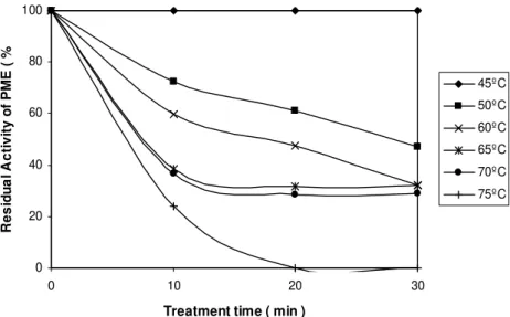
Determination of exogenous PME residual activity in mango juice
After centrifugation of the mango juice, the pH remained at 4.50 and the soluble solids at 16° Brix. Figure 3 shows the thermal stability of the mango juice in the presence of the commercial sample of pectinase (Pectinex 100L Plus). It was observed that for the thermal treatment at 45° C the exposure period (10, 20 and 30 min) did not influence PME activity, as this is the optimum temperature for enzyme activity. However, with increasing temperature there was a decline in activity: at 50° C for 10 min of exposure the residual activity was 72.3%, for 20 min it reduced to 61%, and for 30 min it fell to 47%. At 60° C, for 10, 20 and 30 min the residual activity values were 59.6%, 47.6% and 31.9%, respectively. For treatments at 65° C the residual activity was around 30% for all exposure periods and at 70º C for 20 and 30 min of exposure the
0 20 40 60 80 100
0 10 20 30
Treatment time ( min )
R
e
s
id
u
a
l
A
c
ti
v
it
y
o
f
P
M
E
(
%
)
45ºC
50ºC
60ºC
65ºC
70ºC
75ºC
Figure 2 – Exogenous PME residual activity in mango juice, 5.0 mg mL-1 of apple pectin, at pH 4.0, 45° C and 0.100 mol L-1 NaCl,
activity remained at around 29%. When the temperature was raised to 75° C for 10 min it was observed that the residual activity was 24.1%, however, for 20 min of exposure there was complete inactivation. A study showed that treatment at 66° C for 2.5 min in apple juice with exogenous PME was sufficient for complete inactivation (WILINSKA et al., 2008). Such differences of temperature and time for complete PME inactivation might be due to the distinct composition of Apple and mango juices. According to Seymour et al. (1991) carbohydrates exert a protective effect on the enzyme (PME). This protective effect is dependent on the chemical composition of the juice under study.
The results obtained in the study on PME thermal inactivation in the presence of mango juice, with thermal treatment at 70° C for 10, 20 and 30 min of exposure, show that the residual activity decreased from 36.8 to 29%. In the thermal treatment of the commercial PME, in the absence of mango juice under the same conditions, PME residual activity fell from 29.9% to 10.0% (GONZALEZ; ROSSO, 2011). According to Seymour et al. (1991) this difference in the values for the residual PME activity is due to the presence of mango juice, in which the carbohydrates a protective effect on the enzyme (PME). This protective effect is dependent on the chemical composition of the juice in the study.
CONCLUSION
PME activity in three samples of pectinases (Pectinex 100L Plus, Panzym Clears and Panzym Univers) at 25° C and pH 4.5, was determined by UV-VIS spectroscopy with bromophenol blue indicator. It was concluded that this indicator can be used to determine the PME activity in samples of pectinases in which the optimum pH is located in the acidic range. Data obtained from the use of the reaction of demethoxylated pectin with PME to quantify the methanol and acetyl acetone showed good agreement with those obtained from the procedures used in this study. The determination of methanol with this reagent did not alter the pH of the system, thus it can be affirmed that the methanol detected was produced only by the action of PME. Thermal inactivation of exogenous PME (Pectinex 100L Plus) in mango juice occurred at 75º C with 20 min of exposure.
ACKNOWLEDGMENTS
The authors are grateful for the financial support of the CAPES and Fundação Araucária.
REFERENCES
ANTHON, G.E.; BARRETT, D.M. Comparison of three colorimetric reagents in the determination of methanol with alcohol oxidase: application to assay of pectin methylesterase. Journal of Agricultural and Food Chemistry, Washington,v.52, p.3749-3753, 2004.
ARBAISAH, S.M. et al. Purification and properties of pectinesterase from soursop (Anona muricata) pulp.
Food Chemistry, Chicago, v.59, p.33-40, 1997. ATKINS, P.; JONES, L. Princípios de química: questionando a vida moderna e o meio ambiente. 3.ed. Porto Alegre: Bookman, 2006. 965p.
CECI, L.; LOZANO, J. Determination of enzymatic activities of commercial pectinases for the clarification of apple juice. Food Chemistry, Chicago, v.61, p.237-241, 1998.
DUVETTER, T. et al. Kinetics of papaya pectinesterase. Food Chemistry, Chicago, v.53, p.129-135, 1995. GONZALEZ, S.L.; ROSSO, N.D. Determination of pectin methylesterase activity of commercial pectinases and study of the inactivation kinetics through two potentiometric procedures. Revista de Ciência e
Tecnologia de Alimentos, Campinas, v. 31, p. 412-417 2011. HAGERMAN, A.E.; AUSTIN, P.J. Continuous
spectrophotometric assay for plant pectin methyl esterase. Journal of Agricultural and Food Chemistry, Washington, v.34, p.440-444, 1986.
JACOBSEN, N.W.; DICKINSON, R.G. Spectrometric assay of aldehydes as 6-mercapto-3-substituted-s
-triazolo(4,3-b)-s-tetrazines. Analytical Chemistry, Washington, v.46, p.298-299, 1974.
JAYANI, R.S.; SAXENA, S.; GUPTA, R. Microbial pectinolytic enzymes: a review. Process Biochemistry, London, v.40, p.2931-2944, 2005.
KASHYAP, D.R. et al. Applications of pectinases in the commercial sector: a review. Bioresource Technology, Amsterdam, v.77, p.215-227, 2001.
LY-NGUYEN, B. et al. Strawberry pectin methylesterase (PME): purification, characterization, thermal and high-pressure inactivation. Biotechnology Progress, Washington, v.18, p.1447–1450, 2002.
MANGOS, T.J.; HAAS, M.J. A spectrophotometric assay for the enzymatic demethoxylation of pectin and the determination of pectinesterase activity. Analytical Biochemistry, New York, v.244, p.357-366, 1997. NASH, T. The colorimetric estimation of formaldehyde by means of the Hantzsch reaction. Biochemical Journal, London, v.55, p.416-421, 1953.
NIKOLIC, M.V.; MOJOVIC, L. Hydrolysis of apple pectin by the coordinated activity of pectic enzymes. Food Chemistry, Chicago, v.101, p.1-9, 2007.
OLLÉ, D. et al. Enzymatic degradation of cell wall polysaccharides from mango (Mangifera indica L.)
puree. Journal of Agricultural and Food Chemistry, Washington, v.48, p.2713-2716, 2000.
PLAZA, L. et al. Influence of environmental conditions on thermal stability of recombinant Aspergillus aculeatus pectin methylesterase. Food Chemistry, Chicago,v.111, p.912-920, 2008.
RIAHI, E.; RAMASWAMY, H.S. High-pressure of apple juice: kinetics of pectin methyl esterase inactivation.
Biotechnology Progress, Washington, v.19, p.908-914, 2003.
SEYMOUR, T.A. et al. Stability of pectinesterase of Marsh White grapefruit pulp. Journal of Agricultural and Food Chemistry, Washington, v.39, p.1075-1079, 1991.
SLAVOV, A. et al. Gelation of high methoxy pectin in the presence of pectin methylesterases and calcium. Carbohydrate Polymers,Barking, v.77, p.876-884, 2009. VILARIÒO, C. et al. Spectrophotometric method for fungal pectinesterase activity determination.
Lebensmittel-Wissenchaft und Technologie, London, v.26, p.107-110, 1993.
WILINSKA, A. et al. Thermal inactivation of exogenous pectin methylesterase in apple and cloudberry juices. Journal of Food Engineering, Davis, v.85, p.459-465, 2008.