Persistent papillary membrane in
Wistar
laboratory rats
(
Rattus Norvegicus, Albinus
Variation,
Wistar
)
Membrana pupilar persistente em ratos Wistar de laboratório (Rattus Norvegicus, Variação Albinus, Wistar)
Flor Diana Yokoay Claros Chacaltana1 Karina Kamachi Kobashigawa1
Ivan Ricardo Martinez Padua1 Gisele Pereira Valdetaro1 Marcela Aldrovani1 José Luiz Laus1*
ISSNe 1678-4596
INTRODUCTION
During fetal development, the mammalian eye anterior chamber is partly occupied by the mesodermal tissue supported by primitive blood
vessels composed of fine hyaline fibers and
conjunctival cells. Hyaloid vessels and pupillary membrane (PM) form a temporary capillary network in the anterior chamber, iris diaphragm, and lens. This network nourishes the immature lens, retina, and vitreous humor during morphogenesis (POCHÉ et al., 2015; ZIGLER et al., 2015). The term persistent pupillary membrane (PPM) refers to an alteration in mesodermal development by which the reversal of the normal PM is interrupted at some point in development (GONZÁLEZ ALONSO-ALEGRE & RODRÍGUEZ, 1997).
Regression of PM occurs concomitantly with the formation of the pupillary opening and involves apoptosis and cell necrosis (TARADACH & GREAVES, 1984; BLACKWOOD et al., 2010;
MITCHELL, 2011). In rodents, the process starts around on the day of the birth and continues for 2 weeks (ITO & YOSHIOKA, 1999; POCHÉ et al., 2015). In dogs, it begins at 45 days of gestation and ends at the opening of the eyes, about 14 days after birth (BLACKWOOD et al., 2010; MITCHELL, 2011). In humans, the PM atrophies before birth. In some other mammalian species, like dogs and cats the PM remains after birth for variable periods of time (ITO & YOSHIOKA, 1999; MITCHELL, 2011). Upon complete atrophy, no vascular branches remain in the anterior chamber of the eye (GONZÁLEZ ALONSO-ALEGRE & RODRÍGUEZ, 1997).
PPM represents an incomplete atrophy of the perilenticular vessels. These remnants are
fine strands of pigmented tissue that arise from
the collarette iris and attach to another spot on the iris; they may also extend to the pupillary region (BLACKWOOD et al., 2010; ESSON, 2015). PPM is most commonly manifested as strands extending from the iris collarette to other areas within the collarette,
1Departamento de Clínica e Cirurgia Veterinária, Faculdade de Ciências Agrárias e Veterinárias, Universidade Estadual Paulista (UNESP),
Câmpus de Jaboticabal, Via de Acesso Prof. Paulo Donato Castellane, s/n, 14884-900, Jaboticabal, SP, Brasil. E-mail: [email protected].
*Corresponding author.
ABSTRACT: The aim of this research was to evaluate the presence of persistent pupillary membrane (PPM) in rats. Thirty rats between three and four months of age and weighing 300–500 grams, provided by the biothery section of the General Administration at Universidade Estadual Paulista (UNESP), Botucatu, SP, Brazil, were subjected to ophthalmological examination by slit lamp biomicroscopy, fluorescein eye stain test and rebound tonometry. We found PPM with possible hereditary origin in 15 animals (50%).
Key words: persistent pupillary membrane, Wistar, rats.
RESUMO: Objetivou-se avaliar a presença de membrana pupilar persistente (MPP) em ratos. Foram oftalmicamente avaliados, 30 ratos com idade entre três e quatro meses, e peso entre 300 e 500 gramas, fornecidos pelo Biotério Central da Universidade Estadual Paulista
(UNESP), campus de Botucatu, SP, Brasil. Todos os animais passaram por inspeção à biomicroscopia, teste da fluoresceína e também pela
tonometria de rebote. Encontrou-se MPP em 15 dos animais (50%), cuja origem pode estar associada à herdabilidade.
Palavras-chave: membrana pupilar persistente, Wistar, ratos.
In domestic animals, PPM is a common manifestation of anterior segment dysgenesis (COOK, 2013). It is a congenital anomaly, inherited and manifested in some breeds of dogs, such as the Basenji (ROBERTS & BISTNER, 1968; GRAHN & CULLEN, 2004; MITCHELL, 2011). It has been reported as resulting from inbreeding (YOUNG et al., 1974). PPM may present unilaterally or bilaterally; it has to be emphasized that bilateral manifestation of PPM does not necessarily indicate PPM of the same size or shape in both eyes (ARNBJERG, 1988; GONZÁLEZ ALONSO-ALEGRE & RODRÍGUEZ, 1997; DOYLE & REDDY, 2016). The presence of PPM can cause visual impairments, corneal injury, leukomas, and cataract (STRANDE et al., 1988; GONZÁLEZ ALONSO-ALEGRE & RODRÍGUEZ, 1997; BAYON et al., 2002; GRAHN & CULLEN, 2004; MITCHELL, 2011; SUEDMEYER et al., 2013; ESSON, 2015). Total persistence of the PM, i.e., PPM occupying the entire pupil, is rare and culminates in damage to the vision (COOK, 2013). PPM may also be accompanied by persistent hyperplastic tunica vasculosa lentis, persistent hyperplastic primary vitreous, cataract, microphthalmia (BAYON et al., 2002; MITCHELL, 2011), retinal dysplasia (BAYON et al., 2002), iris hypoplasia (MISK et al., 1998; PINARD & BASRUR, 2011), heterochromia iridis (MISK et al., 1998), angle-closure glaucoma caused by pupillary block, or peripheral anterior synechiae (YOUNG et al., 1974). Another condition that accompanies PPM is the presence of sheets that move away from the iris collarette and remain freely
floating in the anterior chamber without adhering to
any other structure. These sheets can be pigmented or not. They usually do not hinder pupillary activity (GONZÁLEZ ALONSO-ALEGRE & RODRÍGUEZ, 1997); however, if extensive, they can alter the pupillary kinetics (ROPSTAD et al., 2007). The aim of this research was to evaluate the presence of PPM in rats.
MATERIALS AND METHODS
Thirty male rats (Rattus norvegicus) of
the Wistar lineage, between 3 and 4 months of age and weighing 300-500g were evaluated. They were a donated by the biothery section of the General Administration at São Paulo State University (UNESP), Botucatu, SP, Brazil. All animals were housed in appropriate cages in a clean and well-ventilated environment, with alternating light/dark
The biothery section of the UNESP maintains a colony of rats established over a period of 35 years. The approximate number of breeding rats
is 600 females and 100 males.Breeding is performed
using a temporary harem system, with one male for every two females. Males remained with the females for 10-14 days, followed by one week of rest. Subsequently, the same group of males is mated with the other females.
For this study, the rats, which were selected at random, were evaluated by slit lamp biomicroscopy,
the fluorescein eye stain test (Ophthalmos, São
Paulo, Brazil) and rebound tonometry (TonoVet®
-Tiolat, Helsinki, Finland). The intraocular pressure (IOP) values obtained for eyes with PPM were compared with those of “normal” eyes. Differences
were considered significant when P≤0.05. For digital documentation, we acquired photographs using photography equipment (TRC-50DX, Topcon,
Japan) configured to acquire photographs without filters and with a 35-mm focus adjustment and
20°-angle of coverage. Mydriasis was induced with 1% tropicamide (Alcon, São Paulo, Brazil).
RESULTS
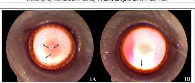
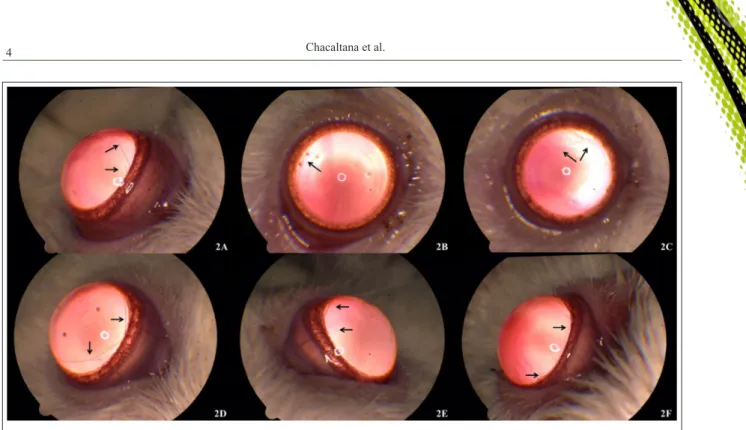
Of the 30 animals evaluated in the present study, 15 (50%) had PPM; which was unilateral in 12 cases (80%) and bilateral in 3 (20%). It was estimated that 18 (30%) of the 60 eyes evaluated in this study exhibited PPM; of these 55.55% (10 eyes) showed strands extending from the collarette iris to the other regions of the collarette (Figure 1A) and 55.55% (10 eyes) showed small sheets originating from and inserted into the collarette iris (Figure 1B). For the
evaluation of the findings, the iris were divided into
presented as small sheets, 44.44% (8 eyes) presented single sheets (Figure 1B), 11.11% (2 eyes) presented two small sheets (Figure 2F).
Mean ± standard deviation (± SD) values for IOP were 9.11 (± 2.47)mmHg for eyes with PPM and 9.42 (± 2.06)mmHg for “normal” eyes (P=
0.609). Examination with fluorescein was negative
for all eyes. No ocular manifestation or discomfort secondary to PPM was observed.
DISCUSSION
In rats, the regression of the PM is observed until 16 days after birth (ITO & YOSHIOKA, 1999; POCHÉ et al., 2015). In the present study, the evaluated animals were between 3 and 4 months of age. Studies on the pattern of regression of the PM
indicated that regression occurs in two stages - the first
stage, dependent on the induction of macrophages and apoptosis of the endothelial cells (LANG & BISHOP, 1993; LANG et al., 1994; DIEZ-ROUX & LANG, 1997; ITO & YOSHIOKA, 1999), and the second one, brought about by the coordinated apoptosis of the capillary endothelial cells caused by the interruption
of plasma flow (MEESON et al., 1996).
The mechanism by which the regression of the PM is interrupted has not been completely elucidated yet (GONZÁLEZ ALONSO-ALEGRE & RODRÍGUEZ, 1997). Among others, the accepted causes include genetic, environmental, and infectious factors (ROBERTS & BISTNER, 1968; ARNBJERG,
1988) as well as heredity (GONZÁLEZ ALONSO-ALEGRE & RODRÍGUEZ, 1997) and inbreeding (YOUNG et al., 1974). PPM of hereditary origin might be present in some dog breeds, in which it manifests with variable degrees of penetration and expression (ROBERTS & BISTNER, 1968). A previous study reported that sibling Poodle dogs with PPM, when crossed, produced offspring without PPM, leading the authors to conclude that hereditary predisposition is not mandatory for the development of PPM
(ARNBJERG, 1980). Nevertheless, this finding does
not rule out the possibility of genetic predisposition to PPM (ARNBJERG, 1980; STRANDE et al., 1988). In purebred horses and other mongrels, heredity has been suggested as a cause of PPM (PINARD & BASRUR, 2011). In view of the high prevalence of PPM in fruit bats, Blackwood et al. (2010) reported that it is possible that genetic predisposition plays a role in the development of PPM in that species.
YOUNG et al. (1974) reported the occurrence of buphthalmos caused by congenital glaucoma because of the interference of PPM with the drainage of aqueous humor in an inbred colony of rats of the WAG strain (YOUNG et al., 1974). SAARI (1975) reported the possibility of circulatory disorder during the development of the eye being an important cause for the incomplete atrophy of the PPM. In the rats evaluated in the present study, these
events were not identified.
Oxygen therapy is often used to prevent the premature infant respiratory distress syndrome. This
treatment has been indicated as a causal factor for the occurrence of PPM (HORNBLASS, 1971). However, a previous study reported that mice challenged with high concentrations of oxygen in the environment did not develop PPM (ARNBJERG, 1988). In the present study, since the affected rats all belonged to the same colony, it is possible that heredity was the probable cause of PPM.
PPMs have been reported in different species of animals used for experimentation (YOUNG et al., 1974; TARADACH & GREAVES, 1984; BOILLOT et al., 2015). Among laboratory animals, PPM has been reported to occur in rats (YOUNG et al., 1974), mice, hamsters, Beagle dogs (TARADACH & GREAVES, 1984), and rabbits (BOILLOT et al., 2015). This condition has also been reported in several dog breeds including Basenji (ROBERTS & BISTNER, 1968; MITCHELL, 2011), Poodle (ARNBJERG, 1980), Doberman Pinscher (BARTOE et al., 2007), English Cocker Spaniel (STRANDE et al., 1988; MITCHELL, 2011), Pembroke Welsh Corgi, Chow Chow (ESSON, 2015), Mastiff (MITCHELL, 2011; ESSON, 2015), Spitz Finnish, Lancashire Heeler, Miniature Wire-Haired Dachshund, Petit Basset Griffon Vendeen, Rottweiler, Siberian Husky, West Highland White
Terrier (MITCHELL, 2011), and Wire-Haired Dachshund (ROPSTAD et al., 2007). It has also been reported in cats (ALARIO et al., 2013), horses (PINARD & BASRUR, 2011), monkeys (BUREK et al., 1974), North American Beavers (CULLEN, 2003), snow leopards (SCHÄFFER et al., 1988), chinchillas (MÜLLER & EULE, 2014), kangaroos (SUEDMEYER et al., 2013), bats (BLACKWOOD et al., 2010), and llamas (GIONFRIDDO, 2013).
The findings of the present study are in
agreement with published studies characterizing the presence of strands or small sheets originating from the iris collarette without touching the lens or cornea and without causing perceptible visual changes (GIONFRIDDO, 2013). Congenital glaucoma associated with PPM has been reported in rats of the WAG strain (YOUNG et al., 1974). Another study reported the occurrence of secondary hyphema in PPM (SAARI, 1975); however, in the present study, we reported no signals of glaucoma ou hyphema. In human patients, amblyopia was reported to be associated with PPM (MILLER & JUDISCH, 1979). In the present study, three animals presented PPM bilaterally not indicated the same size or shape in both eyes like reported before (ARNBJERG, 1988; GONZÁLEZ ALONSO-ALEGRE & RODRÍGUEZ, 1997).
Different tools, notably, biomicroscopy (GONZÁLEZ ALONSO-ALEGRE &
RODRÍGUEZ, 1997) and fluorescein angiography
of the anterior segment (ALARIO et al., 2013) have been used in the evaluation of PPM. In the
present study, the use of equipment for scientific
documentation enabled the recording and characterization of PPM in detail. This emphasizes the importance of inducing cycloplegia for the evaluation of PPM (GONZÁLEZ ALONSO-ALEGRE & RODRÍGUEZ, 1997). However, the induction of cycloplegia should be performed carefully since its excessive use might expand and stretch the strands or sheets of the PM, aggravating the damage to the cornea and the lens in cases where the PPM shows adherence to these structures.
In veterinary medicine, medical or surgical treatment for PPM is not recommended. In cases of corneal opacity, medical treatment is not
beneficial (GONZÁLEZ ALONSO-ALEGRE &
RODRÍGUEZ, 1997). Corneal and lens opacities caused by PPM are generally focal and axial in nature, which allows peripheral vision. Since some PMs are vascularized, in cases with bilateral cataract, the removal of lens, with attention to the risk of bleeding, might be indicated (ESSON, 2015). In the present study, we did not perform therapeutic intervention in any of the cases.
CONCLUSIONS
The findings of evaluation of the cases of
PPM reported in the present study lead to conclude that PPM cannot be a rare condition among Wistar laboratory rats, and it develops because of inbreeding among affected individuals.
ACKNOWLEDGEMENTS
The authors thank the researchers and the laboratory technicians of the biothery section of the General Administration at São Paulo State University (UNESP), Botucatu, SP, Brazil. To the Brazilian Federal Agency for Support and Evaluation of Graduate Education (Capes), for the scholarship, and to the National Council
for Scientific and Technological Development (CNPq), process
number 300833/2010-5 and São Paulo Research Foundation (FAPESP), process number Proc.2009/51773-4.
BIOETHICS AND BIOSSECURITY COMITTEE APPROVAL
This research was accepted by the ethical review committee (protocol number 06174/14 CEUA-UNESP approved on May 14, 2014) and followed the ethical norms of the Association for Research in Vision and Ophthalmology (ARVO) statement for the use of animals in ophthalmic and visual research.
REFERENCES
ALARIO, A.F. et al. Anterior segment fluorescein angiography of
the normal feline eye using a dSLR camera adaptor. Veterinary Ophthalmology, v.16, n.3, p.204-213, 2013. Available from: <http:// onlinelibrary.wiley.com/doi/10.1111/j.1463-5224.2012.01058.x/ epdf>. Accessed: May 21, 2016. doi: 10.1111/j.1463-5224.2012.01058.x.
ARNBJERG, J. Study of ocular abnormalities in Standard Poodles. Canine Practice, v.7, n.6, p.21-23, 1980.
ARNBJERG, J. Persistent pupillary membrane and oxygen therapy: experiments in mice. Journal of Veterinary Medicine, v.35, n.1-10, p.138-140, 1988. Available from: <http://onlinelibrary.wiley. com/doi/10.1111/j.1439-0442.1988.tb00016.x/epdf>. Accessed: May 21, 2016. doi: 10.1111/j.1439-0442.1988.tb00016.x.
BARTOE, J.T. et al. Multiple ophthalmic lesions and melanocytic neoplasia in white Doberman Pinschers. In: ANNUAL MEETING OF THE AMERICAN COLLEGE OF VETERINARY OPHTHALMOLOGISTS, 38., 2007, Kona, Hawaii. Veterinary Ophthalmology, v.10, n.6, p.398-411, 2007. Available from: <http:// onlinelibrary.wiley.com/doi/10.1111/j.1463-5224.2007.00588.x/epdf>. Accessed: May 21, 2016. doi: 10.1111/j.1463-5224.2007.00588.x.
BAYÓN, A. et al. Vítreo primario hiperplásico persistente y anomalías asociadas en un Husky siberiano. Clínica Veterinaria de Pequeños Animales, v.22, n.3, p.257-263, 2002. Available from: <http://ddd.uab.cat/pub/clivetpeqani/11307064v22n3/11307 064v22n3p257.pdf>. Accessed: May 21, 2016.
BLACKWOOD, S.E. et al. Ocular parameters in a captive colony of fruit bats. Veterinary Ophthalmology, v.13, n.1, p.72-79, 2010. Available from: <http://onlinelibrary.wiley.com/doi/10.1111/ j.1463-5224.2010.00816.x/pdf>. Accessed: May 21, 2016. doi: 10.1111/j.1463-5224.2010.00816.x.
BOILLOT, T. et al. Unilateral persistent hyperplastic tunica vasculosa lentis and persistent hyperplastic primary vitreous in a rabbit. Veterinary Ophthalmology, v.18, n.6, p.510-514, 2015. Available from: <http://onlinelibrary.wiley.com/ doi/10.1111/vop.12251/epdf>. Accessed: May 21, 2016. doi: 10.1111/vop.12251.
BUREK, J.D. et al. Persistent pupillary membranes in a rhesus monkey. Journal of the American Veterinary Medical Association, v.164, n.7, p.719, 1974.
COOK, C.S. Ocular embryology and congenital malformations. In: GELATT, K.N.; GILGER, B. C.; KERN, T. J. Veterinary ophthalmology. 5.ed. Ames: Wiley-Blackwell, 2013. p.3-38.
CULLEN, C.L. Normal ocular features, conjunctival microflora
and intraocular pressure in the Canadian beaver (Castor canadensis). Veterinary Ophthalmology, v.6, n.4, p.279-284, 2003. Available from: <http://onlinelibrary.wiley.com/doi/10.1111/ j.1463-5224.2003.00307.x/pdf>. Accessed: May 21, 2016. doi: 10.1111/j.1463-5224.2003.00307.x.
3638, 1997. Available from: <http://dev.biologists.org/content/ develop/124/18/3633.full.pdf>. Accessed: May 21, 2016.
ESSON, D.W. Clinical atlas of canine and feline ophthalmic disease. Tustin: John Wiley & Sons, 2015. 344p.
GIONFRIDDO, J.R. Ophthalmology of new world camelids. In: GELATT, K.N.; GILGER, B. C.; KERN, T. J. Veterinary ophthalmology. 5.ed. Ames: Wiley-Blackwell, 2013. p.1675-1691.
GONZÁLEZ ALONSO-ALEGRE, E.M.; RODRÍGUEZ Á.A. Membrana pupilar persistente. Clínica Veterinaria de Pequeños Animales, v.17, n.1, p.49-54, 1997. Available from: <http://ddd. uab.cat/pub/clivetpeqani/11307064v17n1/11307064v17n1p49. pdf>. Accessed: May 21, 2016.
GRAHN, B.H.; CULLEN, C.L. Iris to lens persistent pupillary membranes. Canadian Veterinary Journal, La revue Vétérinaire Canadienne, v.5, n.7, p.613, 2004. Available from: <http://www. ncbi.nlm.nih.gov/pmc/articles/PMC2751695/pdf/15317396.pdf>. Accessed: May 21, 2016.
HORNBLASS, A. Persistent pupillary membrane and oxygen therapy in premature infants. Annals of ophthalmology, v.3, n.1, p.95-99, 1971.
ITO, M.; YOSHIOKA, M. Regression of the hyaloid vessels and pupillary membrane of the mouse. Anatomy and Embryology, v.200, n.4, p.403-411, 1999. Available from: <http://link.springer. com/article/10.1007/s004290050289>. Accessed: May 21, 2016.
LANG, R.A.; BISHOP, J.M. Macrophages are required for cell death and tissue remodeling in the developing mouse eye. Cell, v.74, n.3, p.453-462, 1993. Available from: <http://www.sciencedirect. com/science/article/pii/009286749380047I>. Accessed: May 21, 2016. doi: 10.1016/0092-8674(93)80047-I.
LANG, R. et al. Apoptosis during macrophage-dependent ocular tissue remodelling. Development, v.120, n.12, p.3395-3403, 1994. Available from: <http://dev.biologists.org/content/120/12/3395. full.pdf>. Accessed: May 21, 2016.
MEESON, A. et al. A relationship between apoptosis and flow
during programmed capillary regression is revealed by vital analysis. Development, v.122, n.12, p.3929-3938, 1996. Available from: <http://dev.biologists.org/content/develop/122/12/3929.full. pdf>. Accessed: May 21, 2016.
MILLER, S.D.; JUDISCH, G.F. Persistent pupillary menbrane: successful medical pmanagement. Archives of ophthalmology, v.97, n.10, p.1911-1913, 1979. Available from: <http://archopht. jamanetwork.com/article.aspx?articleid=633163>. Accessed: May 21, 2016. doi: 10.1001/archopht.1979.01020020359015.
MISK, N.A. et al. Heterochromia iridis in water buffaloes (Bubalus bubalis). Veterinary Ophthalmology, v.1, n.4, p.195-201, 1998. Available from: <http://onlinelibrary.wiley.com/doi/10.1046/ j.1463-5224.1998.00036.x/pdf>. Accessed: May 21, 2016. doi: 10.1046/j.1463-5224.1998.00036.x.
MITCHELL, N. Persistent pupillary membranes in dogs and cats. Veterinary Ireland Journal, v.1, n.11, p.615-618, 2011. Available from: <http://web.a.ebscohost.com/ehost/pdfviewer/
MÜLLER, K.; EULE, J.C. Ophthalmic disorders observed in pet Chinchillas (Chinchilla lanigera). Journal of Exotic Pet Medicine, v.23, n.2, p.201-205, 2014. Available from: <http://www. sciencedirect.com/science/article/pii/S155750631400041X>. Accessed: May 21, 2016. doi: 10.1053/j.jepm.2014.02.007.
PINARD, C.L.; BASRUR, P.K. Ocular anomalies in a herd of exmoor ponies in Canada. Veterinary Ophthalmology, v.14, n.2, p.100-108, 2011. Available from: <http://onlinelibrary.wiley.com/ doi/10.1111/j.1463-5224.2010.00847.x/epdf>. Accessed: May 21, 2016. doi: 10.1111/j.1463-5224.2010.00847.x.
POCHÉ, R.A. et al. Macrophages engulf endothelial cell membrane particles preceding pupillary membrane capillary regression. Developmental Biology, v.403, p.30-42, 2015. Available from: <http:// www.sciencedirect.com/science/article/pii/S0012160615001682>. Accessed: May 21, 2016. doi: 10.1016/j.ydbio.2015.03.017.
ROBERTS, S.R.; BISTNER, S.I. Persistent pupillary membrane in Basenji dogs. Journal of the American Veterinary Medical Association, v.153, n.5, p.533-542, 1968.
ROPSTAD, E.O. et al. Clinical findings in early onset cone-rod
dystrophy in the Standard Wire-haired Dachshund. Veterinary Ophthalmology, v.10, n.2, p.69-75, 2007. Available from: <http:// onlinelibrary.wiley.com/doi/10.1111/j.1463-5224.2007.00503.x/pdf>. Accessed: May 21, 2016. doi: 10.1111/j.1463-5224.2007.00503.x.
SAARI, M. Vascular remnants of pupillary membrane in the albino rat eye. Acta Anatomica, v.91, p.376-379, 1975.
SCHÄFFER, E. et al. Multiple ocular coloboma (MOC) with persistent pupillary membrane in the snow leopard (Panthera uncia). Tierärztliche Praxis, v.16, n.1, p.87-91, 1988.
STRANDE, A. et al. Persistent pupillary membrane and congenital cataract in a litter of English cocker spaniels. Journal of Small Animal Practice, v.29, n.4, p.257-260, 1988.
SUEDMEYER, K. et al. Peters anomaly in a Red Kangaroo (Macropus Rufus). Journal of Zoo and Wildlife Medicine, v.45, n.3, p.715-718, 2013. Available from: <http://www. zoowildlifejournal.com/doi/pdf/10.1638/2013-0308R.1>. Accessed: May 21, 2016. doi: 10.1638/2013-0308R.1.
TARADACH, C.; GREAVES, P. Spontaneous eye lesions in laboratory animals: incidence in relation to age. Critical Reviews in Toxicology, v.12, n.2, p.121-147, 1984. Available from: <http://www.tandfonline. com/doi/pdf/10.3109/10408448409023759#.VxpsWNThDIU>. Accessed: May 21, 2016. doi:10.3109/10408448409023759.
YOUNG, C. et al. Buphthalmos (congenital glaucoma) in the rat. Laboratory Animals, v.8, n.1, p.21-31, 1974. Available from: <http://lan.sagepub.com/content/8/1/21.full.pdf+html>. Accessed: May 21, 2016. doi: 10.1258/002367774780943797.
ZIGLER, J.S. et al. βA3/A1-crystallin and persistent fetal