Mammary fibroadenomatoid hyperplasia in a heifer
Hiperplasia fibroadenomatoide mamária em uma novilha Lorena Ferreira Silva1 Luiz Antonio Franco da Silva2 Rogério Elias Rabelo2 Wanessa Patrícia Rodrigues da Silva2 Geovanni Dantas Cassali3
Aline De Biasi Bassani Gonçalves3 Fabiano José Ferreira de Sant’Ana1*
ISSNe 1678-4596
1Laboratório de Diagnóstico Patológico Veterinário, Hospital Veterinário de Grandes Animais, Universidade de Brasília (UnB), 70636-020, Brasília, DF, Brasil. E-mail: santanafjf@yahoo.com. *Corresponding author.
2Escola de Veterinária e Zootecnia, Universidade Federal de Goiás (UFG), Goiânia, GO, Brasil.
3Instituto de Ciências Biológicas (ICB), Universidade Federal de Minas Gerais (UFMG), Belo Horizonte, MG, Brasil.
ABSTRACT: This manuscript described the anatomopathological and immunohistochemical findings in a rare case of mammary
fibroadenomatoid hyperplasia in a 12-month-old Holstein heifer. A yellow, multilobulated, firm 20cm x 9cm x 6.5cm mass affecting the right quarters of the udder was observed. Total mastectomy was performed. Microscopic evaluation revealed severe hyperplasia of the mammary epithelium and numerous well-differentiated and mildly pleomorphic acini. Additionally, moderate proliferation of the fibrous connective tissue and the myoepithelial cells near the proliferating acini was evident. About 50% of the proliferating epithelial cells showed positive nuclear labeling for estrogen and progesterone receptors, and approximately one-third were positive for Ki-67. In addition, the myoepithelial cells exhibited diffuse nuclear immunoreactivity for p63. Based on the clinical, morphologic, and immunohistochemical findings, a diagnosis of mammary fibroadenomatoid hyperplasia with probable influence of ovarian steroids was made.
Key words:mammary hyperplasia, estrogen, progesterone, diseases of cattle.
RESUMO:Este artigo descreve os achados anatomopatológicos e imuno-histoquímicos de um caso raro de hiperplasia fibroadenomatoide
mamária em uma novilha Holandesa de 12 meses. Observou-se uma massa amarelada, firme, multilobulada, medindo 20 x 9 x 6,5cm e afetando os quartos direitos do úbere. Mastectomia total foi realizada. Microscopicamente, notou-se hiperplasia acentuada do epitélio mamário e numerosos ácinos levemente pleomórficos e bem diferenciados. Proliferação moderada do tecido conjuntivo fibroso e das células mioepiteliais próximos aos ácinos proliferados era também evidente. Aproximadamente 50% das células epiteliais proliferadas mostraram marcação nuclear positiva para receptores de estrógeno e progesterona e cerca de um terço dessas células era positivo para Ki-67. Além disso, as células mioepiteliais exibiram reação nuclear e difusa para p63. Com base nos achados clínicos, morfológicos e imuno-histoquímicos, firmou-se o diagnóstico de hiperplasia fibroadenomatoide mamária com provável influência de esteroides ovarianos.
Palavras-chave: hiperplasia mamária, estrógeno, progesterona, doenças de bovinos.
PATHOLOGY
Progressive lesions of the mammary gland are common in dogs and cats, but are rarely observed in farm animals. Infrequent cases of mammary neoplasms have been reported in cattle (POVEY & OSBORNE, 1969; MINA et al., 1994), mares (HIRAYAMA et al., 2003; BRITO et al., 2008), water buffalo (MANDAL & IYER, 1969), and goats (SINGH & IYER, 1972); whereas, hyperplastic changes have been observed in cattle (HONG et al., 1994) and water buffalo (SANT’ANA et al., 2014). Mammary
epithelial hyperplasia is classified into three distinct
types: lobular (arising from the intralobular ducts), ductal (arising from the extralobular ducts), and adenosis (CASSALI et al., 2011). Another type named
fibroadenomatoid hyperplasia has been reported
in women as a rare benign lesion characterized by prominent hyperplasia of the epithelial cells and
multifocal lobulocentric proliferation of the fibrous
stroma, including the myoepithelial components (WU & CHEN, 2001). Fibroadenomatoid hyperplasia may occur as a localized or diffuse lesion; it is observed in approximately 5-7% of all benign surgical biopsy samples from Japanese and American women (HANSON et al., 1987). In humans, this type of
lesion was formerly known as fibroadenomatosis, sclerosing lobular hyperplasia, and fibroadenomatoid
change or mastopathy (HANSON et al., 1987; WU & CHEN, 2001). This condition was recently observed in three young water buffalo and endocrine
2 Silva et al.
lesion (SANT’ANA et al., 2014). The objective of the current study was to evaluate the clinicopathological
and immunohistochemical findings of a rare case of mammary fibroadenomatoid hyperplasia in a heifer.
A one-year-old, intact Holstein heifer presented with a painless swelling in the udder affecting the right quarters that had progressively evolved over four days (Figure 1A). The animal was from a mixed herd of 40 heifers and cows that were raised in the county of Goiânia, state of Goiás, Brazil. Treatment with
penicillin, anti-inflammatory medications, and showers
with running water for three months was unsuccessful. The entire gland was surgically removed. Grossly, the excised mammary gland contained a yellowish,
homogeneous, firm mass with spongy areas, measuring
20cm x 9cm x 6.5cm (Figure 1B). The cut surfaces of the mammary lesion showed numerous coalescing
lobules separated by fibrous septa (Figure 1B). There
was no evidence of mammary lesion recurrence at eight months after the surgical removal.
Samples of the mammary lesions were
collected, fixed in 10% formalin for 24 h, and
processed using routine methods for histological evaluation. Sections were stained with hematoxylin and eosin, and immunohistochemical staining (IHC) was performed using a biotin-peroxidase system, with labeling of the secondary antibody with a polymer (Advance HRP enzyme, Dako North America Inc., Carpinteria, CA, USA) and diaminobenzidine (DAB) as the chromogen. Antigen retrieval was performed by pressurized heating at 125°C in a citrate-buffered solution at pH 6.0 (Dako North America Inc., Carpinteria, CA) for Ki-67 and p63, or in an ethylenediamine tetra-acetic acid buffer solution at pH 9.0 for the estrogen receptor (ER) and progesterone receptor (PR) (Dako North America Inc., Carpinteria, CA, USA). To block the endogenous peroxidase activity, the slides were incubated in a solution of H2O2 (3%) in methyl alcohol. The reagents were added manually, with 1-hour incubation for the monoclonal primary antibodies and a 30-minute incubation for the other reagents with the exception of DAB, which was incubated for 5min. The IHC antibody panel is described in table 1. The IHC sections were counterstained using Harris hematoxylin. The positive controls for IHC consisted of normal mammary glands. For the negative controls, normal serum (Ultra V Block, NeoMarkers Inc., Fremont, CA, USA) was added instead of the primary antibodies. Nuclear staining (Ki-67, p63, ER, and PR) was assessed by determining the percentage of positive cells among 500 cells (viewed
under 400× magnification).
Microscopic evaluation demonstrated severe proliferation of the mammary epithelium with the formation of numerous, well-differentiated, and mildly pleomorphic tubules and their associated ducts. The acini had 1-4 layers of columnar or cuboidal epithelial cells and were moderately dilated. The cell borders were indistinct. The cytoplasm was
slightly eosinophilic, and the nucleus was oval, finely
granular, and hypochromatic. Mild anisocytosis, anisokaryosis, and rare mitosis were observed.
Moderate proliferation of the fibrous connective tissue
and the myoepithelial cells was observed among the proliferated acini. Few lymphocytes were detected
in the fibrous stroma. Few hyperplasic mammary
tubules had moderate ectasia and were occasionally
filled with amorphous eosinophilic material.
About 50% of the proliferated epithelial cells demonstrated moderate to strong positive nuclear labeling for ER (Figure 1C) and PR (Figure 1D). In addition, approximately one-third of these cells were positive for Ki-67 (Figure 1F). The myoepithelial cells exhibited diffuse, moderate nuclear immunoreactivity for p63 (Figure 1E). Based on the morphologic and immunohistochemical
findings, a diagnosis of mammary fibroadenomatoid hyperplasia with probable influence of ovarian
steroids was made.
Mammary hyperplasia is diagnosed commonly in company animals, but rarely in herbivores (HONG, 1994; MISDORP, 2002). Pathogenesis of this proliferative lesion is not completely understood, but endocrine changes are likely involved in canine and feline females (MISDORP, 2002). Recently, three cases of mammary
diffuse fibroadenomatoid hyperplasia were diagnosed
in water buffalo present in Midwestern and Southern Brazil, and the immunohistochemical evaluation
suggested probable hormonal influence (SANT’ANA
et al., 2014). An isolated case of mammary nodular hyperplasia in a 13-year-old cow was attributed to chronic irritation associated with chronic mastitis (HONG, 1994). This author considered hyperplasia to be a regenerative response to counteract for the affected mammary tissue. In the current case, there
were no significant inflammatory lesions associated with the fibroadenomatoid hyperplasia. Similar to
the previously reported cases in bubaline females (SANT’ANA et al., 2014), ovarian endocrine
influence on the hyperplasia was suspected, given
the IHC-labeling results (positive for estrogen and progesterone receptors). In contrast, the feline
mammary diffuse fibroepithelial hyperplasia
only progesterone receptors (MISDORP, 2002). In the present study, the immunoreactivity tests demonstrated that one-third of the epithelial cells were positive for Ki-67. Similar results have been observed in feline mammary hyperplasia (PEREIRA et al., 2004). However, in cases of benign mammary lesions in humans (GERDES et al., 1986) and canines (PEÑA et al., 1998), the immunoreactivity
for Ki-67 was low. In this study, the positivity for p63 highlighted the proliferation of myoepithelial cells associated with epithelial hyperplasia.
The affected heifer in the current study was young (1 year old). This condition was also detected in young water buffalo (10 to 12 months old) with morphologic changes in the mammary gland very similar to those observed in the present
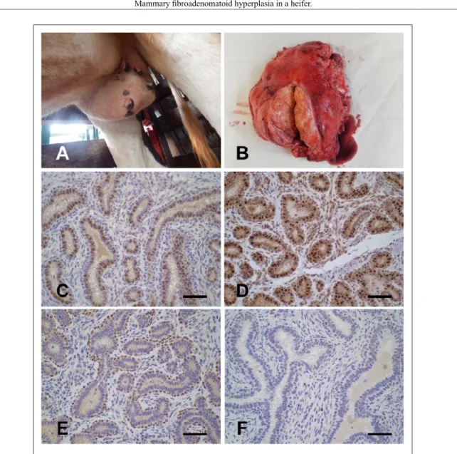
Figure 1 - Mammary fibroadenomatoid hyperplasia in a heifer. A. There is a moderate enlargement of
the right quarters of the udder. B. Cut surface of this mass shows a yellow, multilobulated, and
firm tissue. Moderate and severe positive nuclear labeling for estrogen (C) and progesterone
(D) receptors in the affected epithelium, respectively. Immunohistochemical reaction (IHC)
with 3,3′-diaminobenzidine (DAB) chromogen and hematoxylin counterstain. Bar = 60μm (C) and 80μm (D). E. There is moderate positive nuclear staining for myoepithelial cells (p63).
Immunohistochemical reaction (IHC) with 3,3′-diaminobenzidine (DAB) chromogen and hematoxylin counterstain. Bar = 80μm. F. Few epithelial cells present mild, positive reaction
4 Silva et al.
REFERENCES
BRITO, M.F. et al. Mammary adenocarcinoma in a mare. Ciencia
Rural, v.38, p.556-560, 2008. Available from: <http://www.scielo. br/pdf/cr/v38n2/a45v38n2.pdf>. Accessed: Nov. 18, 2016. doi: 10.1590/S0103-84782008000200045.
CASSALI, G.D. et al. Consensus for the diagnosis, prognosis and treatment of canine mammary tumors. Brazilian Journal
of Veterinary Pathology, v.4, p.153-180, 2011. Available from: <http://bjvp.org.br/wp-content/uploads/2015/07/ DOWNLOAD-FULL-ARTICLE-29-20881_2011_7_11_14_42. pdf>. Accessed: Oct. 11, 2016.
GULBAHAR, M.Y. et al. Mammary fibroadenoma in a lamb. Journal of Veterinary Science, v.8, p. 423-425, 2007. Available from:
<http://www.vetsci.org/journal/view.html?uid=443&vmd=Full>.
Accessed: Oct. 12, 2016. doi: 10.4142/jvs.2007.8.4.423.
HANSON, C.A. et al. Fibroadenomatosis (fibroadenomatoid
mastopathy): a benign breast lesion with composite pathologic features. Pathology, v.19, p.393-396, 1987. Available from: <http://www.pathologyjournal.rcpa.edu.au/article/S0031-3025(16)36694-6/fulltext>. Accessed: Oct. 11, 2016. doi: 10.3109/00313028709103889.
HIRAYAMA, K. et al. Invasive ductal carcinoma of the mammary gland in a mare. Veterinary Pathology, v.40, p.86-91, 2003. Available from: <http://journals.sagepub.com/doi/pdf/10.1354/ vp.40-1-86>. Accessed: Sep. 30, 2016. doi: 10.1354/vp.40-1-86.
HONG, C.B. Mammary nodular hyperplasia in a cow. Journal
of Veterinary Diagnostic Investigation, v.6, p.116-118, 1994. Available from: <http://journals.sagepub.com/doi/pdf/1 0.1177/104063879400600125>. Accessed: Sep. 22, 2016. doi: 10.1177/104063879400600125.
KAMAL, M. et al. Fibroadenomatoid hyperplasia: a cause of
suspicious microcalcification on mammographic screening.
American Journal of Roentgenology, v.171, p.1331-1335, 1998. Available from: <http://www.ajronline.org/doi/pdf/10.2214/ ajr.171.5.9798874>. Accessed: Oct. 15, 2016. doi: 10.2214/ ajr.171.5.9798874.
MANDAL, P.C.; IYER, P.K. Mammary intraductal carcinoma in a buffalo (Bubalus bubalis). Pathologia Veterinaria, v.6, p.534-537, 1969.
case (SANT’ANA et al., 2014). Many cases of proliferative mammary changes (hyperplasia and neoplasms) in dogs and cats have been observed in adult animals, usually after previous therapeutic or spontaneous exposure to ovarian hormones (RUTTEMAN, 1990; MISDORP, 2002). Cases of
mammary fibroadenomatoid hyperplasia in humans have been confirmed in adult women with mean ages
of 32–33 years (HANSON et al., 1987; POULTON et al., 1995) and 58 years (KAMAL et al., 1998) and occasionally in women as young as 24 years (ZHANG et al., 2014).
The main conditions considered in the differential diagnosis in this case included mammary neoplasm and mastitis. Initially, because of the large size of the lesion, a presumptive diagnosis of mammary neoplasm was suggested, but the histopathology associated with the clinical and gross features indicated a diagnosis of mammary fibroadenomatoid hyperplasia. Histological findings observed in the current case are similar to those of a mammary fibroadenoma, which is a frequent neoplasm in young rats and women and has also been reported in a heifer (THIBAULT et al., 1997), a young cow (MINA et al., 1994), and a lamb (GULBAHAR et al., 2007). This benign neoplasm generally forms well-delimited nodules; whereas, fibroadenomatoid hyperplasia is characterized by extensive lesions throughout the mammary glands (HANSON et al., 1987, SANT’ANA et al., 2014), as observed in the current case. This case of bovine mammary hyperplasia appears to have good prognosis because there was no recurrence of the mammary lesions for at least 1 year after surgical excision, as observed in the bubaline mammary fibroadenomatoid hyperplasia (SANT’ANA et al., 2014).
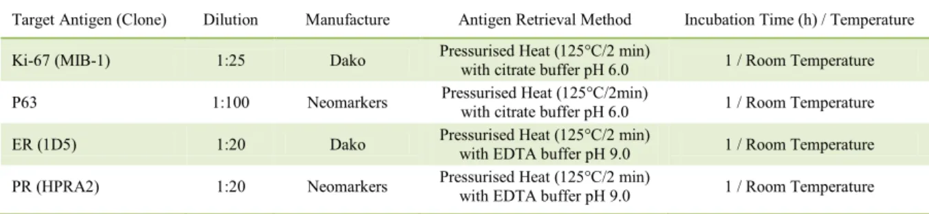
Table 1 - Target antigen and clone, dilution, antigen retrieval method, and incubation time and temperature for immunohistochemical staining for Ki-67, p63, Estrogen Receptor (ER), and Progesterone Receptor (PR).
Target Antigen (Clone) Dilution Manufacture Antigen Retrieval Method Incubation Time (h) / Temperature
Ki-67 (MIB-1) 1:25 Dako Pressurised Heat (125°C/2 min) with citrate buffer pH 6.0 1 / Room Temperature
P63 1:100 Neomarkers Pressurised Heat (125°C/2min)
with citrate buffer pH 6.0 1 / Room Temperature
ER (1D5) 1:20 Dako Pressurised Heat (125°C/2 min)
with EDTA buffer pH 9.0 1 / Room Temperature
PR (HPRA2) 1:20 Neomarkers Pressurised Heat (125°C/2 min)
MINA, R.B. et al. Mammary fibroadenoma in a young Holstein
cow. Journal of Veterinary Medicine Science, v.56, p.1171-1172, 1994.
MISDORP, W. Tumors of the mammary gland. In: MEUTEN, D.J. Tumors in domestic animals. Ames: Iowa State University, 2002. p.575-606.
PEÑA, L.L. et al. Immunohistochemical detection of Ki-67 and PCNA in canine mammary tumors: relationship to clinical and pathologic variables. Journal of Veterinary Diagnostic
Investigation, v.10, p.237-246, 1998. Available from: <http:// journals.sagepub.com/doi/pdf/10.1177/104063879801000303>. Accessed: Sep. 30, 2016. doi: 10.1177/104063879801000303.
PEREIRA, P.D. et al. Cell proliferation in feline normal, hyperplastic and neoplastic mammary tissue – an immunohistochemical study.
Veterinary Journal, v.168, p.180-185, 2004. Available from: <http:// www.sciencedirect.com/science/article/pii/S1090023303002065>. Accessed: Sep. 22, 2016. doi: 10.1016/j.tvjl.2003.10.018.
POULTON, T.B. et al. Sclerosing lobular hyperplasia of the breast: imaging features in 15 cases. American Journal of
Roentgenology, v.165, p.291-294, 1995. Available from: <http://www.ajronline.org/doi/pdf/10.2214/ajr.165.2.7618542>. Accessed: Oct. 15, 2016. doi: 10.2214/ajr.165.2.7618542
POVEY, R.C.; OSBORNE, A.D. Mammary gland neoplasia in
the cow. A review of the literature and report of a fibrosarcoma.
PathologiaVeterinaria v.6, p.502-512, 1969.
RUTTEMAN, G.R. Hormones and mammary tumour disease in the female dog: an update. In Vivo, v.4, p.33-40, 1990.
SANT’ANA, F.J.F. et al. Mammary diffuse fibroadenomatoid
hyperplasia in water buffalo (Bubalus bubalis): three cases.
Journal of Veterinary Diagnostic Investigation, v.26, p.453-456, 2014. Available from: <http://journals.sagepub.com/doi/ pdf/10.1177/1040638714526595>. Accessed: Nov. 30, 2015. doi: 10.1177/1040638714526595.
SINGH, B.; IYER, P.K.R. Mammary intraductal carcinoma in goats (Capra hircus). Veterinary Pathology, v.9, p.441-446, 1972. Available from: <http://journals.sagepub.com/doi/pdf/1 0.1177/030098587200900604>. Accessed: Sep. 12, 2016. doi: 10.1177/030098587200900604.
THIBAULT, S. et al. Mammary fibroadenoma in a heifer.
Canadian Veterinary Journal, v.38, p.785-786, 1997. Available from: <https://www.ncbi.nlm.nih.gov/pmc/articles/PMC1576795/ pdf/canvetj00097-0059.pdf>. Accessed: Aug. 19, 2016.
WU, L.S.; CHEN, A.Y. Ultrasound analysis of fibroadenomatoid
hyperplasia of the breast. Chinese Journal of Ultrasound
Diagnosis, v.2, p.71-73, 2001.
ZHANG, H. et al. Giant fibroadenomatoid hyperplasia of the