rev bras ortop.2017;52(2):238–240
SOCIEDADE BRASILEIRA DE ORTOPEDIA E TRAUMATOLOGIA
w w w . r b o . o r g . b r
Case
report
Pindborg
tumor
in
the
distal
femur
夽
Alex
Oliboni
Sussela
a,∗,
Paulo
Ricardo
Picon
Alves
b,
Vinicius
Duval
da
Silva
c,
Carlos
Daniel
de
Garcia
Bolze
b,
Osvaldo
André
Serafini
baPontifíciaUniversidadeCatólicadoRioGrandedoSul,FaculdadedeMedicina,PortoAlegre,RS,Brazil
bUniversidadeCatólicadoRioGrandedoSul,HospitalSãoLucas,Servic¸odeOrtopediaeTraumatologia,PortoAlegre,RS,Brazil
cUniversidadeCatólicadoRioGrandedoSul,HospitalSãoLucas,LaboratóriodeAnatomiaPatológicaeCitopatologia,PortoAlegre,RS,
Brazil
a
r
t
i
c
l
e
i
n
f
o
Articlehistory: Received20May2016 Accepted30May2016
Availableonline20February2017
Keywords: Femur
Boneneoplasms Biopsy
a
b
s
t
r
a
c
t
TodescribeacaseofpossiblediagnosisofPindborgtumoronthedistalfemur.
A32-years-oldfemalepatient,anativeofBolivia,residentinBrazil,arrivedtothisservice fortumorresearchintherightfemur.Afterbiopsiesandresectionofthelesion,thecasewas referredtoanalysisandconsultancyintheUnitedStates.Inareportofreviewbythe Pathol-ogyLaboratory,itwascharacterizedthehistologicalappearanceandimmunohistochemical profilewerecharacteristicofPindborgtumor.Currently,thepatientisbeingfollowed-upat theOrthopedicsandTraumatologyDepartmentofthisinstitution,andpresentsagood evo-lution.Thisstudypresentsthecaseofapatientwithararetumorthatwasinvestigatedfor anextensiveperiodandthroughmanytests.Pindborgtumorwassuggestedasadiagnostic hypothesisduetothecharacteristicsandbehavioroftheneoplasticlesion.Althoughthis lesionismorecommonlyobservedinodontology,theneoplasiawascompatiblewiththe diagnosis.Therefore,despitethefactthatthistumorhasbenigncharacteristics,long-term monitoringisnecessary,giventhehighrateoftumorrecurrence.
©2017PublishedbyElsevierEditoraLtda.onbehalfofSociedadeBrasileiradeOrtopedia eTraumatologia.ThisisanopenaccessarticleundertheCCBY-NC-NDlicense(http:// creativecommons.org/licenses/by-nc-nd/4.0/).
Tumor
de
Pindborg
em
fêmur
distal
Palavras-chave: Fêmur
Neoplasiasósseas Biópsia
r
e
s
u
m
o
DescreverumcasodepossíveldiagnósticotumordePindborgemfêmurdistal.
Apacientede32anos,naturaldaBolívia,residentenoBrasil,veioaesseservic¸opara investigac¸ãodemassatumoralemfêmurdireito.Apósfeituradebiópsiaseressecc¸ãoda lesão,ocasofoiencaminhadoparaanáliseeconsultorianosEstadosUnidos.Emlaudode revisãodoLaboratóriodePatologia,foicaracterizadoqueoaspectohistológicoeoperfil
夽
StudyconductedatUniversidadeCatólicadoRioGrandedoSul,HospitalSãoLucas,PortoAlegre,RS,Brazil. ∗ Correspondingauthor.
E-mail:[email protected](A.O.Sussela). http://dx.doi.org/10.1016/j.rboe.2017.02.002
rev bras ortop.2017;52(2):238–240
239
imuno-histoquímicoeramcaracterísticosdetumordePindborg.Atualmente,apacienteé acompanhadanoServic¸odeOrtopediaeTraumatologiadanossainstituic¸ãoeapresenta boaevoluc¸ão.Orelatodescreveocasodeumapacientecomumaneoplasiararaquefoi investigadapormuitotempoecomauxíliodemuitosexames.OtumordePindborgfoi sugeridocomhipótesediagnósticadevidoàscaracterísticaseaocomportamentoneoplásico dalesão.Adespeitodeserumalesãomaiscomumenteobservadanoâmbitoodontológico,a neoplasiadapacientemostrou-secompatívelcomodiagnóstico.Assim,mesmosetratando deumtumorcomcaracterísticasbenignas,háanecessidadedeacompanhamentoporlongo tempo,hajavistaoaltograuderecorrênciadotumor.
©2017PublicadoporElsevierEditoraLtda.emnomedeSociedadeBrasileirade OrtopediaeTraumatologia.Este ´eumartigoOpenAccesssobumalicenc¸aCCBY-NC-ND (http://creativecommons.org/licenses/by-nc-nd/4.0/).
Introduction
Pindborgtumor,alsoknownascalcifyingodontogenic epithe-lialtumor,isahighlyrareneoplasm,characterizedbylocal invasivenessandpresentingamyloidmaterial.Thisneoplasm emergesasapainlessmassofslowgrowth,withno predilec-tionforgender. Itaffects patientsbetweenthesecond and sixth decadeoflife,mainly inthe fourthdecade.Thevast majorityofthesetumorsare intraosseousmasses;only6% of them are extraosseous. Upon radiological examination, thetumorisusuallymultilocularandseptatedor,less com-monly,unilocularandradiolucent,andcalcificationswithin thelesionaresometimesobserved.
Case
report
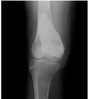
A 32-year-old female patient, originally from Bolivia, was admittedtothishospitalinAugust2009withacomplaintof painintherightkneeforoverayear.Shedeniedprevious sur-geriesonthearea,aswellasusinganymedications.Patient presentedwithrestrictedmobilityoftherightlowerlimband painuponpalpation inthe regionofthe rightfemoral epi-condyle.Onthe13th ofthe samemonth,shewasreferred to a surgical procedure – femoral bone biopsy. The speci-men,whichmeasured1.6cm,waspositiveforamyloidusing Congoredstaining.Inascintigraphy,performedonAugust 19,anirregularradiomarkerconcentrationwasobservedin thedistalthirdoftherightfemur.Anewbiopsy,performed onNovember9th,showedbonefragmentwithdense atypi-calcellinfiltrateandpositivityforamyloidusingCongored staining.Intheradiologicalexamination,aninsufflating oste-olyticlesionwasidentifiedintheinnermarginofthedistal endofthefemur,withseptationsinitsinterior.Withthe sus-picionoflargecelltumor,patientunderwenttumorresection surgeryonDecember21,2009.Microscopicexaminationofthe surgicalspecimenfoundatumoralmasspartiallycoatedby adiposeandmuscletissue,measuring6.5×4.1×3.4cm.When dissected,thelesionwascharacterizedbyits whitish,firm, andmattecolor.Partofthedistalportionofthefemurand tumorwereresected.Patienthadagoodrecoveryaftersurgery. TissuematerialwassenttotheHSLPathologyLaboratory. Ini-tialdiagnosis was unclassifiedneoplasm; subsequently, an immunohistochemicalanalysiswasperformed,whichledto a diagnosis compatible with mesenchymal neoplasm with
Fig.1–Post-surgeryradiographofthelesionindicatingthe presenceofsurgically-insertedorthopediccementmaterial.
240
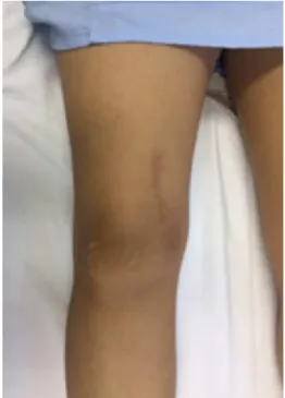
rev bras ortop.2017;52(2):238–240Fig.2–Post-surgicalrecoverylimbflexion.
Fig.3– Post-surgicalrecoverylimbextension.
Discussion
Described in 1958by Jens JorgenPindborg, the tumor that bears hisname isavery rare neoplasm.There are around 200casesdescribedinthecurrentliterature.1Descriptionof this neoplasm shares many features with ameloblastoma, butPindborgtumorislessaggressiveandpresentsaslower growth.2Itisknownthatthistypeoftumorrequiresalong follow-upperiod,becausethereisahighriskoftumor recur-renceifithasbeenincompletelyresected.Accordingtothe literature,tumorrecurrencerateisaround15%;inthese,the frequencyoflesionsthatweretreatedwithcurettageishigh. Inspiteofpresentinghighmitoticactivity,acharacteristicof malignancy,Pindborgtumorisabenignneoplasiawithgood prognosis.3,4Thehistologicalaspectsofthisneoplasiainclude fibrousstroma,islandsofpolyhedralepithelialcells, homoge-neousamyloidcontent,eosinophilia,andpositivereactionto Congoredstaining.Insomecases,itispossibletofindfocal areasofclearcells,calledrarevariantsofclearcells,which presentalessfavorableprognosisforthepatient.3,5Treatment forthis typeofneoplasia consistsofsurgicalremovalwith sufficientsafetymargintopreventrecurrences.1
Conflicts
of
interest
Theauthorsdeclarenoconflictsofinterest.
r
e
f
e
r
e
n
c
e
s
1.PindborgJJ.Acalcifyingepithelialodontogenictumor.Cancer. 1958;11(4):838–43.
2.DahlinDC.Bonetumors–generalaspectsanddataon6221 cases.3rded.Springfield,IL,USA:CharlesC.Thomas;1978. 3.MüllerD,Manojlovi´cS,Luksi´cI,Grgurevi´cJ.Calcifying
epithelialodontogenictumorofthemaxilla(Pindborgtumor). CollAntropol.2012;36Suppl.2:205–8.
4.TakataT,SlootwegPJ.Calcifyingepithelialodontogenic tumour.In:BarnesL,EvesonJW,ReichartP,SidranskyD, editors.Pathologyandgenetics–headandnecktumours. Lyon:IARCPress;2005.p.302–3.