Joana Mendes Lopes de Melo
IL-27-regulated signalling pathways in
Angiogenesis - possible role of MAPKinases.
Tese de Mestrado
Mestrado em Genética Molecular
Trabalho realizado sob a orientação de:
Professora Associada Katerina Tritsaris (Universidade
de Copenhaga)
e co-orientação de:
Professora Auxiliar Paula Sampaio (Universidade do
Minho)
Nome: Joana Mendes Lopes de Melo
Endereço electrónico: [email protected] Número do Bilhete de Identidade: 13742613
Título tese de Mestrado:
IL-‐27-‐regulated signalling pathways in Angiogenesis -‐ possible role of MAPKinases.
Orientadora:
Professora Associada Katerina Tritsaris (Universidade de Copenhaga)
Co-‐Orientadora:
Professora Auxiliar Paula Sampaio (Universidade do Minho)
Ano de conclusão: 2013
Designação do Mestrado: Mestrado em Genética Molecular
É AUTORIZADA A REPRODUÇÃO PARCIAL DESTA TESE/TRABALHO (indicar, caso tal seja necessário, nº máximo de páginas, ilustrações, gráficos, etc.), APENAS PARA EFEITOS DE INVESTIGAÇÃO, MEDIANTE DECLARAÇÃO ESCRITA DO INTERESSADO, QUE A TAL SE COMPROMETE;
Universidade do Minho, ___/___/______
______________________________________________________ Joana Mendes Lopes de Melo
Until today many people crossed the road I have been taking, and influenced in my wishes and decisions. Looking back, I could not be more pleased with the professional I am becoming. Hopefully, I will continue to have the luck to meet new individuals along this road.
I must thank with my entire hearth, the opportunity given by Associate Professor Katerina Tritsaris to join the Angiogenesis Lab in the Faculty of Health Sciences, University of Copenhagen to complete my Master thesis. For all the optimism placed on me and for teaching me so much, my biggest appreciation.
I thank to Professor, Dr. Scient. Steen Dissing all the hours in the fluorescence microscope together and particularly all the patience teaching me how to use it. Additionally, I appreciate all the discussions about the work and the motivation always given.
I also have to thank to my lab colleagues, for such a good working environment, without them going to work every day would not be so pleasing. I specially acknowledge Sebastian, for the work we have done together, for what he taught me and for all the healthy discussions. Not forgetting all the Saturdays’ keeping company to each other in the lab.
Moreover, I would like to recognize everyone in the building 12, 6th floor in the Faculty of Health Sciences, for receiving me in their home and teach me how to become a true Danish.
And naturally, family and friends, my biggest gratitude.
MAPKinases.
Angiogénese é o crescimento de vasos sanguíneos a partir de vasos pré-‐existentes. Sabe-‐se que é um processo envolvido em várias doenças inflamatórias, sobretudo devido a um desequilíbrio entre factores pro-‐ e anti-‐angiogénicos no tecido inflamado. IL-‐27 é uma citocina produzida por células apresentadoras de antigénios, com propriedades pro-‐ e anti-‐ inflamatórias e potencial anti-‐angiogénico. Os sinalizadores de tradução e ativação de transcrição (STATs) são ativados por citocinas e alguns membros da família estão envolvidos em angiogénese, nomeadamente o STAT1. Quando STAT1 é fosforilado no resíduo de serina é induzida a expressão do factor regulatório de interferão-‐1 (IRF-‐1), pelo que se acredita que IRF-‐1 é necessário na angiogénese. Adicionalmente, sabe-‐se que as proteínas de ligação de guanilato (GBPs), são expressas devido à actividade de STAT1 e IRF-‐1. Foi também anteriormente sugerido o envolvimento das MAPKinases na angiogénese patológica, contudo a sua ligação ainda não é clara.
Com o objectivo de elucidar o mecanismo molecular na sinalização de IL-‐27 em células endoteliais, estudámos o mecanismo de fosforilação de STAT1 no resíduo de serina mediada pela IL-‐27, observando-‐se a consequente proliferação/sobrevivência e migração de células endoteliais de veia umbilical humana (HUVEC).
Os dados iniciais demonstraram que IL-‐27 leva à fosforilação do resíduo de serina em STAT1 e à expressão de IRF-‐1 em HUVEC. A inibição de SAPK/JNK reduziu a fosforilação de STAT1 no resíduo de serina, bem como a expressão de IRF-‐1, confirmando o envolvimento desta MAPK na fosforilação de serina em STAT1. Adicionalmente, verificou-‐se que a expressão de GBP1 e GBP2 é estimulada por IL-‐27 através da actividade de STAT1 e IRF-‐1. O papel das GBPs na angiogénese não foi esclarecido, estando a ser alvo de novos estudos.
O segundo objectivo desta tese era compreender o efeito biológico de IL-‐27. Desta forma, foi realizada a optimização de vários ensaios angiogénicos, in vitro e in vivo. Para o estudo in vitro da ramificação de vasos sanguíneos, a migração de células endoteliais e formação de tubos, foram utilizadas as técnicas “Endothelial Spheroid Angiogenesis Assay” e “Endothelial Tube Formation Assay”. Ambos os métodos optimizados revelaram ser ferramentas importantes para investigar sinalização intracelular, pois permitem a utilização de anticorpos, inibidores ou siRNA. Foi ainda testado o modelo de retina de ratinho, observando-‐se a redução do número de ramificações de vasos sanguíneos na retina, quando estimulados com IL-‐27. Este ensaio revelou resultados preliminares encorajadores.
células endoteliais e o seu possível efeito biológico na angiogénese. Espera-‐se ainda contribuir para a identificação de novos alvos terapêuticos para angiogénese patológica. Contudo muitas questões foram levantadas, as quais serão futuramente objeto de estudo.
Angiogenesis is the growth of new blood vessels from pre-‐existing vessels. It is a process known to be involved in many inflammatory diseases, mainly due to imbalance between pro-‐ and anti-‐angiogenic factors in the inflamed tissue. IL-‐27, a cytokine produced by antigen-‐presenting cells with both pro-‐ and anti-‐inflammatory properties, was previously demonstrated to have an anti-‐angiogenic potential. The signalling transducers and activators of transcriptions (STATs) are induced by cytokines and some of the family members take part in angiogenesis, namely STAT1. When STAT1 is serine-‐phosphorylated expression of interferon regulatory factor-‐1 (IRF-‐1) is induced, proposing IRF-‐1 association to angiogenesis. Additionally, is known that the guanilate-‐binding proteins (GBPs) are expressed due to STAT1 and IRF-‐1 activities. Also, it was previously suggested the MAPKinases involvement in pathological angiogenesis, but their connection is still unclear.
In order to clarify the molecular mechanism in IL-‐27 signalling in endothelial cells, was studied the mechanism of STAT1 serine phosphorylation mediated by IL-‐27, detecting subsequent proliferation/survival and migration of Human Umbilical Vein Endothelial Cells (HUVEC).
Initial data showed that IL-‐27 stimulated STAT1 serine phosphorylation and IRF-‐1 expression in HUVEC. Inhibition of SAPK/JNK reduced STAT1 serine phosphorylation and expression of IRF-‐1, thereby establishing the participation of this MAPK in STAT1 serine phosphorylation. Additionally, we observed that GBP1 and GBP2 expression was stimulated by IL-‐27 via STAT1 and IRF-‐1 activity. GBPs role in angiogenesis is now further explored.
The second aim of this thesis was to comprehend the biological effect of IL-‐27. Therefore, I started optimization of different in vitro and in vivo angiogenesis assays. To in
vitro study of sprouting angiogenesis, migration of endothelial cells as well as tube formation,
was used “Endothelial Spheroid Angiogenesis Assay” and “Endothelial Tube Formation Assay”. Both optimized assays are good tools for investigating intracellular signalling since the use of siRNA, inhibitors and antibodies is possible. The mouse Retina assay an in vivo angiogenesis model, showed a reduction in the number of branches in IL-‐27 stimulated eyes, providing encouraging preliminary results.
This study offered a better understanding of IL-‐27 signalling in endothelial cells and possible biological effect in angiogenesis. We hope to contribute for the identification of new therapeutical targets for pathophysiological angiogenesis. However, many new questions have been raised, which we aim to dissolve in the future.
Acknowledgments i Vias de sinalização reguladas por IL-‐27 em Angiogénese – possível envolvimento de MAPKinases. iii IL-‐27-‐regulated signalling pathways in Angiogenesis -‐ possible role of MAPKinases. v
Table of Contents vii
List of Abbreviations ix
CHAPTER I BACKGROUND 1
1 Highlights in Angiogenesis 3
2 Tumor Angiogenesis 4
3 IL-‐27 in Angiogenesis 6
4 Mitogen-‐activated protein kinases (MAPKs) 7
4.1 P38 8
4.2 JNK 10
4.3 ERK 11
5 STAT proteins 11
5.1 Regulation of STAT activity 14
5.2 STAT5 15
6 IRF-‐1 16
7 GBPs 18
8 Angiogenesis Assays 20
8.1 In vitro angiogenesis assays 20
8.2 In vivo angiogenesis assay 22
8.2.1 The mouse retina assay 22
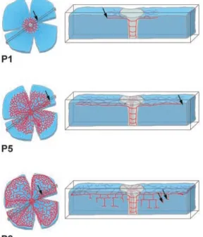
8.2.1.1 Vascular development in the mouse retina 23
CHAPTER II AIMS OF THE WORK 25
CHAPTER III MATERIALS AND METHODS 29
9 Cells lines and Cell culture 31
10 Signal transduction assays 32
10.1.1.1 Sample preparation 32
10.1.1.2 Gel-‐electrophoresis 32
10.1.1.3 Western blot 32
11 RNA purification: 34
11.1 Reverse transcription and Real Time-‐Polimerase Chain Reaction (Q-‐PCR) 34
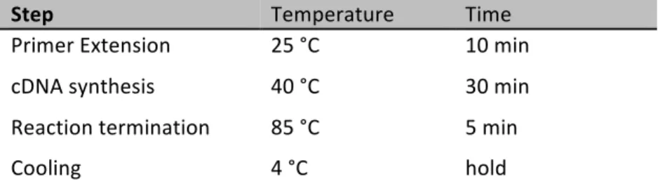
11.1.1.1 Reverse transcription: 35
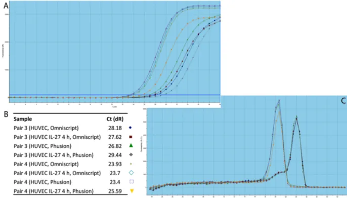
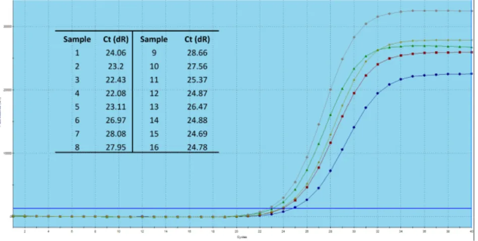
11.1.1.2 Q-‐PCR 35
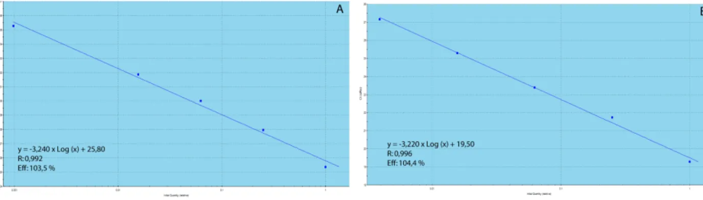
11.1.1.3 Optimization steps 36
14 Endothelial cells Spheroid-‐based Angiogenesis Assay 41
14.1.1.1 Critical steps 41
15 Endothelial Cell Tube Formation Assay 42
16 In vivo mice Retina Angiogenesis 43
17 Cytokines 43
CHAPTER IV RESULTS 45
IL-‐27 induces serine727 phosphorylation of STAT1 47
IRF-‐1 activation is dependent on STAT1 after IL-‐27 stimulation 48 p38 is not responsible for STAT1 serine727 phosphorylation 50
SAPK/JNK is phosphorylated after IL-‐27 stimulation 52
Inhibition of SAPK/JNK inhibits STAT1 serine727 phosphorylation and IRF-‐1 activation 54
GBP2 is activated upon IL-‐27 stimulation 56
Sub-‐project: STAT5 is Tyrosine694 phosphorylated after IL-‐27 stimulation. 58
Angiogenesis Assays 60
Cell Proliferation Assay 60
Endothelial cells Spheroid-‐based Angiogenesis Assay 62
Endothelial Tube Formation 64
In vivo Angiogenesis: The mouse Retina Angiogenesis Assay 66
CHAPTER V DISCUSSION 69
Serine phosphorylation of STAT1 is induced by IL-‐27 71 IRF-‐1 expression is due to IL-‐27-‐induced STAT1 Serine727 phosphorylation 72 IRF-‐1 is localized in the nucleus after IL-‐27 stimulation 72 P38 does not phosphorylate STAT1 at Serine residue after IL-‐27 stimulation 73
SAPK/JNK induces STAT1 Serine727 phosphorylation 74
GBP2 expression is linked to STAT1 and IRF-‐1 activity after IL-‐27 stimulation 76
IL-‐27 induces STAT5 Tyrosine694 phosphorylation 77
Angiogenesis Assays 77
Proliferation assay based on BrdU method 78
Endothelial cells Spheroid-‐based Angiogenesis Assay 78
Endothelial tube formation assay 79
Mouse Retina Angiogenesis assay 80
CHAPTER VI GENERAL CONCLUSIONS AND FUTURE PERSPECTIVES 83
ANG II angiopoietin II ISRE IFN-‐stimulated response element ATP Adenosine triphosphate ISGF3 IFN-‐stimulated gene factor 3 BrdU Bromodeoxyuridine Assay JAK Janus Kinase
BSA Bovine Serum Albumin MAP3Ks/MKKKs MAP Kinase Kinase Kinase CAM Chick Chorioallantoic membrane assay MAPK Mitogen Activated Protein Kinase
cDNA Complementary deoxyribonucleic acid MEKs/MKKs MAPK/extracellular signal-‐regulated kinase (ERK)-‐kinases
Ct Cycle threshold MMP Matrix Metalloproteinase
DMEM Dulbecco’s Modified Eagle’s Medium MYD88 Myeloid Differentiation primary response gene 88
DTT DL-‐Dithiothreitol NGS Normal Goat Serum EBI3 Epstein-‐Barr virus-‐induced gene 3 P5 Post-‐natal day 5
EBM Endothelial Basal Medium PBS Phosphate Buffered Saline ECM Extracellular matrix PDGF Platelet-‐derived Growth Factor EDTA Ethylenediamine Tetraacetic acid PFA Paraformaldehyde
EGF Endothelial Growth Factor PIAS Protein Inhibitors of activated STAT ELISA Enzyme-‐linked immunosorbent assay PLF Proliferin
ERK Extracellular signal-‐regulated kinases PRL Prolactin
FBS Fetal Bovine Serum Q-‐PCR Quantitative-‐ Polymerase Chain Reaction FGF Fibroblast Growth Factor RT Reverse Transcription/ Reverse Transcriptase GAPDH Glyceraldehyde-‐3-‐phosphate dehydrogenase SAPK/JNK Stress-‐Activated Protein Kinases/Jun amino-‐
terminal Kinases
GAS IFN-‐γ activation site SDS-‐PAGE Sodium Dodecyl Sulfate-‐PolyAcrylamide Gel Electrophoresis
GBPs Guanylate-‐Binding Proteins SH2 Src homology-‐2
gDNA Genomic deoxyribonucleic acid siRNA small interference ribonucleic acid GDP/GTP Guanosine di/triphosphate SOCS Suppressor of Cytokine Signalling
gp130 glycoprotein 130 STAT Signalling Transducer and Activator of Transcription
HUVEC Human Umbilical Vein Endothelial Cell TGF Transforming Growth Factor IAD1/2 IRF-‐associated domain 1/2 TLRs Toll like receptors
IFN Interferon TNFα Tumor Necrosis Factor alpha IL-‐27 Interleukine-‐27 TRAF6 TNF-‐receptor-‐associated factor 6 IP-‐10 interferon gamma-‐induced protein 10 UV Ultra Violet
IRF Interferon Regulatory Factor VEGF Vascular Endothelial Growth Factor
Chapter I Background
1 Highlights in Angiogenesis
Angiogenesis is the growth of new blood vessels, from pre-‐existing vessels, which differs from vasculogenesis, the formation of new vessels in an avascular area1 (Figure 1). In the healthy adult blood vessels are very stable and angiogenesis is rare, with the exception of events such as cyclical growth of vessels in the ovarian corpus luteum or during pregnancy2. However, angiogenesis is fundamental in several physiological and pathological conditions, for instance during development, modulation of the nervous system, tissue remodeling and in inflammation and cancer3.
The formation of vessels is a complex process, which requires a precise balance between numerous stimulatory and inhibitory signals. Angiogenesis requires key regulators of vascular endothelial cells sprouting, like VEGF (vascular endothelial growth factor). Presence of VEGF leads endothelial cells begin to proliferate and migrate, originating the branching of new vessels. Other cytokines, such as Platelet-‐derived growth factor (PDGF) and angiopoietin-‐ 1 (ANG I) recruit mural cells around the endothelial channels1,2,4. Pericytes are specialized mesenchymal cells with finger-‐like projections that wrap around endothelial cells to provide structural support for newly formed blood vessels. In normal vessels they deliver paracrine support signals; they can produce low levels of VEGF, operating as trophic function in endothelial homeostasis and collaborate with endothelial cells to synthetize the vascular basal membrane1,2,5.
Angiogenesis has been implicated in more than 70 disorders so far, and the list is ever growing. In many disorders, balance between stimulators and inhibitors is tilted, thereby regulating the angiogenic switch. The best-‐known conditions in which angiogenesis is switched on are malignant, ocular and inflammatory disorders, but many additional processes are affected, such as obesity, diabetes, multiple sclerosis, bacterial infections or autoimmune disease. In other diseases, such as ischemic heart disease or preeclampsia, the angiogenic switch is insufficient, causing endothelial cell dysfunction, vessel malformation or regression, or preventing revascularization, healing and regeneration4.
Figure 1: Development of the vascular system. Endothelial progenitor cells give rise to a primitive vascular network of
arteries and veins, a process known as vasculogenesis. Subsequently, during angiogenesis, the network expands and an organized vascular network, with pericytes and smooth muscle cells covering the endothelial tubes, emerges. Figure withdrawal from Carmeliet (2005).
2 Tumor Angiogenesis
In 1971, Folksman wrote “survival and proliferation of cancer depend on angiogenesis”. Tumors need microenvironment propitious to their growth. Initially, tumors are able to capture oxygen and nutrients by diffusion, however, they only grow until 2mm. In order to tumors to be able to increase in size it is necessary for them to have access to nutrients and oxygen, as well as to be able to excrete metabolic waste products and carbon dioxide. Therefore, a high number of vessels are needed1,5.
Tumor vessels diverge from normal vessels. They are more dilated, tortuous and have low coverage of pericytes, which lead to hyperpermeability, higher interstitial pressure and lower blood flow1. Also, tumor vessels occasionally have cancer cells integrated into the vessel walls6.
The angiogenic switch takes place in tumor development and can be regulated by countervailing factors, which either induce or inhibit angiogenesis. Recent studies, surprisingly shown that angiogenesis can be induced in the beginning of the multistage development of invasive cancers and is an important factor even in the microscopic premalignant phase of neoplastic progression. For this reason, angiogenesis has been considered one of the Hallmarks of Cancer5.
There have been observed different grades of angiogenesis in distinctive tumors. Aggressive tumors, like pancreatic ductal adenocarcinoma, are hypovascularized, in contrast human renal carcinoma, is highly vascularized. These observations led to the hypothesis that
initial switch of angiogenesis is equal in different types of tumor, though this is preceded by variations in intensity of ongoing neovascularization5.
Although tumor vessels present low coverage of pericytes, it has been found that these auxiliary cells are important in tumor neovasculature. The functional meaning of these cells was demonstrated by genetic and pharmacological perturbations, for example, through the inhibition of the PDGF receptor-‐β, expressed in tumor pericytes7, leading to a reduced coverage of tumor vessels and consequently, compromises vascular integrity and function. It is also interesting to refer that pericytes present in normal vessels are not sensitive to pharmacological inhibition, which once more suggests differences in the regulation of normal and tumors vessels5.
Already in 1971, investigators hypothesized that antiangiogenic therapy could be the key in human cancer treatment and began an active search for angiogenesis inducers and inhibitors. However, is now known that tumors vessel structure is thought to affect the drug targeting of cancer through angiogenesis8. Cells of innate immune system have a crucial role in pathological angiogenesis. Macrophages and neutrophils, among others, enter premalignant lesions and tumors and assemble themselves at the margins. Besides providing inducing signals for angiogenesis, they are able to protect the tumor vasculature from the effects of antiangiogenic drugs5.
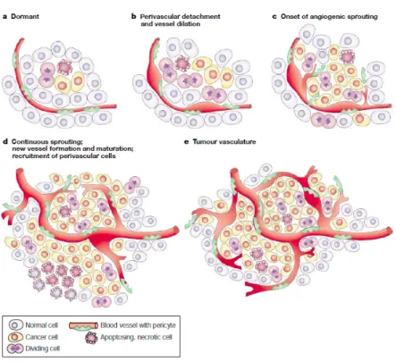
Figure 2: Onset of the angiogenic switch depends on the nature of the tumor and its microenvironment. The angiogenic
switch ensures exponential tumor growth and begins with perivascular detachment and vessel dilation. Image withdrawal from Bergers and Benjamin (2003).
3 IL-‐27 in Angiogenesis
Interleukin 27 (IL-‐27) is a heterodimeric cytokine, belonging to IL-‐6/IL-‐12 family, comprising the cytokine subunit p28 and the soluble receptor Epstein-‐Barr virus-‐induced gene 3 (EBI3)9,10. Both subunits are required for secretion and normal function of IL-‐2711.
The IL-‐27 receptor is composed of WSX-‐1 (also known as T cell cytokine receptor), a type I cytokine receptor, and glycoprotein 130 (gp130), a subunit used by several other IL-‐6 and IL-‐12 family members9. IL-‐27 intracellular signalling only occurs when IL-‐27 interacts with both subunits12.
When IL-‐27 binds, the receptor is phosphorylated by Janus kinase 1 (JAK1), which is constitutively associated with WSX-‐1 via interaction with different cytoplasmic tyrosine residues13. The phosphorylated tyrosine residues function as docking sites for intracellular proteins, including the proteins of the signal transducers and activators of transcription (STAT) family12 (Figure 3).
It is already known that IL-‐27 presents pro-‐ and anti-‐inflammatory properties, although its contradictory effects are not fully understand. It appears that IL-‐27 signalling is dependent on the immunologic context9. Initially, IL-‐27 was reported to induce proliferation of naive CD4+ T cells11, as well as having TH1-‐promoting abilities by up-‐regulating Interleukin 12 receptor b2 on naive T cells14 and synergizing with IL-‐12 to induce production of interferon-‐γ (IFNγ)15.
In vivo studies show that IL-‐27 also possesses immune-‐suppressive properties. IL-‐27R-‐
deficient mice, comparing to wild type mice, developed severe disease in a model of experimental autoimmune encephalomyelitis. This effect was linked to the lack of IL-‐27 suppression of TH17 cells in IL-‐27R-‐deficient mice16.
Recently, IL-‐27 was found to be an important cytokine in angiogenesis. Previous findings, describe its direct role in angiogenesis reduction, however the mechanism of action is still unknown10,17 . Yet, recently it was observed that IL-‐27 had anti-‐proliferative effect in lymphatic endothelial cells viaJAK/STAT1 activation18.
IL-‐27 is involved in the development of arthritis, as this was delayed in IL-‐27R-‐/-‐
mice
comparing with IL-‐27R-‐/-‐ mice19. IL-‐27 also presents potent antitumor activity, which generally
is mediated by CD8+ T cells10 and occurs by indirect mechanisms, like induction of NK cells or inhibition of angiogenesis through induction of IP-‐10 (interferon gamma-‐induced protein 10)
and IP-‐9. Lately, it was shown that IL-‐27 directly inhibits the proliferation of human melanoma cell lines, which express functional IL-‐27R20.
Figure 3: Components of IL-‐27 and its receptor. In the presence of IL-‐27, the IL-‐27 receptor recruits Jak kinases, which in turn
activate the STAT transcription factors. After being phosphorylated STATs enter the nucleus and modulate gene expression. Image withdrawal from Colgan and Rothman (2006)
4 Mitogen-‐activated protein kinases (MAPKs)
Mitogen-‐activated protein kinase (MAPK) signal transduction pathways are extensive mechanisms of eukaryotic cell regulation. Eukaryotic cells have multiple MAPK pathways, each preferentially recruited by different stimuli21.
Mammalian MAPK pathways act through diverse receptor families, including hormones and growth factors that exert their action through receptor tyrosine kinases [e.g., insulin, epidermal growth factor (EGF), platelet-‐derived growth factor (PDGF), and fibroblast growth factor (FGF)] or cytokine receptors (e.g., growth hormone). MAPKs can also be activated by vasoactive peptides that act through G protein-‐coupled receptors (e.g., ANG II, endothelin) or throught transforming growth factor (TGF)-‐related polypeptides, acting through Ser-‐Thr kinase receptors. Environmental stresses also lead to MAPK activation, such as osmotic shock, ionizing radiation and ischemic injury. In turn, MAPK pathways exert a profound effect on cell physiology, namely on gene transcription, protein synthesis, cell cycle machinery, cell death, and differentiation21.
MAPK include ERK, p38 and SAPK/JNK. The extracellular signal–regulated kinases (ERKs) was the first mammalian MAP Kinase to be identified, followed by the stress-‐activated kinases SAPK/JNK and p38. ERK is activated in response to cell cycle progression or cell
differentiation, while both SAPK/JNK and p38 are induced by stress stimuli, for instance, osmotic stress or UV radiation21.
Figure 4: MAPKs pathways. Image Withdrawal from Liu et al.
2007.
All MAPKs are activated by simultaneous Tyrosine and Threonine phosphorylation within a conserved Thr-‐X-‐Tyr motif in the activation loop of the kinase domain subdomain VIII. MAPK phosphorylation and activation are catalysed by a family of dual specificity kinases referred to as MAPK/extracellular signal-‐ regulated kinase (ERK)-‐kinases (MEKs or MKKs). These are themselves regulated by Serine/Threonine phosphorylation, also within a conserved motif in kinase domain subdomain VIII, which is catalysed by any of the protein kinase families referred to as MAPK-‐kinase-‐kinases (MKKKs or MAP3Ks)21.
To date several anti-‐angiogenic compounds have been reported to inhibit the activation of ERK1/2 signal cascade induced by VEGF (vascular endothelial growth factor) and bFGF (basic fibroblast growth factor), indicating an involvement of ERK1/2 in angiogenesis. p38 was likewise reported to be involved in regulating VEGF-‐induced HUVEC cell migration, having an important role in angiogenesis. Furthermore, SAPK/JNK was described having an antiangiogenic role via inhibition of VEGF secretion and metalloproteinase (MMP) from HUVEC cells. Active MAPKs have been detected in many diseases involving angiogenesis like tumor progression and metastasis, diabetic retinopathy and age-‐related macular degeneration, suggesting that MAPKs take part in angiogenesis-‐associated diseases22.
4.1 P38
P38 belongs to mammalian stress-‐activated MAPK family and was originally described as a 38kDa polypeptide, which were regulated by Tyrosine phosphorylation. After the p38 alpha isoform was discovered this mammalian MAPK showed high homology to HOG1, the
osmosensing MAPK of S. cerevisiae, and like this, contained the conserved sequence Thr-‐Gly-‐ Tyr21.
To date, four isoforms of the p38 family, namely p38α, p38β, p38γ, and p38δ, were identified and are encoded by distinct genetic loci, but all share the common Thr-‐Gly-‐Tyr motif in kinase subdomain VIII23,24. Usually all isoforms are inactive in the cytoplasm, but after phosphorylation are translocated to the nuclei, where they phosphorylate diverse substrates24. Among all p38 MAPK isoforms, p38α is the best characterized and it is expressed in most cell types, while p38β is present mostly in the brain, p38γ is most significantly expressed in skeletal muscle and p38δ in endocrine glands25,26. The p38 MAPK subfamily can be divided into two distinct subsets, p38α and p38β on the one hand and p38γ and p38δ on the other. P38α and p38β share a similar amino-‐acid sequence, being 75% identical25. In addition, three-‐dimensional structure of p38β is very similar to that of p38α, with some differences in the orientation of the N-‐ and C-‐terminal domains affecting the size of the p38β ATP-‐binding pocket26. p38γ and p38δ are more identical to each other, about 70% identical25.
P38 pathway is commonly known for tumor suppression, regulation of cell survival and proliferation, differentiation and apoptosis24. The p38 signalling pathway is activated by several stimuli and stresses, like environmental stress, bacterial lipopolysaccharide, inflammatory cytokines, heat shock, UV light, hypoxia, ischemia23,24. Its activation is relative to pro-‐apoptotic stimuli, but the biological outcome is highly dependent on cell type or context23.
P38 is regulated upstream by MKK3 and MKK627. Both MKK3 and MKK6 are highly selective for p38, MKK6 can phosphorylate all p38 family members, whereas MKK3 can not activate p38β26,28. In response to most stimuli p38α is activated by MKK3 or MKK6, although in some conditions, such as ultraviolet radiation, MKK4, an activator of JNK, may contribute to the p38α activation25. However, p38 isoforms are also capable of activating themselves through autophosphorylation, thereby being completely independent of MAPKKs24.
Activation of p38 MAPK occurs within minutes and is transient, suggesting that p38 MAPK functions as a biological switch, probably downregulated under basal conditions and during adaptation. MAPK phosphatases reverse the phosphorylation and MAPK return to their inactive state25. Studies reveal that p38 is activated by several growth factors including FGF-‐227. When FGF-‐2 induces activation of p38 activity in endothelial cells, MKK3/6 negatively
regulates the endothelial cell differentiation through a decrease in expression of proteins involved in differentiation, like Jagged1 (Notch ligand)23.
4.2 JNK
The JNK is one of the major MAPK pathways29. The indication that cells possess several MAPK pathways arises after ERK discovery, since injection of cycloheximide into rats triggered a new Ser/Thr kinase activity with the ability to phosphorylate microtubule-‐associated protein-‐2 (MAP2). JNK was found to be a 54kDa polypeptide, and for that was given the initial name of p54. Similar to ERK, was inactivated with Tyr or Ser/Thr phosphatases, indicating that it required simultaneous Tyr and Ser/Thr phosphorylation for activation21. Afterwards was discovered the ability for p54 to phosphorylate the c-‐Jun transcription factor at two sites (Ser63 and Ser73) in a substantially higher rate than observed for the ERKs21.
JNKs were isolated and characterized as stress-‐activated protein kinases grounded on their activation in response to inhibition of protein synthesis29. JNK is an extremely regulated and complex pathway and is influenced by 13 different MKKKs. Generally, it is identified by two names: stress-‐activated protein kinase (SAPK) due to their regulation by environmental stress and inflammation and c-‐Jun NH2-‐terminal kinase (JNK) referring to the phosphorylation by these kinases of the c-‐Jun NH2-‐terminal trans-‐activation domain affinities21.
The JNK proteins are encoded by three genes (JNK1-‐3), and alternative splicing gives origin to at least 10 different isoforms29. Splicing within the catalytic domain results in type 1 and type 2 SAPKs (b and a JNKs, respectively), while splicing at the COOH terminus bears 54kDa (p54) and 46kDa (p46) polypeptides (type 2 and type 1 JNKs, respectively. The exact significance of the COOH-‐terminal isoforms is still not clear, but was already observed that type 1 and type 2 kinases differ in their substrate binding affinities21. The JNK1 and JNK2 are present in nearly all cells, yet JNK3 is mostly present in the brain29.
JNKs are triggered in response to growth factors, proinflammatory cytokines, microbial components, and a variety of stresses, including oxidative stress30. They are important in controlling programmed cell death31, as well as cell proliferation, migration, survival, and cytokine production30.
JNK has previously linked to cancer, were inhibition of JNK enhanced chemotherapy effect in fighting tumor growth31. Also was recently shown that JNK1 is a critical factor for
VEGF production caused by hypoxia in retina, stimulating hypoxia-‐induced pathological angiogenesis30.
4.3 ERK
Human ERK1 and ERK2 share about 84% similarity and most of the known functions, since all the cellular stimulus lead to parallel activation of both ERK1 and ERK2. Only recently, using gene ablation studies was possible to understand differential functions for these protein kinases. Has shown that ERK1 gene is not necessary for mice development. ERK1-‐ deficient mice were viable, fertile, and of normal size, yet, it appears to play an important role in thymocyte development. ERK2 gene ablation led to embryonic death. It was found that development of placental vasculature is severely impaired in ERK2-‐deficient mice and that embryonic trophoblast development is impaired in ERK2-‐deficient mice32.
ERK1 and ERK2 are widely expressed and participate in the Ras-‐Raf-‐MEK-‐ERK signal transduction cascade. This signalling pathway exert function related with cell adhesion, cell cycle progression, cell migration, cell survival, differentiation, metabolism, proliferation, and transcription32. Many different stimuli activate the ERK1 and ERK2 pathways, like growth factors, cytokines, virus infection, transforming agents, and carcinogens31.
There are common oncogenic mutations that convert Ras to an activated oncogene in many human tumors31, and this occurs in about 30% of all human cancers32. Oncogenic Ras persistently activates ERK1 and ERK2 pathways, contributing to increased proliferation of tumor cells. Therefore, inhibitors of ERK pathways are in clinical trials as potential anticancer agents31. Activating mutations in Raf occur in near 7% of all human cancers32.
Recently, there are reports of several anti-‐angiogenic compounds capable of inhibiting activation of ERK1/2 signal cascade induced by VEGF and bFGF, indicating that ERK pathway might play a role in angiogenesis22.
5 STAT proteins
Signal Transducer and Activator of Transcription (STAT) family is a group of transcription factors that are present in their inactive form in the cytoplasm, but move to the nucleus once they are activated. The first two members of the family, STAT1 and STAT2 were
identified more than a decade ago and were acknowledged as mediators of cellular response to interferons33.
There are seven different STAT proteins, which can be divided in two main groups. STAT2, 4 and 6 are activated by a low number of cytokines and have a role in T-‐cells development and IFNγ signalling. STAT1, 3, 5a and 5b are activated in several different tissues, with an important role in cell-‐cycle progression and apoptosis. This last group also contributes to oncogenesis, which makes them extensively studied34.
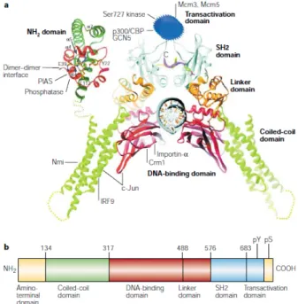
All STATs proteins share similar structural domains (Figure 5). The NH2-‐terminal domain highly conserved and mediates protein-‐protein interactions and stabilizes binding of two STAT dimers to DNA; as for the coiled-‐coil domain, this establishes association with other important proteins. The DNA-‐binding domain determines the specificity of each STAT protein to DNA and the linker domain establishes a bridge between DNA-‐binding and Src-‐homology-‐2 (SH2) domains. The SH2 domain is necessary for dimerization of two STAT monomers through phosphotyrosine interactions. In this domain there is a critical tyrosine residue, when STAT proteins are phosphorylated, the SH2 domain binds to another STAT protein. At the C-‐ terminal of STAT, there is the Transcriptional activation domain (TAD), which interacts with the transcriptional machinery to promote gene expression. Inside this domain some STAT proteins have a conserved serine, another phosphorylation site that regulates transcriptional activity34,35.
Figure 5: Signal Transducer and Activator of Transcription structure. a: STAT domain structure and protein biding sites.
b: STAT domains and phosphorylation sites. Image withdrawal from Levy and Darnell (2002)
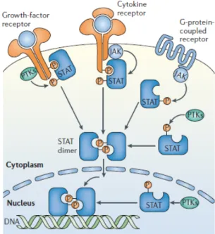
Currently several mechanisms to induce tyrosine phosphorylation in STATs are known (Figure 6). The well-‐known JAK-‐STAT pathway is initiated by a cytokine, which binds to a cell-‐ surface cytokine receptor. Commonly, the intracellular domains of these receptors are
associated with tyrosine kinases from the Janus kinase (JAK) family. The receptor-‐associated JAKs phosphorylate the cytoplasmic tails of the cytokines receptors upon activation, leading to recruitment and after that phosphorylation of STATs proteins. JAKs are also involved in phosphorylation of STATs via G-‐protein coupled receptors. Usually hormones or chemokines bind to this type of receptors and phosphorylation of STATs occurs. There is a third mechanism of STATs activation, which involves Growth-‐factor receptors; these possess an intrinsic tyrosine kinase activity33,34.
Figure 6: STAT tyrosine phosphorylation by receptor and non-‐ receptor kinases. There are multiple mechanism used by JAKs or
other PTKs (protein tyrosine kinases), which are intrinsic in the receptor or present in cytoplasm or nucleus. After tyrosine phosphorylation, there is STAT dimerization and binding to DNA. Image withdrawal from Reich and Liu (2006)
Via the SH2 domain, the STAT proteins become phosphorylated themselves by Jak on the C-‐terminal tyrosine residue, which leads to dissociation from the receptor and enables the phosphorylated STAT proteins to form homodimers or heterodimers. STAT dimerization is followed by translocation to the nucleus, where the STAT dimer binds to specific promoter elements in the DNA and activates gene transcription. Different STAT proteins are able to activate transcription of different genes, since they can recruit distinct co-‐activators, and so bind to different DNA sequences36.
A second phosphorylation of STAT proteins can take place on a serine residue in the C-‐ terminal end. In both STAT1 and STAT3, this serine residue is found in position 727 within a conserved proline-‐methionine-‐serine-‐phenylalanine (PMSP) motif. Several results suggest that the phosphorylation of this serine enhances the activity of STAT proteins, since mutation of the serine to alanine results in a reduced cytokine-‐stimulated transcription activity37.
Serine phosphorylation of STATs can occur through different signal transduction pathways, depending on the signal received by the cell. Until now, the involvement of ERK pathway in serine phosphorylation of STAT3 by receptor tyrosine kinases and IL-‐2 has been
reported38,39, as well as ERK involvement in serine725 phosphorylation of STAT540. ERK2 was also connected to serine phosphorylation of STAT141. SAPK/JNK pathway so far is only linked to STAT3 serine phosphorylation caused by stress signals, like UV irradiation and hyperosmolarity or by inflammatory signals, like TNFα42,43. STAT1 serine phosphorylation, also in situations of stress and inflammation seems to occur via p3844. However, further studies are required to fully understand STATs serine phosphorylation.
STATs proteins have an important role in immune responses. STAT1 knockout mice are highly susceptible to infection by microbial agents and viruses, since they have compromised response to IFNs. Animals lacking STAT3 dies in an early embryonic state, indicating that this protein might have a crucial role in normal development33. In addition, STAT1 and STAT3 have both been implicated in the biological functions of the vasculature. STAT1 is reported to mediate anti-‐angiogenic signalling of IFNγ in human umbilical vascular endothelial cells (HUVEC)45 and STAT3 to mediate pro-‐angiogenic signaling46.
5.1 Regulation of STAT activity
STATs proteins are temporarily activated. After a few hours, activating signals decay and STATs are exported from the nucleus back to cytoplasm34.
To decrease STAT DNA-‐binding activity dephosphorylation of STAT and/or dephosphorylating of the binding sites on the cytokines receptor is required. In both cases this is occurs by action of protein phosphatases (Figure 7a and c)34,47. SHP-‐2, a protein phosphatase, was identified as a regulator of STAT1 in the nucleus, dephosphorylating both Tyrosine701 and Serine727 48.
STATs proteins are also regulated by the suppressor of cytokine signalling (SOCS) protein family (Figure 7b)34. SOCS proteins are generally expressed at low levels in unstimulated cells and become rapidly induced by cytokines to inhibit STAT signaling49. Interestingly, some SOCS proteins are transcriptionally regulated by STAT activity, working as a negative feedback loop34.
SOCS proteins via the SH2-‐domains, to phosphorylated tyrosine residues on the cytoplasmic part of receptor chains or to phosphorylated proteins associated with the receptors, such as Jak proteins50.
Moreover, protein inhibitors of activated STATs (PIAS) are thought to inhibit STAT signalling by blocking STAT DNA-‐binding (Figure 7d) PIAS1 and PIAS3 interact with STAT1 and STAT3, respectively, only when their tyrosines are phosphorylated. There are also natural occurring truncated forms of STATs, which act as dominant-‐negative proteins, either forming dimers with wild-‐type STATs (Figure 7e) or binding to DNA as non-‐functional protein34.
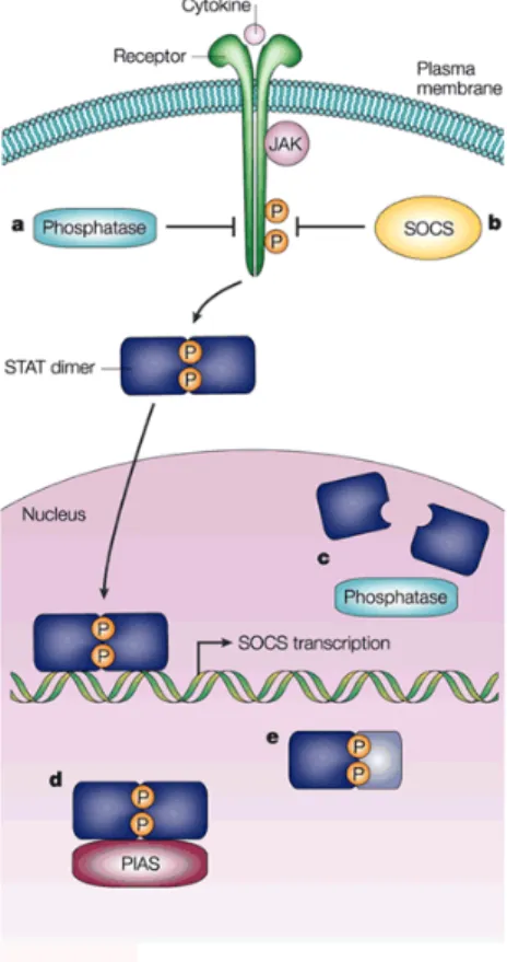
Figure 7: Regulation of JAK-‐STAT signalling
(A) Cytoplasmic phosphatases regulate STAT signalling by dephosphorylation of the phosphorylated tyrosine residues in the cytoplasmic tails of the receptor to prevent STAT-‐binding to the receptor. (B) SOCS proteins regulate STAT signalling by binding to the phosphorylated tyrosine residues in the receptor and inhibiting the enzymatic activity of Jak.
(C) Nuclear phosphatases regulate STAT signalling by dephosphorylation of STAT proteins, which results in STAT dimer dissociation.
(D) PIAS proteins regulate STAT signalling by binding to STAT dimers in the nucleus and blocking their DNA-‐binding ability.
(E) Endogenous truncated STAT proteins regulate STAT signalling by forming dimers with phosphorylated STAT proteins and functioning like dominant negative proteins.
Figure withdrawal from Levy and Darnell (2002)
5.2 STAT5
STAT5a was identified originally as a transcription factor that regulates the β-‐casein gene in response to prolactin (PRL), however it is now known that it is also activated by numerous cytokines and growth factors51. STAT5a and STAT5b are two members of the STATs family, which share 96% identity and diverge basically in the C-‐terminal transactivation domain52. Their expression pattern is also different, STAT5a is mainly present in the mammary gland, but STAT5b is predominantly expressed in liver51.
STAT5 has been implicated in several signalling events associated to the immune system, mammary epithelial cells or hepatocytes52. In addition, it has been found to be involved in a few malignances. In the case of hematopoietic cells, STAT5 regulates apoptosis, proliferation and cell cycle progression51.
Initial studies suggested that STAT5a and STAT5b possessed similar functions, however, after STAT5a and STAT5b single knockout mice showed different phenotypes. Knockout of STAT5a results in compromised mammary gland development owing to loss of prolactin response. Knockout of STAT5b leads to sexually dimorphic growth retardation. Moreover, STAT5a/b double knockout showed a severe phenotype, many mice died a few days after birth were infertile and presented compromised immune system, with absence of NK-‐cells and a reduced number of B-‐cell precursors51. Yet, is interesting to see upon STAT5 deletion that many cells appear to adapt instead of undergoing apoptosis. Other members of the family are recruited to their receptor-‐binding sites, in fact, deletion of STAT5 from hepatocytes caused an abnormal activation of both STAT1 and STAT352.
STAT5 has recently been implicated in Angiogenesis. Researchers have reported that STAT5 and to a minor degree STAT1 were activated by FGF2 and FGF8b in mouse brain endothelial cells, inducing migration, invasion and tube formation in collagen. However STAT5 had no effect on proliferation53. The same group further investigated, and discovered that STAT5 is necessary on secretion of proliferin (PLF, an autocrine factor from the prolactin family) for STAT5-‐mediated angiogenesis54.
6 IRF-‐1
The first Interferon-‐Regulatory Factor (IRF-‐1) was identified in 1988, and since then, nine members of the family have been recognized55.
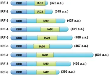
Structurally, all members present a well-‐conserved N-‐terminal DNA-‐binding domain (DBD) that recognizes a DNA sequence known as IFN-‐stimulated response element (ISRE). It is characterized by a set of five tryptophan-‐rich repeats. The IRF C-‐terminal domain is less conserved among the members of this family. This domain is responsible for the specificity of each member, since it mediates interactions with other proteins and co-‐factors. Within the C-‐ terminal there is an IRF-‐associated domain 1 (IAD1, in IRF members 3-‐9) and IAD2 (only shared by IRF-‐1 and 2), necessary for DNA binding55 (Figure 8).