A ro le fo r angio ge ne sis in
rhe um ato id arthritis
Departments of Immunology and Vascular Biology, The Scripps Research Institute, La Jolla, CA, USA D.G. Stupack,
C.M. Storgard and D.A. Cheresh
Abstract
Rheumatoid arthritis (RA) is a chronic debilitating disease character-ized by distinct autoimmune, inflammatory and fibrovascular compo-nents which lead to synovial proliferation and joint destruction. However, existing treatments specifically target only autoimmune and inflammatory components despite the fact that neovascularization of the inflamed synovium is a hallmark of rheumatoid arthritis. Angio-genesis may contribute to synovial growth, leukocyte recruitment and tissue remodeling, thus potentiating disease progression. Although no therapies currently target angiogenesis, several existing therapies have anti-angiogenic activity. Recent advances in anti-angiogenic strate-gies in oncology, including the identification of integrin avß3 as a crucial effector of angiogenesis, suggest a means to assess the role of angiogenesis in rheumatoid arthritis. Synovial endothelial cells have been shown to express integrin avß3, suggesting that these cells may be targeted for angiogenesis inhibition. Prior studies in rat arthritis models have shown benefit after the addition of broad spectrum integrin antagonists. However, formal assessment of integrin-targeted anti-angiogenic activity is now underway. These controlled studies will be important in assessing the efficacy of therapies which target angiogenesis in RA.
Co rre spo nde nce
D.A. Cheresh
The Scripps Research Institute IMM24
10550 N. Torrey Pines Rd. La Jolla, CA 92037 USA
Fax: + 1-619-784-8926 E-mail: [email protected]
Presented at the 5th Brazilian Symposium on Extracellular Matrix - SIMEC, Angra dos Reis, RJ, Brasil, September 7-10, 1998.
Research supported in part by the Jeanette Hennings Foundation and NIH (Nos. CA50286, CA 45726 and HL54444). D.G. Stupack and C.M. Storgard are recipients of fellowships from the Joseph Drown Foundation and the Arthritis Foundation, respectively.
Received O ctober 19, 1998 Accepted November 12, 1998
Ke y wo rds ·Angiogenesis ·Endothelium ·Arthritis ·Integrin
Intro ductio n
Rheumatoid arthritis (RA) is a complex chronic inflammatory disease which affects approximately 1-3% of the general popula-tion. The disease has several complementary yet distinct components, including autoim-mune, inflammatory and fibrovascular re-sponses, which contribute to the significant morbidity and mortality associated with ad-vanced disease (1,2). Currently, a conclusive etiology is lacking, but after an initiating insult, autoimmunity is triggered in a geneti-cally predisposed individual. The resultant
articular pathology is characterized by in-flammation and proliferation of the synovial lining, resulting in the generation of inter-digitating folds of tissue, termed pannus (3). The pannus is a major site of ongoing in-flammation and protease production, and is thought to be central to the development of cartilage and bone-erosive disease which leads to the destruction of joint architecture (3-5).
which subdue but ultimately fail to stop pro-gression to erosive joint destruction. Clearly, further modes of intervention for RA are needed. In this regard, the extensive syn-ovial neovasculature, which is one of the earliest histopathologic findings and a hall-mark of RA (6,7), has been suggested as an attractive target for therapy. Importantly, many of the therapies currently in use in-cluding gold (8), sulfonamides (9), chloro-quine (10), methotrexate (11), penicillamine (12), and COX inhibitors (13) have retro-spectively been found to possess anti-angio-genic activity either in vivo or in vitro.
How-ever, previous examination of angiogenesis inhibition as a primary therapeutic approach in RA is limited (14,15), and presently there are no drugs specifically targeted at prevent-ing the subsynovial vascularization which characterizes this disease.
Angio ge ne sis as a pro pagating facto r in RA
Angiogenesis, the coordinated growth of new blood vessels from pre-existing vascu-lature, occurs physiologically during repro-ductive and developmental processes as well as during the late phases of wound healing following tissue damage. However, inappro-priate or aberrant angiogenesis is associated with a variety of diseases, including tumor growth and metastasis as well as fibrovascu-lar disorders such as psoriasis, diabetic reti-nopathy and RA (16). In fact, the invasive growth of pannus tissue in RA has been compared to that of neoplastic tumors, and it has been suggested that the pannus itself may be considered a form of benign tumor (17). The recent successes of anti-angio-genic therapies in controlling tumor growth provide a strong rationale to investigate these agents as potential new strategies to limit pannus growth and joint destruction in RA. Angiogenesis has been suggested to be central to the pathophysiology of RA, con-tributing to disease progression at multiple
levels (6,7). The most obvious role of vascu-larization during RA is an increased capacity to sustain the nutritional and metabolic re-quirements of the hyperproliferating syno-vium and invading pannus (3-5). However, it is clear that the neovascularization which is ultimately achieved is not completely suf-ficient to relieve the intra-articular hypoxia associated with RA (18), resulting in a chronic angiogenic response.
In part, this is likely due to the distinction that in RA angiogenesis occurs within the context of an ongoing autoimmune reaction, where leukocyte extravasation into the tis-sues serves to maintain a local inflammatory response. Neovascularization, and the re-sulting increased vascular bed volume, di-rectly permit increased recruitment of blood-borne leukocytes into the synovial tissue. In turn, these activated leukocytes release an-giogenic cytokines (see below) but also cause local microvascular occlusion and injury. Further damage to endothelial cells occurs directly via the release of high levels of reactive oxygen species and proteolytic en-zymes (19). In turn, local vessel damage will subsequently induce a reparative angiogenic response from adjacent or contiguous ves-sels. Thus, the synovial vasculature is not static, but undergoes dynamic reorganiza-tion in response to cumulative cycles of en-dothelial proliferation and death (18).
A dire ct ro le fo r e ndo the lial ce lls in articular re mo de ling
angio-genic, providing a direct autocrine mecha-nism to sustain neovascularization of the subsynovial tissue (20). Alternatively, many cytokines present in synovial tissues play an indirect role in neovascularization by stimu-lating the secretion of directly angiogenic cytokines from either inflammatory or by-stander cell populations, while selected cytokines have both activities (Table 1). For example, in addition to angiogenic effects on endothelial cells, the production of VEGF and bFGF also serves to activate cells of the macrophage (21) and fibroblast (22) lineages, respectively. Interestingly, these two lineages are the predominant components of the hyperproliferative synovial lining, and com-prise a significant proportion of the pannus in RA.
Early responses of the endothelium to angiogenic cytokines include the initiation of vascular permeability and subsequently the upregulation of protease production. These events result in significant alteration of the composition of the local extracellular matrix (ECM). Numerous proteases includ-ing urokinase, tissue plasminogen activator and the metalloproteinases MMP-1, 2, 3, and 9 are produced by activated endothelial cells (5-8,23). The metalloproteinases are capable of digesting subendothelial
base-ment matrix, particularly collagen types IV and V, which act to suppress the angiogenic phenotype (14,24,25). The combined pro-teolysis and vascular permeability not only eliminates the capacity of these collagens to inhibit angiogenesis, but additionally pro-vides a new provisional ECM to sustain endothelial survival (26) and migration (27). Plasma glycoproteins including fibrinogen and fibronectin polymerize extravascularly (28) and probably also on the luminal face of the endothelium (29), similar to normal wound healing responses. However, unlike wound healing, the deposited provisional matrix is not effectively remodeled and re-placed in RA. In fact, evidence suggests that the deposition of this provisional ECM is sustained (30,31), and actually contributes to arthritic disease severity (31).
In this respect, components of the provi-sional ECM are potent cell activators. Fibronectin, vitronectin and fibrinogen can be chemotactic or haptotactic substrates for endothelial cells (32-35). The principal re-ceptors involved in endothelial attachment to, and migration on, these substrates are integrins, a family of heterodimeric (a/ß) transmembrane adhesion receptors (36). Al-though the integrins have no intrinsic kinase activity, it has become clear that
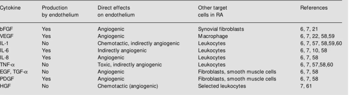
integrin-Table 1 - Selected angiogenic cytokines present in RA.
Selected cytokines found in rheumatoid arthritis synovial tissues w hich have been associated w ith angiogenesis in vivo. Each cytokine listed is classified w ith respect to possible production by, and characterized effects on, endothelial cells. References discussing the potential role of each cytokine in RA are cited.
Cytokine Production Direct effects Other target References by endothelium on endothelium cells in RA
bFGF Yes Angiogenic Synovial fibroblasts 6, 7, 21 VEGF Yes Angiogenic M acrophage 6, 7, 22, 58,59 IL-1 No Chemotactic, indirectly angiogenic Leukocytes 6, 7, 57, 58,59,60 IL-6 Yes Indirectly angiogenic Leukocytes 6, 7, 10, 58
IL-8 Yes Angiogenic Leukocytes 6, 7, 58
mediated adhesion events are sufficient to activate cellular signaling through a variety of nonreceptor tyrosine, serine/threonine and lipid kinases (36). In leukocytes, integrin-mediated interaction with provisional ECM proteins can facilitate mitogenic, chemotac-tic and oxidative responses, depending upon the specific subpopulation of cells studied (36-39). Integrin-mediated signaling also provides a crucial mechanism for adhesion-dependent survival in endothelial cells and fibroblasts (36). The importance of this integrin-mediated event is demonstrated by the observation that starved, but adherent endothelial cells, resist apoptosis better than starved or growth factor-stimulated cells maintained in suspension (26). Moreover, recent investigations show that endothelial cell signaling events elicited by angiogenic growth factors, including activation of the downstream effector MAP kinase, are dra-matically augmented in vitro and in vivo by
integrin-mediated interactions with the ECM (40,41).
Vascular inte grin avß3 in angio ge ne sis
Integrin avb3 has recently been charac-terized as a marker of angiogenic endothe-lium and as a central effector of this process (42). It is clear that ECM composition plays a determining role in endothelial cell re-sponses, and in this respect integrin avß3 is known to be a receptor for a variety of ECM proteins, including denatured collagen, fibronectin, fibrinogen and vitronectin (43). Further, avß3 can bind to the metallopro-teinase MMP-2, localizing it to the endothe-lial cell surface and subsequently potentiat-ing its activation from precursor zymogen to active enzyme (44). Therefore, expression of integrin avß3 expands the range of ligands which endothelial cells can interact with and also modifies cellular interactions with pre-existing ligands.
Inhibition of integrin ligation by selec-tive antagonists not only blocks the migra-tion of endothelial cells stimulated by
angio-Potential sites of action for avb3 antagonists in angiogenesis
Deposition of provisional ECM
(avb3 ligands) (
Digestion of basement ECM
Inhibition of survival signals from the provisional ECM
Prevention of avb 3-dependent migration and invasion
Angiogenic gradient Integrin avb3
M M P-2 zymogen Active M M P-2 Proteolyzed collagen (avb3 ligand) Angiogenic cytokine Figure 1 - Potential mechanisms
of angiogenesis inhibition by an-tagonists of integrin avß3. An-giogenic grow th factors induce vascular perm eabilit y, provi-sional extracellular matrix (ECM ) deposition, degradation of the subendothelial basement matrix proteins and expression of inte-grin avß3. The presence of an-tagonists of integrin avß3 may prevent angiogenesis by block-ing migratory or invasive pro-cesses associated w ith endothe-lial sprouts, thus limiting endo-thelial penetration to target ar-eas. How ever, antagonists of in-tegrin avß3 can also block ECM -dependent cell survival signals (grey arrow s), including activa-tion of NF-kB and suppression of p53-mediated transcription of the apoptosis-inducing genes bax and p21w af. Blockade of
genic cytokines (35), but also induces apop-tosis of angiogenic endothelial cells in vivo
(45). Recent investigations have demon-strated that lack of avß3 ligation in a vß3-expressing endothelial cell effects changes in p53 tumor suppressor gene activity, re-sulting in alterations in downstream effector expression (including p21cip1/waf1 and bax)
and apoptotic cell death (46). Conversely, ligation of avß3 leads to translocation of the transcription factor NF-kB to the cell nucleus, promoting endothelial cell survival (47). Thus, antagonism of integrin avß3 on endo-thelial cells may block angiogenic responses through complementary mechanisms, sum-marized in Figure 1.
Anti-angiogenic effects of avß3 antago-nists have been demonstrated in several in vivo models, including the chick
chorio-al-lantoic membrane (CAM) model, where a vß3-selective antagonists inhibit both cytokine-(bFGF) and tumor cell- (melanoma) induced angiogenesis (43,46), and the rabbit corneal micropocket assay where bFGF-induced neovascularization is also blocked by local administration of avß3 antagonists (48). Sys-temic administration of these agents inhibited neovascularization and tumor growth in nude mice (45) as well as tumor growth in human skin in a chimeric human/mouse xenograft model (49). These results support a conserved role for integrin avß3 as a crucial angiogenic mediator under a variety of circumstances.
Given the role of this integrin in promot-ing angiogenesis and modulatpromot-ing endothe-lial cell responses, it is not surprising that the expression of avß3 is tightly regulated. Stud-ies within our laboratory have demonstrated that avß3 expression is specifically induced on activated endothelial cells, and is one of several proteins coordinately induced by the Hox D3 gene program. Expression of Hox D3 results in the acquisition of an invasive phenotype in quiescent endothelial cells (24). The invasive phenotype appears to pro-vide a selective target for antagonist activity
in vivo. Immunohistochemical examination
of tumor fragments implanted in either hu-man skin xenograft or on chick CAM re-vealed necrotic areas associated with reduced vascularity and obvious disruption of the tumor-associated vasculature (45). TUNEL staining revealed the presence of apoptotic blood vessels in tumor-associated vascula-ture, yet no apoptosis was observed in adja-cent uninvolved tissues. Thus, although inte-grin antagonists are potent inhibitors of an-giogenesis, no effect is apparent on quies-cent endothelium.
Targe ting inte grins in arthritis
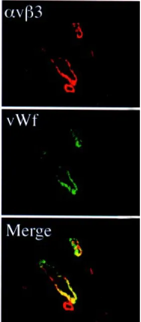
Integrin avß3 is highly expressed on the synovial endothelium in RA (50,51), as iden-tified by costaining for von Willebrand fac-tor, an endothelial cell marker (Figure 2). Studies in our laboratory have revealed that integrin avß3 is also upregulated on vascular cells in a rabbit antigen-induced arthritis (AIA) model (52). This model re-sembles human disease histopathologically, including neovascularization, synovial hy-pertrophy and subsynovial inflammatory in-filtrate, and presented a unique opportunity to demonstrate specifically the impact of integrin-based anti-angiogenic therapy in ar-thritis.
Previously, integrin antagonists derived from fragments of the fibronectin cell-bind-ing domain have been demonstrated to in-hibit the development of arthritis in a rat model of disease (53). The activity of these peptides has been attributed to their capacity to block the activity of ß1 integrins, and therefore it was proposed that decreased ar-thritis resulted from the prevention of au-toimmune/proinflammatory cell recruitment
into the synovium. Similar peptides prevent inflammation in delayed-type hypersensitiv-ity reactions through this mechanism (54). However, it is noteworthy that these linear peptide antagonists have broad specificity, and actually inhibit integrin a vß3-depen-dent adhesion at lower concentrations than, for example, integrin a5ß1-mediated adhe-sion (55). This raises the possibility that some of the observed anti-arthritic activity resulted from anti-angiogenic effects. In or-der to address these types of questions, an-tagonists with higher specificity/selectivity will be required. Since the principal target of novel cyclic av antagonists used in tumor studies appears to be endothelial cells ex-pressing avß3, these compounds may be amenable to future studies on RA (46). The success of alternative, toxin-based anti-an-giogenic strategies such as taxol (14) or de-rivatives of the fungal metabolite fumagillin (15) validates targeting endothelial cell-me-diated pathology in RA. Together, these stud-ies provide an excellent rationale to pursue development of integrin-based anti-angio-genic strategies as a viable approach to con-trol RA.
Untreated angiogenesis Blockade by excess of PEX
Angiogenic gradient
M M P-2 zymogen
Active M M P-2
PEX
Collagen type IV,V
Interstitial collagens
Proteolyzed collagen Figure 3 - An alternative
inhibi-tory mechanism for angiogen-esis. The protease M M P-2 is produced as a nonact ive zy-mogen. Binding to integrin avß3 via the noncatalytic hemopexin (PEX) domain facilitates the pro-cessing of M M P-2 to its active form, and also localizes the ac-tivity of the protease to the sur-face of invasive cells (left panel). An excess of the recombinant PEX domain blocks neovascular-ization by competitively inhibit-ing the bindinhibit-ing of unactivated M M P-2 to endothelial cells, pre-venting activation of proteolytic activity (right panel).
Inco rpo rate d the m e s in future anti-angio ge nic strate gie s
Since cellular responses to cytokines and other soluble factors occur within the gov-erning context of the ECM, modulation of these responses through the antagonism of adhesion receptors is becoming increasingly attractive as a therapy. Increased efficacy may be achieved by alternative approaches, including systemic administration of stable antagonists or possibly local gene delivery allowing regional production of anti-angio-genic proteins for extended periods. Angio-genesis is influenced by an ever-growing list of proteins, protein fragments, peptides, sug-ars and lipids, but a common element in the angiogenic response induced by any means is the local reorganization of the ECM. To-ward this end, a second strategy developed for the control of angiogenesis combines antagonism of avß3 with prevention of ECM alteration. The recent discovery that integrin
avß3 binding to MMP-2 is mediated via the hemopexin domain of MMP-2 (PEX) (56) has led to the development of recombinant PEX as a coordinate inhibitor of integrin function and MMP-2 activation in vitro. As
might be predicted from these results, PEX also blocks angiogenesis in vivo (Figure 3)
(56), suggesting that future identification of key sequences in PEX may provide addi-tional multifuncaddi-tional angiogenesis inhibi-tors.
Enhanced control of angiogenic responses may be possible by coordinately inhibiting complementary potentiating events involved in neovascularization. In the case of RA, the ongoing inflammatory component presents an obvious and important target (57), since angiogenesis and inflammation are comple-mentary events contributing to disease pro-gression. This type of logical combination therapy, in which both ongoing neovascular-ization and inflammation are targeted as dis-tinct but inter-related components, may of-fer the most effective means of treatment for rheumatoid arthritis. Recent studies focus-ing on the interactions of cells with the ECM have led to advances in both angiogenesis and inflammation research, providing valu-able tools for future investigations. Ulti-mately, an increased understanding of the pathological mechanisms in RA will permit development of new and innovative thera-pies.
Re fe re nce s
1. Pincus T (1995). The underestimated long t erm m edical and econom ic conse-quences of rheumatoid arthritis. Drugs, 50 (Suppl 1): 1-14.
2. M cCabe CJ (1997). Health economics in rheumatology. Baillieres Clinical Rheuma-tology, 11: 145-156.
3. Zvaifler NJ & Firestein GS (1994). Pannus and pannocytes. Alternative models of joint destruction in rheumatoid arthritis.
Arthritis and Rheumatism, 37: 783-789. 4. Shiozaw a S & Tokuhisa T (1992).
Contri-bution of synovial mesenchymal cells to the pathogenesis of rheumatoid arthritis.
Seminars in Arthritis and Rheumatology, 21: 267-273.
5. Kimball ES & Gross JL (1991). Angiogen-esis in pannus formation. Agents and Ac-tions, 34: 329-331.
6. Colville-Nash PR & Scott DL (1992). An-giogenesis and rheumatoid arthritis: path-ogenic and therapeutic implications. An-nals of the Rheumatic Diseases, 51: 919-925.
7. Koch AE (1998). Angiogenesis: implica-tions for rheumatoid arthritis. Arthritis and Rheumatism, 41: 951-962.
8. M atsubara T & Ziff M (1987). Inhibition of endothelial cell proliferation by gold com-pounds. Journal of Clinical Investigation, 79: 1440-1446.
9. M adhok R, Wijelath E, Smith J, Watson J, Sturrock RD & Capell HA (1991). Is the beneficial effect of sulfasalazine due to inhibition of synovial neovascularization?
Journal of Rheumatology, 18: 199-202. 10. Pot vin F, Pet it clerc E, M arceau F &
Poubelle PE (1997). M echanisms of
ac-tion of antimalarials in inflammaac-tion: in-duction of apoptosis in human endothelial cells. Journal of Immunology, 158: 1872-1879.
11. Hirata S, M atsubara T, Saura R, Tateishi H & Hirohata K (1989). Inhibition of in vitro
vascular endothelial cell proliferation and
in vivo neovascularization by low dose methotrexate. Arthritis and Rheumatism, 32: 1065-1073.
12. M atsubara T, Saura R, Hirohata K & Ziff M (1989). Inhibition of human endothelial cell proliferation in vitro and neovascular-ization in vivo by D-penicillamine. Journal of Clinical Investigation, 83: 158-167. 13. Tsujii M , Kaw ano S, Tsuji S, Saw aoka H,
14. Peacock DJ, Banquerigo M L & Brahn E (1995). A novel angiogenesis inhibitor suppresses rat adjuvant arthritis. Cellular Immunology, 160: 178-184.
15. Oliver SJ, Banquerigo M L & Brahn E (1994). Suppression of collagen-induced arthritis using an angiogenesis inhibitor, AGM 1470, and a microtubule stabilizer, Taxol. Cellular Immunology, 157: 291-299. 16. Folkman J (1995). Angiogenesis in can-cer, vascular, rheumatoid and other dis-ease. Nature M edicine, 1: 27-31. 17. M uller-Ladner U, Kriegsmann J, Gay RE &
Gay S (1995). Oncogenes in rheumatoid arthritis. Rheumatic Diseases Clinics of North America, 21: 675-690.
18. Walsh DA, Wade M , M app PI & Blake DR (1998). Focally regulated endothelial pro-liferation and cell death in human syno-vium. American Journal of Pathology, 152: 691-702.
19. Varani J, M ulligan M S & Ward PA (1994). The vascular endothelium and acute in-flammation. In: Klippel JH & Dieppe PA (Editors), Rheumatology. M osby, St Louis. 20. Gualandris A, Rusnati M , Belleri M , Nelli EE, Bast aki M , M olinari-Tosat t i M P, Bonardi F, Parolini S, Albini A, M orbidelli L, Ziche M , Corallini A, Possati L, Vacca A, Ribatti D & Presta M (1996). Basic fibro-blast grow th factor overexpression in en-dothelial cells: an autocrine mechanism for angiogenesis and angioproliferative diseases. Cell Grow th and Differentiation, 7: 147-160.
21. Barleon B, Sozzani S, Zhou D, Weich HA, M antovani A & M arme D (1996). M igra-tion of human monocytes in response to vascular endothelial grow th factor (VEGF) is mediated via the VEGF receptor flt-1.
Blood, 87: 3336-3343.
22. Eguchi K, M igita K, Nakashima M , Ida H, Terada K, Sakai M , Kaw akami A, Aoyagi T, Ishimaru T & Nagataki S (1992). Fibroblast grow th factors released by w ounded en-dothelial cells stimulate proliferation of synovial cells. Rheumatology, 19: 1925-1932.
23. M ignatti P & Rifkin DB (1996). Plasmino-gen activators and matrix metalloprotein-ases in angiogenesis. Enzymes and Pro-teins, 49: 117-137.
24. Ziats NP & Anderson JM (1993). Human vascular endothelial cell attachment and grow th inhibition by type V collagen. Jour-nal of Vascular Surgery, 17: 710-718. 25. Boudreau N, Andrew s C, Srebrow A,
Ravanpay A & Cheresh DA (1997). Regu-lation of the angiogenic phenotype by Hox D3. Journal of Cell Biology, 139: 257-264. 26. M eredith Jr JE, Fazeli B & Schw artz M A
(1993). The extracellular matrix as a cell survival factor. M olecular Biology of the Cell, 4: 953-961.
27. Cornelius LA, Nehring LC, Roby JD, Parks WC & Welgus HG (1995). Human dermal microvascular endothelial cells produce matrix metalloproteinases in response to angiogenic factors and migration. Journal of Investigative Dermatology, 105: 170-176.
28. Dvorak HF, Brow n LF, Det m ar M & Dvorak AM (1995). Vascular permeability factor/vascular endothelial grow th factor, microvascular hyperpermeability, and an-giogenesis. American Journal of Pathol-ogy, 146: 1029-1039.
29. Reininger AJ, Heinzmann U, Reininger CB, Friedrich P & Wurzinger LJ (1994). Flow mediated fibrin thrombus formation in an endothelium-lined model of arterial branching. Thrombosis Research, 74: 629-641.
30. Postigo AA, Garcia-Vicuna R, Laffon A & Sanchez-M adrid F (1993). The role of ad-hesion molecules in the pathogenesis of rheumatoid arthritis. Autoimmunity, 16: 69-76.
31. Busso N, Peclat V, Van Ness K, Kolodzi-esczyk E, Degen J, Bugge T & So A (1998). Exacerbation of antigen-induced arthritis in urokinase-deficient mice. Journal of Clinical Investigation, 102: 41-50. 32. Leavesley DI, Schw artz M A, Rosenfeld M
& Cheresh DA (1990). Integrin ß1- and ß3-mediated endothelial cell migration is trig-gered through distinct signaling mecha-nisms. Journal of Cell Biology, 121: 163-170.
33. Lampugnani M G, Giorgi M , Gaboli M , Dejana E & M archisio PC (1990). Endo-thelial cell motility, integrin receptor clus-tering, and microfilament organization are inhibited by agents that increase intracel-lular cAM P. Laboratory Investigation, 63: 521-531.
34. Bull DA, Seftor EA, Hendrix M J, Larson DF, Hunter GC & Putnam CW (1993). Pu-tative vascular endothelial cell chemotac-tic factors: comparison in a standardized migration assay. Journal of Surgical Re-search, 55: 473-479.
35. Saiki I, M urata J, M akabe T, Nishi N, Tokura S & Azuma I (1990). Inhibition of tumor angiogenesis by a synthetic cell-adhesive polypeptide containing the Arg-Gly-Asp (RGD) sequence of fibronectin, poly(RGD). Japanese Journal of Cancer Research, 81: 668-675.
36. Aplin AE, How e A, Alahari SK & Juliano RL (1998). Signal transduction and signal modulation by cell adhesion receptors:
the role of integrins, cadherins, immuno-globulin-cell adhesion m olecules and selectins. Pharmacological Review s, 50: 197-263.
37. Shimizu Y & Shaw S (1991). Lymphocyte interactions w ith extracellular m atrix.
FASEBJournal, 5: 2292-2299.
38. Gillis S, Furie BC & Furie B (1997). Interac-tions of neutrophils and coagulation pro-teins. Seminars in Hematology, 34: 336-342.
39. Simms H & D’Amica R (1994). M atrix pro-tein regulation of PM N oxidative metabo-lism during ischemia. American Journal of Physiology, 266: C637-C647.
40. Eliceiri BP, Strömblad S, Klemke R & Cheresh DA (1998). Integrin requirement for sustained M AP kinase activity during angiogenesis. Journal of Cell Biology, 140: 1255-1263.
41. Short SM , Talbott GA & Juliano RL (1998). Integrin-mediated signaling events in hu-man endothelial cells. M olecular Biology of the Cell, 9: 1969-1980.
42. Brooks PC, Clark RA & Cheresh DA (1994). Requirement of vascular integrin
avß3 for angiogenesis. Science, 264: 569-571.
43. Varner JA, Brooks PC & Cheresh DA (1995). The integrin avß3: Angiogenesis and apoptosis. Cell Adhesion and Com-munication, 3: 367-374.
44. Brooks PC, Stromblad S, Sanders LC, von Schalscha T, Aimes RT, Stetler-Stevenson WG, Quigley JP & Cheresh DA (1996). Localization of matrix metalloproteinase M M P-2 to the surface of invasive cells by interaction w ith integrin avß3. Cell, 85: 1-20.
45. Brooks PC, M ontgomery AM P, Rosenfeld M , Reisfeld RA, Hu T, Klier G & Cheresh DA (1994). Integrin avß3 antagonists pro-mote tumor regression by inducing apop-tosis of angiogenic blood vessels. Cell, 79: 1157-1164.
46. Strömblad S, Becker JC, Yebra M , Brooks PC & Cheresh DA (1996). Suppression of p53 and p21WAF1/CIP1 expression by vascular cell integrin avß3 during angio-genesis in vivo. Journal of Clinical Investi-gation, 98: 426-433.
47. Scat ena M , Alm eida M L, Faust o N, Nicosia RE & Giachelli CM (1998). NF-kB mediates avß3 integrin-induced endothe-lial cell survival. Journal of Cell Biology, 141: 1083-1093.
49. Brooks PC, St röm blad S, Klem ke R, Visscher D, Sarkar FH & Cheresh DA (1995). Anti-integrin avß3 blocks human breast cancer grow th and angiogenesis in human skin. JournalofClinical Investiga-tion, 96: 1815-1822.
50. Johnson BA, Haines GK, Harlow LA & Kock AE (1993). Adhesion molecule ex-pression in human synovial tissue. Arthri-tis and RheumaArthri-tism, 36: 137-146. 51. Nikkari L, Haapasalmi K, Aho H, Torvinen
A, Sheppard D, Larjava H & Heino J (1995). Localization of the av subfamily of integrins and their putative ligands in syn-ovial lining cell layer. Journal of Rheuma-tology, 22: 16-23.
52. Storgard CM , Stupack DG, Jonczyk A, Goodm an SL, Fox RI & Cheresh DA (1999). Decreased angiogenesis and ar-thritic disease in rabbits treated w ith an
avß3 antagonist. Journal of Clinical Inves-tigation, 103: 1-8.
53. Wahl SM , Allen JB, Hines KL, Imamichi T, Wahl AM , Furcht LT & M cCarthy JB (1994). Synthetic fibronectin peptides suppress arthritis in rats by interrupting
leukocyte adhesion and recruitment. Jour-nal of Clinical Investigation, 94: 655-662. 54. Hershkoviz R, Greenspoon N, M ekori YA,
Hadari R, Alon R, Kapustina G & Lider O (1994). Inhibition of CD4+ T lymphocyte binding to fibronectin and immune-cell accumulation in inflammatory sites by non-peptidic mimetics of Arg-Gly-Asp.
Clinical and Experimental Immunology, 95: 270-276.
55. Pfaff M , Tangemann K, M uller B, Gurrath M , M uller G, Kessler H, Timpl R & Engel J (1994). Selective recognition of cyclic RGD peptides of NM R defined conforma-tion by aIIßß3, avß3, and a5ß1 integrins.
Journal of Biological Chemistry, 2692: 20233-20238.
56. Brooks PC, Silletti S, von Schalscha TL, Friedlander M & Cheresh DA (1998). Dis-ruption of angiogenesis by PEX, a non-catalytic metalloproteinase fragment w ith integrin binding activity. Cell, 92: 391-400. 57. Paleolog E (1997). Target effector role of vascular endothelium in the inflammatory response: insights from the clinical trial of anti-TNFalpha antibody in rheumatoid
ar-thritis. M olecular Pathology, 50: 225-233. 58. Sunderkotter C, Steinbrink K, Goebeler M , Bhardw aj R & Sorg C (1994). M acroph-ages and angiogenesis. Journal of Leuko-cyte Biology, 55: 410-422.
59. Jackson JR, M inton JA, Ho M L, Wei N & Winkler JD (1997). Expression of vascular endothelial grow th factor in synovial fibro-blasts is induced by hypoxia and interleu-kin 1 beta. Journal of Rheumatology, 24: 1253-1259.
60. Ruegg C, Yilmaz A, Bieler G, Bamat J, Chaubert P & Lejeune FJ (1998). Evidence for the involvement of endothelial cell integrin avß3 in the disruption of the tu-mor vasculature induced by TNF and
IFN-g. Nature M edicine, 4: 408-414. 61. Koch AE, Halloran M M , Hosaka S, Shah