Inês Viana de Paula Monteiro
A importância da abordagem
multidisciplinar nas anomalias
vasculares congénitas / Congenital
Vascular Anomalies: The Importance of a
Multidisciplinary Approach
Mestrado Integrado em Medicina
Área: Pediatria Tipologia: Monografia
Trabalho efetuado sob a Orientação de: Doutor Tiago Henriques Coelho
Trabalho organizado de acordo com as normas da revista: Porto Biomedical Journal
multidisciplinar nas anomalias
vasculares congénitas / Congenital
Vascular Anomalies: The Importance
of a Multidisciplinary Approach
Ao Doutor Tiago Henriques Coelho, que orientou este projecto, pela disponibilidade, prontidão, paciência, compreensão e colaboração, e pelo contributo para a minha formação profissional.
À minha família e amigos e, em particular, ao meu pai, pelas orientações e encorajamento, na busca da excelência.
1 Artigo Revisão
A importância da abordagem multidisciplinar nas anomalias vasculares congénitas Inês Monteiro
Abstract
As anomalias vasculares congénitas (AVaC´s) são patologias prevalentes, com uma complexa abordagem diagnóstica e terapêutica. Um dos principais obstáculos para os avanços do conhecimento científico nesta área advém da inconsistência na utilização de uma nomenclatura estandardizada. O progressivo conhecimento etiopatogénico destas patologias, o novo sistema de classificação, o surgimento de novas técnicas diagnósticas e de tratamento e a incorporação de conhecimentos de outras especialidades, tem salientado a importância de uma abordagem multidisciplinar, específica e adequada. Esta revisão tem como objectivo a análise bibliográfica da importância e do impacto da abordagem multidisciplinar na identificação da patologia, na planificação de uma intervenção e acompanhamento terapêutico e no outcome. Apesar dos poucos estudos comparativos relativos às diferentes abordagens, a estratégia multidisciplinar tem-se revelado vantajosa no diagnóstico, tratamento e seguimento dos doentes com anomalias vasculares, potenciando um conhecimento holístico das diferentes anomalias.
Definição
As anomalias vasculares congénitas (AVaC´s) são um grupo heterogéneo de lesões com uma prevalência de cerca de 4,5% (1). São um grupo de patologias endoteliais resultantes de erros (difusos ou localizados), da embriogénese vascular, as quais se podem manifestar como tumores ou malformações (2) (3). Ocorrem numa fase precoce da gestação, normalmente, entre a 3ª e a 7ª semanas de desenvolvimento embrionário, podendo afectar o sistema arterial, capilar, venoso e linfático (4). Estas anomalias englobam várias alterações possíveis, que ocorrem de forma isolada ou combinada: i) ausência de vasos que, normalmente, se encontram presentes; ii) presença de vasos anómalos; iii) presença de vasos de morfologia e tamanho anormal, com alterações nas suas paredes. (5) (6) Palavras-chave: Multidisciplinar, Abordagem; Classificação; Especialidades; AVaC´s Abreviações: AVaC´s – Anomalias Vasculares Congénitas
2 As anomalias vasculares simples são patologias comuns nas crianças e estima-se que cerca de 1/3 dos recém-nascidos nascem com uma “marca”. (7) Dado a sua natureza, têm um largo espectro de apresentações, mas são geralmente diagnosticadas durante a infância e adolescência, uma grande imprevisibilidade de progressão e resposta terapêutica, bem como elevada recorrência e morbilidade quer psicológica, quer algumas vezes, funcional. (5) (3) (8) (9) (10) (11) (6) .
Etiopatogenia
A etiologia das AVaC´s permanece por explicar mas recentemente, os avanços na área da genética permitiram a identificação de inúmeras mutações genéticas quer germinativas quer somáticas. (2) (12) Alguns estudos sugerem uma possível ligação etiológica à diminuição da capacidade de regular o processo responsável pela sinalização, maturação, adesão e apoptose das células vasculares. (2)
Classificação
A classificação das AVaC´s tem sofrido inúmeras alterações, ao longo dos anos. (13) As AVaC´s englobam um grande espectro de lesões, o que dificulta a sua sistematização. (3) (1) (14) O uso de uma inadequada nomenclatura continua a ser a causa de diagnósticos incorrectos e de falsos negativos. (14) Inicialmente, as classificações baseavam-se na aparência patológica das lesões. (13) Foi apenas em 1982, com Mulliken e Glowacki, que foi criado um sistema de classificação binário que permitiu uma melhor categorização destas patologias. (13) (14) Este sistema baseava-se na clínica, na radiologia, no comportamento biológico e nas características endoteliais e dividia as AVaC´s em duas grandes categorias: Tumores vasculares (Hemangiomas) e Malformações vasculares. (13) (15) (16) Em 1993, Jackson e colaboradores surge com uma classificação radiológica que tinha por base o fluxo e a hemodinâmica das anomalias, subdividindo-as em alto e baixo fluxo. (17) A classificação binária de Mulliken e Glowacki foi a base estrutural adotada pela International Society for the Study of Vacular Anomalies (ISSVA). Esta nova abordagem dividia as anomalias em duas categorias principais, nomeadamente tumores vasculares ou vasoproliferativos e malformações vasculares, sendo que a principal diferença assentava na presença ou ausência de um aumento do turnover das células endoteliais, identificado histologicamente pela presença ou ausência de mitoses. Desta forma, os tumores vasoproliferativos apresentam um aumento do turnover das células enquanto que as malformações vasculares não apresentam turnover celular, sendo consideradas anormalidades estruturais cujo crescimento acompanha o crescimento da criança. (14) (18) Este sistema de classificação é dinâmico e tem sofrido várias atualizações, nomeadamente pela inserção de novos tumores, e a subclassificação das malformações vasculares em simples e combinadas. (13) (12) (14) (17) A classificação do ISSVA, permitiu avanços consideráveis, nomeadamente nos estudos genéticos, na patofisiologia, no diagnóstico, no tratamento e no prognóstico. (16)
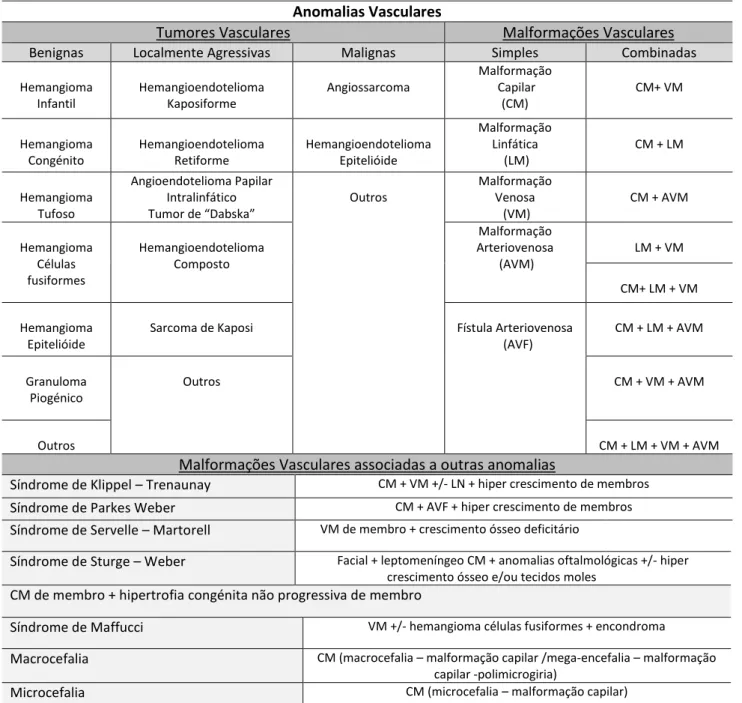
3 Em 2014, a classificação preconizada pela ISSVA, sofreu algumas alterações, nomeadamente, na subclassificação dos Tumores e na associação com outras anomalias ou síndromes. (17) (Tabela 1)
A utilização de uma classificação universalmente aceite como a da ISSVA, permite uma constante evolução e adaptação ao conhecimento científico, nomeadamente aos achados genéticos e histológicos que vão sendo descobertos. (12)
Tabela 1 Classificação das Anomalias Vasculares congénitas (Adaptada da ISSVA (a))
Anomalias Vasculares
Tumores Vasculares Malformações Vasculares
Benignas Localmente Agressivas Malignas Simples Combinadas
Hemangioma Infantil Hemangioendotelioma Kaposiforme Angiossarcoma Malformação Capilar (CM) CM+ VM Hemangioma Congénito Hemangioendotelioma Retiforme Hemangioendotelioma Epitelióide Malformação Linfática (LM) CM + LM Hemangioma Tufoso Angioendotelioma Papilar Intralinfático Tumor de “Dabska” Outros Malformação Venosa (VM) CM + AVM Hemangioma Células fusiformes Hemangioendotelioma Composto Malformação Arteriovenosa (AVM) LM + VM CM+ LM + VM Hemangioma Epitelióide
Sarcoma de Kaposi Fístula Arteriovenosa (AVF) CM + LM + AVM Granuloma Piogénico Outros CM + VM + AVM Outros CM + LM + VM + AVM
Malformações Vasculares associadas a outras anomalias
Síndrome de Klippel – Trenaunay CM + VM +/- LN + hiper crescimento de membros
Síndrome de Parkes Weber CM + AVF + hiper crescimento de membros
Síndrome de Servelle – Martorell VM de membro + crescimento ósseo deficitário
Síndrome de Sturge – Weber Facial + leptomeníngeo CM + anomalias oftalmológicas +/- hiper crescimento ósseo e/ou tecidos moles
CM de membro + hipertrofia congénita não progressiva de membro
Síndrome de Maffucci VM +/- hemangioma células fusiformes + encondroma
Macrocefalia CM (macrocefalia – malformação capilar /mega-encefalia – malformação capilar -polimicrogiria)
Microcefalia CM (microcefalia – malformação capilar)
Síndrome de CLOVES LM + VM + CM +/- AVM + hiper crescimento lipomatoso
Síndrome de Proteus Cm, VM e/ou LM + hiper crescimento somático assimétrico
4
Paradigma do Hemangioma
Apesar da etiologia, progressão e tratamento, os termos hemangiomas ou angiomas são utilizados de modo frequente para se referir a qualquer tipo de AVaC, quer tumores quer malformações. (3) (12) (14) (19) Segundo alguns estudos, mais de 70% dos autores utilizam a palavra hemangioma de forma indiscriminada para descrever uma anomalia vascular. Este erro pode-se repercutir num tratamento desadequado em cerca de 20% dos casos. (6) Dentro das AVaC´s, o erro de nomenclatura estende-se por exemplo às malformações vasculares, que não são patologias hipercelulares, mas sim compostas de endotélio normal, possuindo um grande crescimento na infância, com tendência à expansão na puberdade e não à sua regressão, contrariamente ao hemangioma infantil. (3) A adopção de uma nomenclatura correta é fulcral para a diferenciação e categorização das diferentes patologias.
Abordagem isolada - Limitações
Apesar dos esforços, nos últimos anos, as AVaC´s constituem patologias de difícil diagnóstico e terapêuticas. (8) (10) A adopção de uma nomenclatura e abordagem inadequada tem resultado em altas taxas de insucesso com consequências desastrosas, quer ao nível de terapêuticas desajustadas, quer ao nível da morbilidade de tratamentos desnecessários ou invasivos. (3) (10)
Felizmente, nas últimas décadas, a abordagem às AVaC´s têm sofrido progressos consideráveis. Esta mudança de paradigma só foi possível após o surgimento de uma nova classificação, de técnicas diagnósticas adequadas e de novas modalidades terapêuticas. (3) (18) O surgimento de novas técnicas diagnósticas, sobretudo não invasivas, veio potenciar o conhecimento destas patologias, por forma a realizar um diagnóstico mais preciso. (3) Dada a raridade de algumas patologias, a natureza heterogénea e a complexidade das AVaC´s, é fulcral que a abordagem englobe um conjunto de profissionais de várias especialidades, capacitados de conhecimentos e visões de diferentes áreas, por forma a alcançar um conhecimento holístico da patologia e individualizado do doente. (2) (16) (18) (19)
Abordagem multidisciplinar
Na realidade, algumas das AVaC´s, nomeadamente os hemangiomas infantis assintomáticos, possuem um diagnóstico e tratamento simples. Porém, outras patologias, nomeadamente as malformações, necessitam do contributo de diferentes especialidades para a sua avaliação e planificação de tratamento. (5) (20) (21) Alguns centros multidisciplinares referem que cerca de 47% dos casos que lhes são referenciados possuem um diagnóstico inicial errado, sendo que as malformações (54,4%) têm maior probabilidade de erro, quando comparadas com os tumores (29,6%). (6) (20) (22) Noutro estudo comparativo, cerca de 42% dos casos referenciados para uma equipa multidisciplinar, possuíam um diagnóstico inicial incorreto, tendo os tumores uma taxa de erro diagnóstico inicial de 11% e as malformações de 68%. (5)
5 Salienta-se, ainda, que os hemangiomas foram corretamente identificados em mais de 90% dos casos. Após referenciação para estes centros, a maior parte dos casos acaba por ser correctamente diagnosticada após a revisão da história clínica e do exame físico. (22) Alguns centros têm-se debruçado no estabelecimento de guiões orientadores para a referenciação dos casos suspeitos de AVaC´s, por defenderem que, nos centros especializados, o diagnóstico é mais preciso, particularmente em tumores raros, havendo melhor acesso ao conhecimento e terapêutica mais actualizados. (22)
Uma equipa multidisciplinar vai permitir reunir o conhecimento relativo à patologia, às estratégias terapêuticas e aos resultados das mesmas, permitindo um melhor acompanhamento. (5) Esta abordagem permite o acesso do doente a um conjunto de serviços, nomeadamente, a terapias médicas, imagiologia, intervenção e cirurgia. (19) Uma equipa coordenada e multidisciplinar experiente, que tenha contacto com o maior número destas patologias, das suas diferentes manifestações e evolução, acaba por reunir em si, como grupo, o conhecimento necessário para tratar os casos mais complexos, com uma elevada taxa de sucesso. (5) (23)
A abordagem multidisciplinar deve iniciar-se logo no diagnóstico, devendo permitir que o diagnóstico seja mais célere e adequado, e poupando a criança a exames desnecessários e invasivos e a procedimentos terapêuticos desnecessários. (19) De facto, como mencionado anteriormente, as AVaC´s constituem patologias de difícil reconhecimento e classificação. (8) Esta complexidade diagnóstica advém, muitas vezes, da coexistência, na mesma ou em diferentes regiões corporais, de mais do que uma anomalia vascular, ou a mesma anomalia, em diferentes formas e estádios de desenvolvimento. (3) Deste modo, só uma compreensão holística dos diferentes tipos de anomalias permite a sua correta caracterização. Idealmente, a avaliação clínica do paciente deve ser realizada, individualmente, por cada interveniente do processo e as conclusões individuais devem ser partilhadas e discutidas, posteriormente, para uma melhor planificação da estratégia terapêutica. (2) (8) A existência de um responsável pela coordenação entre os elementos da equipa multidisciplinar é fundamental, por forma a avaliar o processo, as suas necessidades e a ligação com o doente e a sua família. (2) Uma história clínica completa e detalhada, bem como um exame físico minucioso são fundamentais. Na maioria das vezes é também necessário o recurso a exames complementares, idealmente o menos invasivos possível. (2) A escolha da melhor combinação de exames diagnósticos só é possível após uma recolha eficiente e completa dos achados clínicos prévios, realizados pelas diferentes áreas envolvidas. (3) Desde já, o contributo dos especialistas das áreas da Pediatria e da Radiologia torna-se indispensável. (2)
Com o aumento das opções terapêuticas, a comunicação entre os elementos da equipa multidisciplinar torna-se ainda mais importante. (24) (25) A abordagem multidisciplinar decide, inicialmente, sobre a necessidade ou não de intervenção médica. (3) Caso essa se venha a comprovar, a equipa estabelece um plano de tratamento individualizado, que facilita a integração e discussão das melhores opções terapêuticas, sejam elas farmacológicas, de intervenção ou cirúrgicas. (3) (2) (5) (26)
6 Para o planeamento terapêutico, é necessário reunir a informação relativa à natureza da anomalia, à região anatómica envolvida, à profundidade, dimensão e ritmo de crescimento da lesão, ao grau de envolvimento do tecido circundante e à idade do paciente. Desta forma, é possível estabelecer uma estratégia terapêutica ao longo do tempo, com eventual associação de diferentes terapias. (3) (27)
Outra das vantagens das equipas multidisciplinares diz respeito às possíveis complicações da doença e/ou do tratamento, cujo contributo das diversas especialidades, para a sua previsão e gestão é crucial. As complicações mais comumente encontradas nas malformações vasculares, são a dor, a anemia, a trombose local, a hipertensão, as coagulopatias, a sepsis e a celulite. Outros estudos referem ainda morbilidades sistémicas de maior gravidade, nomeadamente insuficiência cardíaca congestiva, coagulação disseminada intravascular, embolia pulmonar e a trombocitopenia. (6) (20) Em contraste com os hemangiomas, as malformações vasculares não têm tendência para regredir e, como tal, o tecido que envolve as mesmas pode sofrer processos isquémicos e necróticos, por hipertensão venosa ou dermatite por estase. (7) Além das complicações inerentes às patologias, é necessário que os intervenientes conheçam os potenciais riscos das próprias intervenções. (7) Um plano terapêutico deve prever e antecipar as complicações a curto e a longo prazo, como eventuais deformidades residuais. (20)
No que diz respeito ao seguimento, também é necessário uma abordagem multidisciplinar, em relação à recorrência e à morbilidade, nomeadamente das malformações arteriovenosas. (7) (20) Em relação às comorbilidades associadas, deve-se promover atitudes e medidas profiláticas adequadas, por forma a minimizar o risco do surgimento de complicações. (27) Por exemplo, em relação às malformações das extremidades, as atitudes profiláticas podem passar pela compressão e profilaxia antibiótica. Esse seguimento multidisciplinar deve ainda identificar atempadamente complicações, como a trombose venosa e o embolismo pulmonar. (27)
Especialidades Médicas envolvidas
As malformações vasculares englobam uma variedade de diagnósticos individuais que necessitam do contributo de diferentes especialidades médicas. (11)
Pediatria
Os Hematologistas / Oncologistas pediátricos desempenham um papel fulcral, quer no diagnóstico, nomeadamente, na análise da necessidade da realização de biópsias, quer no tratamento, nomeadamente, na seleção e monitorização de fármacos, bem como em todo o processo de coordenação no que diz respeito à eventual necessidade de intervenções médicas e cirúrgicas, para além das inicialmente planeadas. (11) A Hematologia é adicionalmente necessária, devido à alta prevalência de coagulopatias e ao risco aumentado de eventos trombo-embólicos e hemorrágicos. (2) (11)
7 Radiologia
O contributo da imagiologia é fundamental para o planeamento e tratamento das anomalias vasculares congénitas. Tem-se assistido à diminuição de intervenções diagnósticas invasivas. (1) (25) Desde logo, os avanços da tecnologia imagiológica, quer diagnóstica quer de intervenção, permitiram um importante contributo a nível do diagnóstico, do planeamento e da terapêutica. (26) (25) Numa perspectiva diagnóstica, a radiologia pode avaliar o tipo de vascularização e o respetivo fluxo por ecodopler, a dinâmica vascular e o envolvimento dos tecidos periféricos por ressonância ou angio-ressonância magnética (RM) ou, menos frequentemente por angio-tomografia computorizada. (1) (26) Ao nível de tratamento, a escleroterapia, a radiologia de intervenção através da escleroterapia, da embolização ou do LASER endovascular, posicionam-se na primeira linha de tratamento de inúmeras AVaC´s. (24) (25) Com base em diferentes métodos de imagem é ainda possível monitorizar a eficácia de terapêuticas farmacológicas, percutâneas e/ou cirúrgicas. (26)
Cirurgia
No campo da cirurgia, nomeadamente a cirurgia pediátrica, o seu contributo vai desde a excisão do defeito vascular em si, até à correcção de alguns efeitos tardios das AVaC´s, como as distorções anatómicas. (8) (23) (26) (28) A Ortopedia é também necessária, no caso do envolvimento muscular e ósseo que algumas AVaC´s apresentam. (20) (21) Perante anomalias combinadas, como a Síndrome de Klippel-Trenaunay e a Síndrome de Parkes Weber, o contributo da ortopedia pode ser necessário, uma vez que estas patologias estão associadas a um sobre-crescimento e assimetria dos membros. Nestes casos, terapias conservadoras ou de ressecção e amputação podem ter que ser contempladas. (28) (29) Dado que grande parte dos hemangiomas e malformações vasculares ocorrem na região da cabeça e do pescoço, o contributo dos otorrinolaringologistas e oftalmologistas pode ser necessário para prevenir, por exemplo, a ambliopia e o estigmatismo. (30)
Dermatologia
Os dermatologistas são, muitas vezes, a especialidade para a qual os doentes com AVaC´s são referenciados. (1) (31) Desta forma, assume um papel importante no diagnóstico e na identificação de associações ou síndromes, para que possam ser alvo de referenciação posterior para as equipas multidisciplinares. (1)
Anatomia Patológica
No campo da Anatomia Patológica, as biópsias podem ter um contributo definitivo, quando a sua análise é realizada por um especialista experiente e com conhecimento da classificação actual da ISSVA. (5)
8 Psicologia
É importante não negligenciar a componente psicológica destas anomalias. Doentes com AVaC´s em áreas como a face, são confrontados diariamente com uma imagem corporal desfigurada. Alguns estudos referem que doentes com AVaC´s possuem menor qualidade de vida, quando comparados com indivíduos saudáveis, apresentando baixa auto-estima, ansiedade, depressão, angústia, medo e insatisfação perante a sua aparência. (19) (24) (32) A identificação e a gestão atempadas deste sentimento, vai permitir promover o empowerment do doente, em vista à obtenção do melhor outcome possível. (24) A importância desta vertente psíquica é corroborada pela existência, de grupos de ajuda presenciais e online, associados a diferentes patologias, por forma a apoiar a estigmatização que os doentes vivenciam, perante determinada patologia. (24)
A abordagem multidisciplinar pode mesmo ter influência a nível prenatal, com recurso aos conhecimentos da obstetrícia, da radiologia e da cirurgia pediátrica. As anomalias vasculares podem ser diagnosticadas, a partir do segundo e terceiro trimestres. (27) (33) Os hemangiomas congénitos, por exemplo, podem ser detetados a partir da 12º semana gestacional, através de ecografias e RM. Normalmente, trata-se de lesões que não se associam a complicações durante o parto; porém, em alguns casos, estão descritas complicações cardíacas e pulmonares, em que é necessário intervir de forma multidisciplinar. (27) (33) O diagnóstico pré-natal incorreto de algumas malformações linfáticas, foi associado ao aumento de morbilidade e ao aumento do número de cirurgias desnecessárias. (33) Lesões linfáticas císticas, cervicais e linguais e lesões que comprimam a via respiratória podem e devem ser diagnosticadas, in utero, durante o segundo trimestre, sendo que o diagnóstico prenatal vai influenciar o tipo, a data e o local do parto e as possíveis intervenções necessárias para corrigir as condições que afetam a viabilidade do recém-nascido. (27)
A maior parte destas patologias surge de forma esporádica, enquanto outras podem surgir associadas a síndromes. Em alguns casos, a AVaC é a causa principal da morbilidade; porém, quando em associação sindrómica, esta pode ser a causa patológica principal. (13) Um exemplo da necessidade da multidisciplinariedade é a Síndrome de PHACE, que consiste na associação de malformações cerebrais, hemangiomas, arteriais, cardíacas, oftalmológicas ou endócrinas, cuja gestão vai envolver a intervenção adicional da cardiologia, da oftalmologia e da neurologia. Outro síndrome associado é o Síndrome de Sturge-Weber, que se caracteriza por malformações capilares da pele, oculares e cerebrais, e que pode potenciar convulsões, atraso mental, glaucoma e alterações dermatológicas. Perante tais alterações, é crucial o contributo adicional de áreas como a oftalmologia e a neurologia. (1)
9
Conclusão
São poucos os estudos comparativos entre as abordagens realizadas de forma isolada e através de equipas multidisciplinares das AVaC´s. Na maior parte dos estudos que comparam estas abordagens, o grau de maior complexidade referenciados às equipas multidisciplinares é maior, o que dificulta a comparação. O surgimento de uma nomenclatura universal é fundamental. Novas técnicas diagnósticas e terapêuticas e o contributo das diversas especialidades, vieram potenciar o conhecimento da etiopatogénese das diferentes AVaC´s. A estratégia de abordagem multidisciplinar tem vindo a alcançar resultados significativamente superiores, sobretudo a nível do diagnóstico e terapêutico. Dada a natureza imprevisível destas patologias, as equipas multidisciplinares permitiram a partilha de conhecimento científico, possibilitando quer a discussão diagnóstica, quer das opções terapêuticas mais apropriadas, quer do acompanhamento holístico do doente, por forma à obtenção do melhor resultado possível. O futuro advinha-se promissor devido aos avanços tecnológicos quer no campo diagnóstico, quer no campo terapêutico, sendo que o progressivo conhecimento destas anomalias vai permitir: a intervenção cada vez mais precoce e adequada nas patologias que já se conhecem, bem como de novas entidades que vão surgindo à medida que o conhecimento vai aumentando.
10
Referências
1. Nguyen, TA, et al. Imaging Pediatric Vascular Lesions. J Clin Aesthet Dermatol. 2015, Vols. 8(12): 27-41.
2. Markovic, JN & Shortell, CEK. Multidisciplinary treatment of extremity arteriovenous malformations. JOURNAL OF VASCULAR SURGERY: Venous and Lymphatic Disorders. 2015.
3. Lee, BB. Critical issues in Management of Congenital Vascular Malformation . Annals of Vascular Surgery lnc. April de 2004, pp. 380-392.
4. PascuaI-Castroviejo, I& PascuaI-Pascual, SI. Congenital Vascular Malformations in Childhood. Seminars in Pediatric Neurology. 2002, Vols. Vol 9, No 4 pp 254-273.
5. Mattila, KA, et al. An interdisciplinary specialist team leads to improved diagnostics and treatment for paediatric patients with vascular anomalies . Acta Pædiatrica. 2015, Vols. 104, pp. 1109–1116.
6. Greene, AK Vascular Anomalies: Current Overview of the Field. Clin Plastic Surg. 2011, Vols. 38 pp 1-5.
7. Kim, J, et al. Morbidity and healthcare costs of vascular anomalies: a national study. Pediatr Surg Int. 2016.
8. Lee, BB. New Approaches to the Treament of Congenital Vascular Malformations (CVMs) – A Single Centre Experience 2004. Eur J Vasc Endovasc Surg. 2005, Vol. 30.
9. Lee, BB. New Classification of congenital vascular malformations (CVMs) . VascularMedicine. 2015, Vols. 3 pp 1-5.
10. Lee, BB, et al. Management of arteriovenous malformations: A multidisciplinary approach . Journal Of Vascular Surgery. 2004, Vols. 39, number 3.
11. Margolin, JF, Soni, HM & Pimpalwar, S. Medical therapy for Pediatric Vascular Anomalies. Seminars in Plastic Surgery. 2014, Vols. 28 pp79-86.
12. Wassef, M et al. Vascular Anomalies Classification: Recommendations From the International Society for the study of Vascular Anomalies. PEDIATRICS. 1, 2015, Vol. 136.
13. Nosher, JL, et al. Vascular anomalies: A pictorial review of nomenclature, diagnosis and treatment. World Journal Radiology. 2014, Vols. 28; 6(9): 677-692.
14. Lowe, LH, et al. Vascular malformations: Classification and Terminology the Radiologist Needs to Know . Semin Roentgenol. 2012, Vols. 47(2) pp106-17.
11 15. Ballah, D, et al. Vascular Anomalies: What They Are, How to Diagnose Them, and How to Treat Them . Curr Probl Diagn Radiol. 2011, Vols. 40 pp233-247.
16. Pozo, JD, Gómez-Tellado, MG e López-Gutiérrezc, JC. Vascular malformations in Childhood. Actas Dermosifiliogr. 2012, Vols. 103(8) pp 661---678.
17. Sierre, S, Teplisky, D & Lipsich, J. Vascular malformations: an update on imaging and management . Arch Argent Pediatr. 2016, Vols. 114(2) pp 167-176.
18. Maksimovic, Z, et al. Second Look at congenital vascular malformations: current classification, diagnostic and treatment principles . International Angiology. 2013, Vol. 32.
19. Donnelly, LF, Adams, DM & Bisset III, GS. Vascular malformations: A Practical Approach in a Multidisciplinary Clinic. Am J Roentgenol. 2000, Vols. 174(3) pp 597-608.
20. Greene, AK. Current Concepts of Vascular Anomalies. The Journal of Craniofacial Surgery. 2012, Vols. 23 pp 220-224.
21. Brude, E, et al. Vascular lesions of bone in children, adolescents. Virchows Arch. 2009, Vols. 454 pp 161–179.
22. Greene, AK, et al. Vascular anomalies in 5621 patients: guidelines for referral. Journal of Pediatric Surgery. 2011, Vols. 46 pp 1784-1789.
23. Lidsky, ME, et al. 24. Analysis of the treatment of congenital vascular malformations using a multidisciplinary approach. JOURNAL OF VASCULAR SURGERY. 2012, Vols. 56 pp 1355-62.
24. Kenny, SA, et al. Psychological comorbidities and compliance to interventional treamente of patients with cutaneous vascular malformations . Interventional Neuroradiology. 2016.
25. MULLIGAN, PR, et al. Vascular anomalies classification, imaging characteristics and implications for interventional radiology tretament approaches. Br J Radiol. 2014.
26. Ernemanna, U, et al. Current concepts in the Classification, diagnosis and treatment of vascular anomalies . European Journal of Radiology. 2010, Vols. 75 pp 2-11.
27. Azizkhan, RG. Complex vascular anomalies . Pediatr Surg Int. 2013, Vols. 29 pp 1023-1038.
28. Marler, JJ & Mulliken, JB. Current management of hemangiomas and vascular malformations . Clinics in Plastic Surgery. 2005, Vols. 32 pp 99-116.
29. Foley, LS & Kulungowski, AM Vascular Anomalies in Pediatrics. Advances in Pediatrics. 2015, Vol. 62 pp.227.255.
30. Buckmiller, LM. Update on hemangiomas and vascular malformations . Curr Opin Otolaryngol Head Neck Surg. 2004, Vols. 12 pp 476–487.
12 31. Miguel, R, López-Gutierrez,JC & Boixeda, P. Arteriovenous Malformations: A Diagnostic and Therapeutic Challenge . Actas Dermosifiliogr. 2014, Vols. 105(4) pp 347-358.
32. Fahrni, JO, et al. Quality of life in patients with congenital vascular malformations . Journal of VascularSurgery: venous and Lymphatic Disorders. 2014, Vols. 2 pp 46-51.
33. Marler, JJ, et al. Prenatal Diagnosis of Vascular anomalies . J Pediatr Surg. 2002, Vols. 37 pp 318-326.
Referências Web
a. SSVA Classification of Vascular Anomalies ©2014 International Society for the Study of Vascular Anomalies Available at "issva.org/classification" Accessed [19/01/2017]
1
Congenital Vascular Anomalies: The Importance of a Multidisciplinary Approach
Inês Monteiro
A Abstract
Congenital vascular anomalies (AVaC's) are prevalent pathologies, with a complex diagnostic and therapeutic approach. One of the main obstacles to the advance of scientific knowledge in this area occurs as a consequence of inconsistency in the use of a standardized nomenclature. The progressive etiopathogenic knowledge of these pathologies, the new classification system, the emergence of new diagnostic and treatment techniques and the incorporation of knowledge from other specialties, have emphasized the importance of a specific and adequate multidisciplinary approach. This review aims to make a bibliographic analysis of the importance and impact of the multidisciplinary approach in identifying the pathology, in planning an intervention and therapeutic follow-up and in the outcome. Despite the few comparative studies regarding the different approaches, the multidisciplinary strategy has been proved advantageous in the diagnosis, treatment and follow-up of patients with vascular anomalies, enhancing a holistic knowledge of the different anomalies.
Definition
Congenital vascular anomalies (AVaC’s) are a heterogeneous group of lesions with a prevalence of 4,5%. (1) They are a group of vascular endothelium disorders resulting from errors (diffuse or located) of vascular embryogenesis, and can manifest as tumors of malformations. (2) (3) They occur in an early gestation phase, normally, between the 3ª and 7ª week of embryonic development, and can affect arteries, veins, capillaries and the lymphatic systems. (4) These anomalies comprise a large range of possible alterations that can occur in a isolated or in combining form, namely: i) the absence of vessels, that normally are present; ii) the presence of anomalous vessels; iii) the presence of vessels with abnormal morphology and size, with alterations in their walls. (5) (6) Simple vascular anomalies are common pathologies in children, and it estimates that a birth mark occur in about 1/3 of births. (7) Given its nature and pathogeneses, they have a wide range of presentations, being normally diagnosis during childhood and adolescence, and they have unpredictable behavioral and therapeutic progression, and also a high recurrence and psychological and sometimes functional morbidity. (5) (3) (8) (9) (10) (11) (6) Key Words: Multidisciplinary; Approach; Classification; Specialties; AVaC´s Abbreviations: AVaC´s – Congenital Vascular anomalies
2
Etiopathogenesis
The etiology of AVaC´s remains unknown, however, in recent years, advances in the genetic field were made, allowing the identification of hereditary and somatic genetic mutations. (2) (12) Some genetic studies suggest an etiological link with the reduction of the capacity of regulating the process for signaling, maturing, adhesion and apoptosis of the vascular cells. (2)
Classification
Throughout the years the AVaC´s classification has suffered numerous alterations. (13) AVaC´s comprises a variety of different injuries which makes it difficult for its systematization. (3) (1) (14) The use of an inadequate nomenclature continues to be the genesis of incorrect diagnostics and false negatives. (14) Initially, the classifications were based on the appearance of the anomalies. (13) It was only, in 1982, with Mulliken and Glowacki, that a binary system of classification was created, allowing a better categorization. (13) (14) This system was based on the clinic and radiology aspects, the biological behavior and the endothelial characteristics, and divided AVaC´s in two categories: Vascular tumors (Hemangiomas) and vascular Malformations. (13) (15) (16) In 1993 Jackson and his collaborators appeared with a radiologic classification based on the flow and the hemodynamics components, subdividing them in high and low flow. (17) The binary classification of Mulliken and Glowacki was the structural base adopted by the International Society for the Study of Vascular Anomalies (ISSVA). This new approach divided the anomalies in two main categories, namely vascular or vasoproliferative tumors and vascular malformations, in which the main difference was based in the presence or absence of an increase turnover of the endothelial cells, histologically identified with the presence or absence of mitoses. Therefore, the vasoproliferative tumors exhibit increase turnover of the cells as opposed to vascular malformations in which this turnover is absence, being considered structural abnormality’s, whose growth accompanies the growth of the child. (14) (18)
This classification system is dynamic, and has suffered alterations, namely the insertion of other tumors and the subdivision of the vascular malformations in simple and combined. (13) (12) (14) (17) The ISSVA classification allowed considerable advances, namely in the genetic, pathophysiology, diagnostic and therapeutic fields. (16) In 2014, the ISSVA classification, suffered some changes, namely in the subclassification of the tumors and the association with other anomalies and syndromes. (17) (Table 1) The use of a universal and unified classification such as the ISSVA, allows the constant evolution and the adaptation of the scientific knowledge, namely the genetic and histological findings that have been discovered. (12)
3 Table 1. Congenital Vascular Anomalies Classification (ISSVA adaptation (a))
Vascular Anomalies
Vascular Tumors Vascular Malformations
Benign Locally Aggressive Malignant Simple Combined
Infantile Hemangioma Kaposiform Hemangioendothelioma Angiosarcoma Capillary Malformation (CM) CM+ VM Congenital Hemangioma Retiform Hemangioendothelioma Epitheloid Hemangioendothelioma Lymphatic Malformation (LM) CM + LM Tufted Hemangioma
Papillary intralymphatic angi angioendothelioma “Dabska” tumor Others Venous Malformation (VM) CM + AVM Spindle-cell Hemangioma Composite Hemangioendothiome Arteriovenous Malformation (AVM) LM + VM CM+ LM + VM Epithelioid Hemangioma
Kaposi Sarcoma Arteriovenous Fistula (AVF) CM + LM + AVM Pyogenic Granuloma others CM + VM + AVM Others CM + LM + VM + AVM
Vascular malformations associated with other anomalies
Klippel – Trenaunay syndrome CM + VM +/- LN + limb overgrowth
Parkes Weber syndrome CM + AVF + limb overgrowth
Servelle – Martorell syndrome Limb VM + bone undergrowth
Sturge – Weber syndrome Facial + leptomeningeal CM + ophthalmology anomalies +/- bone and/or soft tissue overgrowth
Limb CM + congenital non progressive limb hypertrophy
Maffucci syndrome VM +/- spindle-cell hemangioma + enchondroma
Macrocephaly CM (macrocephaly - capillary malformation /megalencephally – capillary malformation -polymicrogyria)
Microcephaly CM (microcephaly – capillary malformation)
CLOVES syndrome LM + VM + CM +/- AVM + lipomatous overgrowth
Proteus syndrome Cm, VM and/or LM + asymmetrical somatic overgrowth
Bannayan – Riley – Ruvalcaba syndrome AVM + VM + macrocephaly, lipomatous overgrowth
Hemangioma Paradigm
Despite the etiology, progression and treatment, the terms hemangiomas or angiomas are frequently use to refer to any AVaC´s, either tumors or malformations. (3) (12) (14) (19) According to some studies, more than 71% of the specialists use the word hemangioma indiscriminately to describe a vascular anomaly. This mistake is reverberated in inappropriate therapeutic, in about 20% of the cases. (6)
4 The nomenclature error encompasses, for example, vascular malformations, that are not hypercellular pathologies, but instead composed of normal endothelium, with a vast growth in infancy, with tendency to expand in puberty, as opposed to the infantile hemangioma. (3) The adoption of the right nomenclature is essential for the differentiation and categorization of the different pathologies.
The isolated approach
Despite the efforts in the last years, AVaC´s still constitute difficult diagnostic and therapeutic entities. (8) (10) The adoption of an inadequate nomenclature and approach has resulted in high rates of failure and disastrous consequences, product of therapeutic maladjustment or high rates of morbidity due to unnecessary and invasive treatments. (3) (10)
Fortunately, in the last few decades, the AVaC´s approach has suffered substantial progress. This paradigm change was only possible after the appearance of a new classification, diagnostic techniques and new therapeutic modalities. (3) (18) The emergence of new diagnostic techniques, especially noninvasive ones, came to improve the knowledge of these pathologies, allowing a more precise diagnosis. (3) Given the uncommonness of some of these anomaly’s, the heterogeneity nature and the complexity of the AVaC´s, it is vital that its approach incorporates a group of professionals of multiple specialties, skilled with contents and visons from multiple areas, in order to reach holistic knowledge of the anomaly, and individualize of the patient. (2) (16) (18) (19)
The multidisciplinary approach
In reality, some of AVaC´s, namely asymptomatic infantile hemangiomas, have simple diagnosis and treatment; however, other pathologies, namely malformations, need the contribute of different specialties for its evaluation and therapeutic planning strategies. (5) (20) (21) Some multidisciplinary centers state that about 47% of the cases that are referred to them have an incorrect initial diagnosis, in which malformations have greater error probability (54.4%), when compared with the tumors (29.6%). (6) (20) (22) In another comparative study, 42% of the referral cases for the multidisciplinary team had an incorrect initial diagnosis, with tumors having an initial diagnostic error of 11% comparatively to 68% of the malformations. (5) In addition, hemangiomas had been correctly identified, in more than 90%. After the referral for these centers, most of the cases are correctly diagnosed, subsequently the revision of clinical history and the physical examination. (22) Some centers have been focused in the establishment of guiding guides for the referral of suspicious cases of AVaC´s, for defending that, in specialized centers, the diagnosis is more accurate, particularly in rare tumors, enabling the access to a more updated knowledge and therapy. (22)
5 A multidisciplinary team will allow gather the knowledge related to the pathology, the therapeutic strategies and their results, enabling a better follow-up. (5) This approach provides to the patient a set of services, namely, medical and imagological therapy, interventions and surgery. (19) A coordinate and experience multidisciplinary team, that has contact with a large number of these anomalies, its different manifestations and evolution, has, in itself, as group, the necessary knowledge to treat the most complex cases, with a high rate of success. (5) (23)
The multidisciplinary approach must be initiated early in the diagnosis, allowing the diagnosis to be as prompt and adequate as possible, sparing the child to unnecessary and invasive examinations and needless therapeutic procedures. (19) In fact, as previously mentioned, AVaC’s constitute a difficult recognition and classification pathology. (8) This diagnostic complexity emerges from the coexistence, in the same one or different corporal regions, of one or more vascular anomaly, or the same anomaly in different forms and stadiums of development. (3) Only a holistic understanding of the different types of anomalies allows the correct characterization. Ideally, the clinical evaluation of the patient must be carried through individually, by each intervening element of the process, and individual conclusions must be later shared and discuss, for the planning of the best therapeutic strategy. (2) (8)
The existence of a coordinator of the team elements is fundamental, someone who can evaluate the process and its needs and can made the link between the multidisciplinary team and the patient and his family. (2) A complete and detail clinical history, and a thorough physical examination are fundamentals. In most of the times, it is necessary to resort to complementary exams, as least invasive as possible. (2) Choosing the best combination of diagnostic exams is only possible after an efficient and complete gathering of previous clinical findings, carried through by the different areas involved. (3) It´s clear that Pediatric and a Radiology contribution becomes essential. (2)
With the proliferation of treatment options, the communication between the multidisciplinary team elements becomes even more important. (24) (25) Initially, the multidisciplinary approach decides on the necessity or not of medical intervention. (3) In the inevitability of medical intervention, the team establish an individualize treatment plan, that facilitates the integration and discussion of the best therapeutic options, whether pharmacological, interventional or surgical. (3) (2) (5) (26) For the therapeutic planning, it is necessary to assemble the information related to the nature of the anomaly, the anatomic region involved, the depth, dimension and growth rhythm, the degree of surrounding tissue involvement and the age of the patient. Thus, it is possible to establish a therapeutic strategy throughout time with the eventual combination of the different therapies. (3) (27)
6 Another advantage of multidisciplinary team regards to the potential complications of the disease and/or the treatment, and so, the contribution of the different specialties is crucial, for its forecast and management. The most common complications encounter in the vascular malformations, are pain, anemia, local thrombose, hypertension coagulopathy, sepsis e cellulite. Other studies refer systemic morbidity of greater severity, namely, heart congestive failure, intravascular disseminated coagulation, pulmonary embolism and thrombocytopenia. (6) (20) In contrast with hemangiomas, vascular malformations do not tend to regress and so, the surrounding tissue, can suffer ischemic and necrotic processes, due to venous hypertension and dermatitis due to stasis. (7) Besides the inherent pathologies complications, it´s necessary that, the intervening ones, have knowledge of the potential risks of the future interventions. (7) A therapeutic plan should anticipate and prevent the complications that can occur in a short and long period of time as well as possible residual deformities. (20)
Concerning the follow-up, a multidisciplinary approach it´s also necessary, regarding the recurrence and morbidity, namely arteriovenous malformations. (7) (20) Concerning the co-morbidity associated, it’s essential to promote adequate attitudes and prophylactic procedures for minimizing the risk of the developing of complications. (27) For example, regarding malformations of the extremities, the prophylactic attitudes are often compression and broad-spectrum antibiotic. This multidisciplinary follow-up must also timely identify complications such venous thrombosis and pulmonary embolism. (27)
Medical specialties
The vascular malformations comprehend a variety of individual diagnostics that need contribute of the different specialties. (11)
Pediatrics
The pediatric Hematologists and oncologists play a central role, both in the diagnosis, namely, in the analysis of the necessity for biopsies, and in the treatment, specifically in the selection and monitoring of the use of pharmacology, as well as in the whole coordination process regarding to the necessity of medical and surgical interventions additionally to those initially planned. (11) The Hematology is additionally necessary, due to the high prevalence of coagulopatias and to the increased risk of thromboembolic and hemorrhagic events. (2) (11)
Radiology
The imaging contribute is fundamental to the planning and treatment of the AVaC´s. We have witnessed the decrease of the invasive diagnostic interventions. (1) (25) The advances of the technology imaging, diagnostic and/or interventional, have given an important diagnostic, planning and therapeutic contribute. (26) (25)
7 In a diagnostic perspective, radiology evaluates the type of vascularization and the respective flow by ecodoppler, the vascular dynamics and the involvement of surrounded tissues using magnetic resonance imaging (MRI) or angio-MR or, less frequently by angio-computed tomography (CT). (1) (26) Regarding the treatment, interventional radiology through sclerotherapy, embolization or endovascular LASER, are considered first line treatment in innumerous AVaC’s. (24) (25) Based on different imaging methods, it is still possible to monitor the efficacy of pharmacological, percutaneous and / or surgical therapies. (26)
Surgery
In the surgery field, namely pediatric surgery, its contribution ranges from the excision of the vascular defect in itself, to the correction of some late effects of AVaC´s, such as anatomical distortions. (8) (23) (26) (28) Orthopedics is also necessary in the case of muscular and bone involvement that some AVaC´s present. (20) (21) In the presence of combined anomalies such as Klippel-Trenaunay Syndrome and Parkes Weber Syndrome, the contribution of orthopedics may be necessary, since these pathologies are associated with limb overgrowth and asymmetry. In these cases, conservative or resection and amputation therapies have to be considered. (28) (29) Since most hemangiomas and vascular malformations occur in the head and neck region, the contribution of otolaryngologists and ophthalmologists may be necessary to prevent, for example, amblyopia and astigmatism. (30)
Dermatology
Dermatologists are often the specialty for which patients with AVaC´s are referenced. (1) (31) Therefore, they play an important role in the diagnosis and identification of possible associations or syndromes, so that they can be a target for later referral for multidisciplinary teams.(1)
Pathology
In the field of pathology, biopsies can make a definitive contribution when their analysis is performed by an experienced specialist with knowledge of the current ISSVA classification. (5)
Psychology
It is important not to neglect the psychological component of these anomalies. Patients with AVaC´s in areas such as the face are confronted daily with their disfigured body image. Some studies report that patients with AVaC´s have lower quality of life when compared to healthy individuals, presenting low self-esteem, anxiety, depression, anguish, fear and dissatisfaction with their appearance. (19) (24) (32) The timely identification and management of these feelings will allow the empowerment of the patient in order to achieve the best outcome possible. (24) The importance of this psychic aspect is corroborated by the existence of face-to-face and on-line help groups associated with different pathologies, in order to support the stigmatization that patients experience in the face of certain pathology. (24)
8 The multidisciplinary approach may even have an influence at prenatal level, by using the knowledge of obstetrics, radiology and pediatric surgery. Vascular anomalies can be diagnosed as early as the second and third trimesters. (27) (33) Congenital hemangiomas, for example, can be detected from the 12th gestational week, through ultrasound and MRI scans. Usually, these lesions are not associated with complications during childbirth; however, in some cases, cardiac and pulmonary complications are described, in which it is necessary to intervene in a multidisciplinary way. (27) (33) The incorrect prenatal diagnosis of some lymphatic malformations was associated with increased morbidity and in the number of unnecessary surgeries. (33) Cystic, cervical, and lingual lymphatic lesions and lesions that compress the respiratory tract can and should be diagnosed in utero during the second trimester, these prenatal diagnosis can influence the type, date and place of birth and possible necessary interventions to correct conditions affecting the viability of the newborn. (27)
The majorities of these pathologies emerge sporadically, while others may arise associated with syndromes. In some cases, AVaC is the main cause of morbidity; however, when in syndromic association, the syndrome may be the main pathological cause. (13) An example of the need for multidisciplinarity is PHACE Syndrome, which consists of the association of cerebral malformations, hemangiomas, arterial, cardiac, ophthalmological or endocrine, whose management will involve the additional intervention of cardiology, ophthalmology and neurology. In the case of Sturge-Weber Syndrome, which is characterized by capillary malformations of the skin, ocular and cerebral, that can potentiate seizures, mental retardation, glaucoma and dermatological alterations. Faced with such changes, the additional contribution of areas such as ophthalmology and neurology is crucial. (1)
Conclusion
There are few comparative studies between the approaches carried out in isolation approach and through multidisciplinary teams of AVaC´s. In most studies comparing these approaches, the degree of greater complexity referred to multidisciplinary teams is greater, which makes comparison difficult. The development of a universal nomenclature is fundamental. New diagnostic and therapeutic techniques and the contribution of the different specialties came to potentiate the knowledge of the etiopathogenesis of the different AVaC´s. The strategy of multidisciplinary approach has been achieving significantly greater results, especially at a diagnostic and therapeutic level. Given the unpredictable nature of these pathologies, the multidisciplinary teams allowed the sharing of scientific knowledge, boosting and allowing both diagnostic discussion, the most appropriate therapeutic options and the patient's holistic follow-up, in order to obtain the best possible result. The future is promising due to the technological advances in the field of diagnosis and therapeutic; the progressive knowledge of these anomalies will allow: an increasingly precocious and adequate intervention in pathologies already known, as well as new entities which are emerging as knowledge grows.
9
References
1. Nguyen, TA, et al. Imaging Pediatric Vascular Lesions. J Clin Aesthet Dermatol. 2015, Vols. 8(12): 27-41.
2. Markovic, JN & Shortell, CEK. Multidisciplinary treatment of extremity arteriovenous malformations. JOURNAL OF VASCULAR SURGERY: Venous and Lymphatic Disorders. 2015.
3. Lee, BB. Critical issues in Management of Congenital Vascular Malformation . Annals of Vascular Surgery lnc. April de 2004, pp. 380-392.
4. PascuaI-Castroviejo, I& PascuaI-Pascual, SI. Congenital Vascular Malformations in Childhood. Seminars in Pediatric Neurology. 2002, Vols. Vol 9, No 4 pp 254-273.
5. Mattila, KA, et al. An interdisciplinary specialist team leads to improved diagnostics and treatment for paediatric patients with vascular anomalies . Acta Pædiatrica. 2015, Vols. 104, pp. 1109–1116.
6. Greene, AK Vascular Anomalies: Current Overview of the Field. Clin Plastic Surg. 2011, Vols. 38 pp 1-5.
7. Kim, J, et al. Morbidity and healthcare costs of vascular anomalies: a national study. Pediatr Surg Int. 2016.
8. Lee, BB. New Approaches to the Treament of Congenital Vascular Malformations (CVMs) – A Single Centre Experience 2004. Eur J Vasc Endovasc Surg. 2005, Vol. 30.
9. Lee, BB. New Classification of congenital vascular malformations (CVMs) . VascularMedicine. 2015, Vols. 3 pp 1-5.
10. Lee, BB, et al. Management of arteriovenous malformations: A multidisciplinary approach . Journal Of Vascular Surgery. 2004, Vols. 39, number 3.
11. Margolin, JF, Soni, HM & Pimpalwar, S. Medical therapy for Pediatric Vascular Anomalies. Seminars in Plastic Surgery. 2014, Vols. 28 pp79-86.
12. Wassef, M et al. Vascular Anomalies Classification: Recommendations From the International Society for the study of Vascular Anomalies. PEDIATRICS. 1, 2015, Vol. 136.
13. Nosher, JL, et al. Vascular anomalies: A pictorial review of nomenclature, diagnosis and treatment. World Journal Radiology. 2014, Vols. 28; 6(9): 677-692.
14. Lowe, LH, et al. Vascular malformations: Classification and Terminology the Radiologist Needs to Know . Semin Roentgenol. 2012, Vols. 47(2) pp106-17.
10 15. Ballah, D, et al. Vascular Anomalies: What They Are, How to Diagnose Them, and How to Treat Them . Curr Probl Diagn Radiol. 2011, Vols. 40 pp233-247.
16. Pozo, JD, Gómez-Tellado, MG e López-Gutiérrezc, JC. Vascular malformations in Childhood. Actas Dermosifiliogr. 2012, Vols. 103(8) pp 661---678.
17. Sierre, S, Teplisky, D & Lipsich, J. Vascular malformations: an update on imaging and management . Arch Argent Pediatr. 2016, Vols. 114(2) pp 167-176.
18. Maksimovic, Z, et al. Second Look at congenital vascular malformations: current classification, diagnostic and treatment principles . International Angiology. 2013, Vol. 32.
19. Donnelly, LF, Adams, DM & Bisset III, GS. Vascular malformations: A Practical Approach in a Multidisciplinary Clinic. Am J Roentgenol. 2000, Vols. 174(3) pp 597-608.
20. Greene, AK. Current Concepts of Vascular Anomalies. The Journal of Craniofacial Surgery. 2012, Vols. 23 pp 220-224.
21. Brude, E, et al. Vascular lesions of bone in children, adolescents. Virchows Arch. 2009, Vols. 454 pp 161–179.
22. Greene, AK, et al. Vascular anomalies in 5621 patients: guidelines for referral. Journal of Pediatric Surgery. 2011, Vols. 46 pp 1784-1789.
23. Lidsky, ME, et al. 24. Analysis of the treatment of congenital vascular malformations using a multidisciplinary approach. JOURNAL OF VASCULAR SURGERY. 2012, Vols. 56 pp 1355-62.
24. Kenny, SA, et al. Psychological comorbidities and compliance to interventional treamente of patients with cutaneous vascular malformations . Interventional Neuroradiology. 2016.
25. MULLIGAN, PR, et al. Vascular anomalies classification, imaging characteristics and implications for interventional radiology tretament approaches. Br J Radiol. 2014.
26. Ernemanna, U, et al. Current concepts in the Classification, diagnosis and treatment of vascular anomalies . European Journal of Radiology. 2010, Vols. 75 pp 2-11.
27. Azizkhan, RG. Complex vascular anomalies . Pediatr Surg Int. 2013, Vols. 29 pp 1023-1038.
28. Marler, JJ & Mulliken, JB. Current management of hemangiomas and vascular malformations . Clinics in Plastic Surgery. 2005, Vols. 32 pp 99-116.
29. Foley, LS & Kulungowski, AM Vascular Anomalies in Pediatrics. Advances in Pediatrics. 2015, Vol. 62 pp.227.255.
30. Buckmiller, LM. Update on hemangiomas and vascular malformations . Curr Opin Otolaryngol Head Neck Surg. 2004, Vols. 12 pp 476–487.
11 31. Miguel, R, López-Gutierrez,JC & Boixeda, P. Arteriovenous Malformations: A Diagnostic and Therapeutic Challenge . Actas Dermosifiliogr. 2014, Vols. 105(4) pp 347-358.
32. Fahrni, JO, et al. Quality of life in patients with congenital vascular malformations . Journal of VascularSurgery: venous and Lymphatic Disorders. 2014, Vols. 2 pp 46-51.
33. Marler, JJ, et al. Prenatal Diagnosis of Vascular anomalies . J Pediatr Surg. 2002, Vols. 37 pp 318-326.
Web References
a. SSVA Classification of Vascular Anomalies ©2014 International Society for the Study of Vascular Anomalies Available at "issva.org/classification" Accessed [19/01/2017]
AUTHOR INFORMATION PACK 18 Mar 2017 www.elsevier.com/locate/pbj 1 AUTHOR INFORMATION PACK
TABLE OF CONTENTS . XXX . • Description • Editorial Board • Guide for Authors
p.1 p.1 p.4 ISSN: 2444-8664 DESCRIPTION .
Porto Biomedical Journal (PBJ) is an online free-to-submit and open-access journal devoted to the publication of top quality original research conducted in the biomedical fields, especially within the clinical and basic medical settings. The project aims to provide a valuable collection of generalist biomedical literature freely accessible to the international community, in order to become a reference in the current scientific landscape.
In addition, to ensure the quality and scientific relevance of PBJ, the journal counts with a diversified and international editorial board, and only accepts original research and review articles that undergo a strict revision process in a double-blind refereeing system, a procedure that safeguards the fairness of the article selection process.
As a generalist journal, PBJ accepts both original works and reviews in all biomedical areas, be they basic or clinical research. If you believe in a free and open scientific community and want to take your work one step further and closer to your peers, please consider submitting your work to Porto Biomedical Journal, the place "where Science meets Knowledge".
EDITORIAL BOARD
.
Editor in Chief
André Moreira, Porto, Portugal
Managing Editor
Bárbara Peleteiro, Porto, Portugal
Associate Editors
Idalina Beirão, Porto, Portugal John Preto, Porto, Portugal Rafaela Rosário, Porto, Portugal Raquel Soares, Porto, Portugal Rita Ferreira, Porto, Portugal Susana Irving, London, UK
Statistic Editor
AUTHOR INFORMATION PACK 18 Mar 2017 www.elsevier.com/locate/pbj 2 Albertino Damasceno, Maputo, Mozambique
Álvaro Almeida, Porto, Portugal
Amândio Rocha Sousa, Porto, Portugal Amélia Ricon Ferraz, Porto, Portugal Ana Azevedo, Porto, Portugal
Carla Lopes, Porto, Portugal Carla Sá Couto, Porto, Portugal Carlos Martins, Porto, Portugal Carlos Monteiro, São Paulo, Brasil Carmen Lisboa, Porto, Portugal
Catarina Aguiar Branco, Porto, Portugal Duarte Nuno Vieira, Coimbra, Portugal Eduardo Muñiz Diaz, Barcelona, Spain Elísio Costa, Porto, Portugal
Fátima Carneiro, Porto, Portugal Fernando Abelha, Porto, Portugal Fernando Araújo, Porto, Portugal Fernando Magro, Porto, Portugal Filipe Almeida, Porto, Portugal Filippo M. Santorelli, Pisa, Italy
Francisco Baldaque, Stockholm, Sweden Fred Witjes, Nijmegen, The Netherlands Guilherme Macedo, Porto, Portugal José Paulo Andrade
João Bernardes, Porto, Portugal João Frazão, Porto, Portugal João Massano, Porto, Portugal Joana Marques, Braga, Portugal Joaquim Murta, Coimbra, Portugal José Artur Paiva, Porto, Portugal José Melo Cristino, Porto, Portugal Laura Vilarinho, Porto, Portugal Liane Costa, Porto, Portugal Marcus Zanetti, Porto, Portugal Mário Santos, Porto, Portugal Marta Drummond, Porto, Portugal Matteo Bonini, Rome, Italy
Miguel Gonçalves Ferreira, Porto, Portugal Nélson Puga, Porto, Portugal
Patrícia Padrão, Porto, Portugal Paulo Bettencourt, Porto, Portugal Paulo Diniz, Porto, Portugal
Pedro Granja, Porto, Portugal Pedro Martins, Lisbon, Portugal Rajesh Kumar, India
Rute Santos, Wollongong, Australia Sandra Martins, Braga, Portugal Satish Tulsidás, Maputo, Mozambique Sérgio Sampaio, Porto, Portugal Tiago Reis Marques, London, UK Vicente Riambau, Barcelona, Spain Walter Frontera, Nashville, USA
Advisory Board
Adelino Leite Moreira, Porto, Portugal Agostinho Marques, Porto, Portugal Alcino Silva, California, USA
Altamiro Costa Pereira, Porto, Portugal António Ferreira, Porto, Portugal
António Sousa Guerreiro, Lisbon, Portugal Celso Carvalho, São Paulo, Brasil
Henrique Barros, Porto, Portugal Javier Belda Nacher, Valencia, Spain
AUTHOR INFORMATION PACK 18 Mar 2017 www.elsevier.com/locate/pbj 3 Maria Carmo Fonseca, Lisbon, Portugal
Pedro Moreira, Porto, Portugal Tari Haahtela, Helsinki, Finland Vanessa Garcia Larsen, London, UK
AUTHOR INFORMATION PACK 18 Mar 2017 www.elsevier.com/locate/pbj 4 This document contains complete guidelines for the preparation of your manuscript. For instructions regarding online submission, please visit http://ees.elsevier.com/pbj. Technical support is available by email at support@portobiomedical.com. In any correspondence, please provide the corresponding author's name, title of the manuscript, manuscript number (if assigned), and a clear description of the problem.
Types of article
Original articles
These should describe fully, but as concisely as feasible, the results of original clinical, laboratory or biomedical research. Special note regarding case studies: Case studies will be considered for publication only in the Letters to the Editor section of the Journal.The average Original Article fills 7 pages in the printed journal, although manuscripts that exceed this may be occasionally accepted for publication at the Editors' discretion. In general, an Original Article should not exceed 3500 words, not including the abstract, figure legends, and references. Abstracts should be 250 words or less. If possible, each figure legend should be held to 60 words or less. Each Original Article may be accompanied by no more than 8 graphic presentations (tables and/or figures)-for example, 3 tables + 5 figures. (Additional text, tables, or figures can be designated as "supplemental" material, which will be included in the PBJS Online Repository. Please note: Original Article manuscripts that are determined to significantly exceed these limits, or that do not include all of the elements listed below, may be returned to the authors for revision prior to review.
Letters to the Editor
Letters to the Editor are brief reports of clinical or laboratory observations, substantiated by controlled data but limited in scope, and without sufficient depth of investigation to qualify as Original Articles. Like Original Articles, these manuscripts are subject to peer review. A Letter to the Editor must: 1) Be brief. The average Letter to the Editor fills 2 pages in the printed journal, although manuscripts that exceed this may be occasionally accepted for publication at the Editors' discretion. In general, a Letter to the Editor should not exceed 1000 words, not including the figure legend(s) and references. If possible, the figure legend(s) should be held to 60 words or less. Please note: Letter to the Editor manuscripts that are determined to significantly exceed these limits may be returned to the authors for shortening prior to review.
2) Have a short, relevant title. Please see the suggestions that appear above (under "A. Original Articles").
3) Have a complete title page (see section A1).
4) Be accompanied by a short summary that encapsulates the report's findings for a clinically oriented audience (see above).
5) Begin with the salutation "To the Editor:"
6) Close with the author's name(s), academic degree(s), institutions(s), and location(s). 7) Have no more than nine references.
8) List the references as complete bibliographic citations following the closure of the letter (see section above for formatting).
9) Present lists of Key words, as relevant (see sections above).
10) Be limited to a total of 2 figures and/or tables. (Additional figures or tables may be placed in the article's Online Repository; please see the relevant section below.)
Correspondence and replies
Correspondence concerning recent publications in the Journal will be considered for publication and accepted based on their pertinence, their scientific quality, and available space in the Journal. If the correspondence is considered acceptable, a response will be requested from the authors of the referenced PBJ article. Upon review and approval by the Editor, the Correspondence and relevant Reply will both be published together. Both Correspondence and Reply manuscripts must:
1) Be no longer than 500 words.
2) Have a short, relevant title, distinct from the title of the referenced article. Please note that all Replies should have the title "Reply to [Corresponding author's name]."