Asymmetric post-translational modifications regulate the intracellular
distribution of unstimulated STAT3 dimers
Beatriz Joana Marques Cardoso
Orientadores:
Federico Herrera
Sandra Vaz
Dissertação especialmente elaborada para obtenção do grau de Mestre em
Neurociências
Asymmetric post-translational modifications regulate the intracellular
distribution of unstimulated STAT3 dimers
Beatriz Joana Marques Cardoso
Orientadores:
Federico Herrera
Sandra Vaz
Dissertação especialmente elaborada para obtenção do grau de Mestre em
Neurociências
A impressão desta dissertação foi aprovada pelo Conselho Científico da
Faculdade de Medicina de Lisboa em reunião de 25 de Junho de 2019.
Acknowledgements
In the first place, I would like to thank Dr. Federico Herrera for giving me the opportunity to work with him. Since our first meeting I knew I was in need for someone like him to teach me how to become a better and more complete professional and I was not wrong. During this time I learnt more that I could ever have imagined. To you, my most sincere and honest thank you.
A big acknowledgment to our lab group and those who have come and gone, for welcoming and helping me in whatever I needed. Thank you, ITQB-NOVA, for making me feel welcome and for providing me an outstanding environment to perform my thesis. I would also like to express my gratitude for Imaging and Flow Cytometry Facility of Instituto Gulbenkian de Ciência for their services and assistance. A sincere thank you to my co-advisor, Sandra Vaz, for her availability and kind advice throughout my thesis.
To Ricardo and Fernanda, I could not have asked for better partners in crime! Your support has been restless right from the start, and I cannot thank you enough for that. You have been much more than colleagues to me. Above all, you have become my friends and I will always remember the good times we spent in that little corner of ours.
To my dear Tony, there are no words to express how much you mean to me. You have always been there for me and I couldn’t have done this thesis without you. There were times that you had to remind me to keep believing in myself, that everything was going to be alright and that gave me strength. I will never forget your wise advices. With you I can be the best version of myself and for that, I am thankful for having you in my life. I have never felt happier.
Mom, Dad and Bro, three people that will always have my back, and I’ll have theirs. We are bounded for life and I couldn’t have asked for a better family. Thank you for dealing with me and supporting me whenever I needed. Your love warms my heart and I know that I will never be cold.
Babs, my twin, there is a space in my heart that it will be forever yours. I love the way we deal with each other. Always joking around but when in times of seriousness, we are there right away. Thank you for showing me that life is better enjoyed with a smile on your face.
And finally, to Art of Dance, thank you for always reminding me that even in the hardest times, the show must go on.
Resumo
Os astrócitos são um tipo de células nervosas essenciais para um bom funcionamento do Sistema Nervoso Central (SNC). Normalmente, são responsáveis pela regulação da corrente sanguínea, manutenção da barreira hematoencefálica, fornecimento de energia através dos metabólicos, participação na função e plasticidade sináptica e manutenção do equilíbrio de iões extracelulares, fluídos e transmissores.
Estas células são também os primeiros elementos a responderem após um insulto do SNC. Os astrócitos perante traumas apresentam uma variedade de mecanismos de defesa e um deles é a via do JAK/STAT3. A ativação desta via ocorre durante a astrogliose reativa, um processo pelo qual os astrócitos sofrem grandes alterações, principalmente na expressão dos seus genes, na morfologia e proliferação. Em casos mais graves, a ativação de STAT3 permite a formação da cicatrização glial, uma barreia física que protege as células saudáveis das danificadas e da própria inflamação.
STAT3 (signal transducer and activator of transcription 3) no geral participa em inúmeras funções biológicas em forma de homo-dímero e hetero-dímero (com STAT1, outro membro da família das proteínas STAT). Esta proteína é um fator de transcrição citoplasmático que tem como principal função a regulação da expressão de genes específicos envolvidos no crescimento, sobrevivência e diferenciação celular, assim como no desenvolvimento e na inflamação, entre outros processos biológicos. No entanto, tem sido demonstrado que a desregulação da ativação de STAT3 pode contribuir para o desenvolvimento de várias doenças. Por exemplo, STAT3 aberrante ou constitutivamente ativo está associado a uma grande variedade de cancros e malignidades hematológicas.
A clássica via de sinalização de STAT3 é a JAK/STAT3. Esta começa quando citocinas ou fatores de crescimento se ligam a recetores permitindo que proteínas como JAK fosforilem o domínio citoplasmático do mesmo. Dímeros de STAT3 são recrutados para o recetor e fosforilados na tirosina na posição 705 também por JAK, ativando-os. Estes dímeros ativos entram para o núcleo e regulam a expressão de genes alvo. Inicialmente pensava-se que a formação de dímeros ocorria apenas depois da ativação de STAT3, ou seja, depois da fosforilação da tirosina 705. No entanto, nos últimos anos tem sido demonstrado que STAT3 encontra-se maioritariamente no citoplasma em forma de dímero não-fosforilado (U-STAT3) em células não estimuladas. Para além disso, dímeros de U-STAT3 conseguem entrar no núcleo e na mitocôndria para regular a atividade transcricional e aumentar a respiração celular, respetivamente. Esta descoberta levantou bastantes questões na comunidade científica pois STAT3 está a revelar ser uma proteína muito mais complexa do que se pensava.
STAT3 apresenta seis domínios, cada um com funções específicas, e é alvo de inúmeras alterações como mutações e modificações pós-traducionais em certos resíduos. Estas alterações são capazes de influenciar o comportamento de STAT3 e, por conseguinte, a sua função. Por esta razão, neste projeto foram estudados vários resíduos. Cinco deles estão relacionados com modificações pós-traducionais: K49, K140, K685, Y705 e S727. K49 pode ser acetilado ou dimetilado, podendo regular positivamente a transcrição de genes e a ligação do STAT3 ao ADN. A dimetilação de K140 atenua a expressão de um regulador negativo de STAT3. Também ocorre acetilação do resíduo K685 que participa na dimerização e permite amplificar o efeito da fosforilação de Y705. A fosforilação de Y705 é, por sua vez, a modificação pós-traducional mais estudada, pois ela é responsável por ativar STAT3 para a sua função transcricional. Outra fosforilação acontece no resíduo S727 que tanto aumenta o efeito da fosforilação de Y705 ao ajudar na regulação da expressão de genes, como está envolvida na respiração mitocondrial. Para além destes resíduos, ainda foi estudado um resíduo relacionado com adenomas hepatocelulares inflamatórios quando está mutado (L78); outro relacionado com a estabilidade estrutural dos dímeros (R609); e o terminal carboxílico que inclui os dois últimos domínios de STAT3, SH2 e TAD. Para avaliar a contribuição relativa destes resíduos na dimerização e na distribuição intracelular, foram geradas combinações simétricas (com a mesma mutação nos dois monómeros) e assimétricas (com mutações diferentes em cada monómero). Ao longo do projeto foi usado o sistema de complementação bimolecular de fluorescência (BiFC) em células não estimuladas. Venus foi a proteína fluorescente usada neste sistema. Ambas metades não fluorescentes desta proteína (Venus 1 e Venus 2) foram fundidas com o terminal amínico de STAT3 em dois plasmídeos independentes. Quando duas moléculas de STAT3 dimerizam, as metades de Venus são capazes de interagir diretamente uma com a outra tornando-se numa proteína funcional e emitir fluorescência. O sinal que é emitido é diretamente proporcional à quantidade de dímeros formados.
Na primeira parte deste projeto foi focada a dimerização. Através da técnica de citometria de fluxo, conseguiu-se perceber que esta não foi afetada pelos mutantes simples e duplos (Y705F e S727A) resistentes à fosforilação. No entanto, foi parcialmente diminuída pelo mutante que não apresentava os domínios SH2 e TAD, por um inibidor de STAT3 e pelas combinações assimétricas e simétrica do mutante L78R. Isto significa que a dimerização ocorre independentemente da fosforilação, e que o resíduo L78 e o domínio SH2 apresentam um papel crucial na formação e estabilização da mesma, respetivamente.
Na segunda parte, foi avaliada a distribuição intracelular de STAT3 através da técnica de microscopia de fluorescência. Os resultados obtidos revelaram que os resíduos estudados afetam de maneira diferente a distribuição intracelular de STAT3. K49, Y705 e S727 são os que mais afetam a localização de STAT3. Os mutantes K49R e S727A aumentam a translocação de STAT3
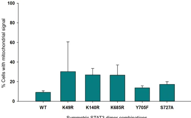
entre o núcleo e o citoplasma apenas quando são combinados assimetricamente. Em contraste, o mutante Y705F aumenta este movimento apenas quando está acoplado com ele próprio. Enquanto que o mutante L78R aumenta a localização nuclear e a formação de agregados com praticamente todas as combinações (simétrica e assimétricas), a combinação simétrica de R609Q aumenta apenas a localização mitocondrial. Por fim, os pares contendo as mutações K140R e K685R apresentam uma tendência de alterar a localização citoplasmática dos dímeros de STAT3 para o núcleo, mas só com um par é que foi significativo. No entanto, vendo noutra perspetiva, as combinações com duas mutações K-R foram as que mais alteraram a translocação entre citoplasma e núcleo. As combinações com apenas uma mutação K-R ou com um mutante resistente à fosforilação conseguiram aumentar a quantidade de STAT3 no núcleo.
Em suma, a ocorrência de diferentes modificações pós-traducionais nos dois monómeros do mesmo dímero influenciam a distribuição intracelular de STAT3, assim como o resíduo L78. Os resultados obtidos sugerem que as modificações pós-traducionais que ocorrem nas lisinas são as que mais regulam a circulação de STAT3 entre o citoplasma e o núcleo mesmo combinadas com outras modificações. O STAT3 que vai para a mitocôndria parece também ser regulado por estas mesmas modificações. Para além disso, estes resultados também indicam que é bastante importante que o resíduo L78 se mantenha intacto durante o funcionamento de STAT3. Esta mutação pode causar grandes alterações no comportamento de STAT3 tanto combinado com ele próprio como com outros mutantes. Estas combinações conseguiram diminuir a formação de dímeros e alterar a distribuição intracelular.
Concluindo, há a possibilidade de haver um novo nível da regulação da atividade de STAT3 e, consequentemente, novos alvos terapêuticos. Para além disso, estes resultados revelaram ser importantes para outros complexos de proteínas que também sejam regulados por modificações pós-traducionais. Sendo STAT3 uma proteína envolvida no desenvolvimento, imunidade e em várias doenças, o comportamento destes resíduos estudados e das modificações pós-traducionais podem ter implicações relevantes para o diagnóstico, tratamento e o estudo de uma grande variedade de patologias humanas.
Palavras chave: STAT3, complementação bimolecular de fluorescência, dimerização, modificações pós-traducionais, distribuição intracelular
Abstract
STAT3 is a transcription factor involved in many biological functions, such as cell proliferation, differentiation and survival, development and immunity, among others. STAT3 is functional when it dimerizes with itself or with STAT1. These dimers can be phosphorylated and become active, but inactive STAT3 dimers can influence its activity as well. Dysregulation of STAT3 activation initiates, contributes and sustains a variety of human diseases, including cancer. The aim of this project was to study specific residues/domains that had been described to influence STAT3 activity in the literature: K49, L78, K140, R609, K685, Y705, S727 and the SH2 domain. A Venus-STAT3 BiFC system was used in order to study the dimerization and intracellular distribution of STAT3 dimers in unstimulated cells. Asymmetric post-translational modifications change the intracellular distribution of STAT3 homodimers more strikingly than symmetric ones. The symmetric combinations carrying the L78R, R609Q and Y705F mutations were the only ones to affect intracellular distribution. Meanwhile, combinations carrying one or more K-R substitutions affected the nucleocytoplasmic shuttling. The L78 residue is also important for dimerization, mostly when combined with K49 and R609, as well as C-terminal of STAT3. This could mean a new level of regulation of STAT3 activity, and therefore a new possible therapeutic target. These results could be highly relevant for other protein complexes regulated by post-translational modifications beyond STAT3. Given the essential roles of STAT3 in development, immunity, tissue stress and cancer, our findings could have important implications for the diagnosis, treatment and understanding of a wide spectrum of human pathologies.
Key-words: STAT3, bimolecular fluorescence complementation, dimerization, post-translational modifications, intracellular distribution
Index
Acknowledgements ... i
Resumo ... ii
Abstract ... v
Index ... vi
List of acronyms and abbreviations ... viii
List of figures ... x
List of tables ... xii
1. Background ... 1
1.1. The Discovery of STATs ... 1
1.2. STAT structure ... 1
1.3. Activation of STATs and their regulation ... 2
1.4. JAK/STAT signaling pathway ... 3
1.5. Roles of STATs in biological processes ... 4
1.6. STAT3 ... 5
1.6.1. Post-translational modifications of STAT3 ... 8
1.6.2. Unphosphorylated STAT3 ... 9
2. Objectives ... 13
3. Material and Methods ... 13
3.1. The Venus-STAT3 bimolecular fluorescent complementation system ... 13
3.2. Generation of Venus-STAT3 BiFC constructs ... 14
3.3. Cell Culture ... 17
3.3.1. Transfection ... 18
3.4. Fluorescence Microscopy ... 18
3.5. Flow Cytometry ... 19
3.6. Protein Analysis ... 19
3.6.2. SDS-PAGE and Western Blot ... 20
3.7. Statistical analysis ... 20
4. Results ... 21
4.1. High levels of U-STAT3 are found in the cytoplasm in HeLa cells ... 21
4.2. Venus-STAT3 BiFC constructs are able to dimerize ... 21
4.3. L78R STAT3 mutant affects dimerization in unstimulated cells ... 22
4.4. Phosphoresistant mutations affect STAT3 intracellular localization in unstimulated HeLa cells ... 23
4.5. Asymmetric combinations of STAT3 PTM mutants affect intracellular distribution of STAT3 homodimers ... 26
4.6. The STAT3-L78R mutation has a dominant effect in intracellular distribution ... 28
5. Discussion ... 31
6. Concluding remarks ... 35
List of acronyms and abbreviations
A Alanine
ABL Abelson leukemia protein
APRE/F Acute phase response element/factor BiFC Bimolecular fluorescence complementation
BSA Bovine serum albumin
C Cysteine
CCD Coiled-coil domain
CNS Central nervous system
DBD DNA binding domain
DMEM Dulbecco’s Modified Eagle’s Medium
EGF Epidermal growth factor
F Phenylalamine
FBS Fetal bovine serum
GAPDH Glyceraldehyde-3-phosphate dehydrogenase
GAS IFNγ-activated sequence
Gp130 Glycoprotein 130
GRIM-19 Retinoid interferon induced cell mortality 19 HELA Human cervical cancer cells
I/R Ischemia/reperfusion
IFN Interferon
IHCA Inflammatory hepatocellular adenoma
IL Interleukin
ISGF3 Interferon-stimulated gene factor 3
JAB JAK-binding protein
JAK Janus kinase
JH JAK homology
K Lysine
L Leucine
LIF Leukemia inhibitor factor
NES Nuclear-export sequence
NLS Nuclear-import sequence
NRTK Non-receptor tyrosine kinase
NTD Amino-terminal domain
P/S Penicillin/Streptomycin solution
P-STAT Phosphorylated STAT
PBS Phosphate buffered saline
PCR Polymerase chain reaction
PIAS Protein inhibitor of activated STAT
PPI Protein-protein interaction
PTM Post-translational modification
PY Phosphotyrosine
Q Glutamine
R Arginine
ROS Reactive oxygen species
S Serine
SH2 Scr-homology 2
SIE Sis-inducible element
SOCS Suppressor of cytokine signaling
SSI STAT-induced STAT inhibitor
STAT Signal transducer and activator of transcription
TAD Transactivation domain
TK Tyrosine kinase
U-STAT Unphosphorylated STAT
V1 Venus 1
V2 Venus 2
WT Wild-type
List of figures
Figure 1 – The STAT family of proteins ... 2
Figure 2 – JAK structure ... 4
Figure 3 – A BiFC cellular model for the visualization of STAT3 dimers in living cells ... 13
Figure 4 – Generation of the STAT3 constructs. ... 16
Figure 5 – Generation of Venus-STAT3ΔCT. ... 17
Figure 6 – Representative fluorescence microscopy pictures of the qualitative classification in HeLa cells. ... 19
Figure 7 – Unphosphorylated STAT3 is found expressed mostly in the cytoplasm. ... 21
Figure 8 – The Venus-STAT3 BiFC system allows the visualization and study of STAT3 dimers in living cells ... 22
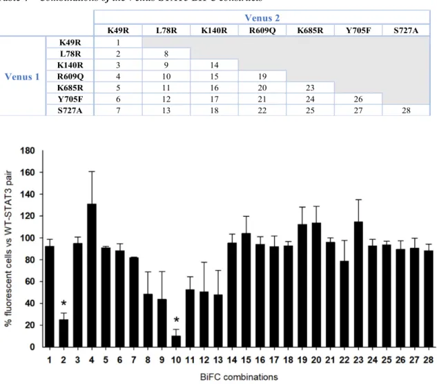
Figure 9 – Venus-STAT3 symmetric and asymmetric dimers do not regulate STAT3 homodimerization in unstimulated cells ... 23
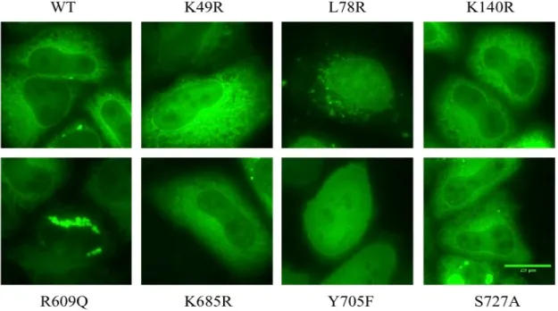
Figure 10 – Representative microscopy pictures of intracellular distribution of each symmetric combination ... 24
Figure 11 –Nuclear, cytoplasmic and nucleocytoplasmic localization of symmetric STAT3 dimers ... 25
Figure 12 – Mitochondrial localization of symmetric STAT3 dimers ... 25
Figure 13 – Presence of aggregates in cells with symmetric STAT3 dimers. ... 26
Figure 14 – Asymmetric STAT3 PTMs regulate intracellular localization of STAT3 homodimers. ... 26
Figure 15 – The intracellular distribution of STAT3 dimers is highly regulated by PTM mutations and asymmetric pairs. ... 27
Figure 16 – Nuclear, cytoplasmic and nucleocytoplasmic localization of STAT3 dimer combinations with L78R and R609Q mutants. ... 28
Figure 17 – Mitochondrial localization of STAT3 dimer combinations with L78R and R609Q mutants ... 29
Figure 18 – Presence of aggregates in cells with STAT3 dimer combinations with L78R and R609Q mutants ... 29
Figure 19 – Intracellular distribution of STAT3-L78R and R609Q symmetric and asymmetric dimers ... 30
List of tables
Table 1 – Role of STAT proteins ... 5
Table 2 - Gene Regulation by STAT3 ... 7
Table 3 – Primers used for mutagenesis and PCR cloning. ... 14
1. Background
1.1.
The Discovery of STATs
Signal transducer and activator of transcription (STAT) proteins belong to a family of cytoplasmic transcription factors constituted by seven members: STAT1, 2, 3, 4, 5a, 5b and 6. Genetic mapping of mouse and human genes revealed that STAT genes are chromosomally localized in only three clusters: STAT1 and STAT4, STAT2 and STAT6, STAT3 and STAT5a/b. This suggests the existence of a common ancestral gene which was initially duplicated, followed by dispersion of the linked loci to different chromossomes1.
The first STAT family members, STAT1 and STAT2, were discovered in interferon (IFN) signaling pathways in mammalian cells during the 1990s. In response to IFNα and IFNβ, a DNA-binding complex is formed consisting of STAT1, STAT2 and the p48 DNA-DNA-binding protein, which binds to the IFN-stimulated response element. On other hand, in response to IFNγ, a DNA-binding complex is formed by STAT1 homodimers that binds to the IFNγ-activated sequence (GAS)2,3. Following these studies, it became clear that STATs could dimerize with each other and their activities could be activated by various cytokines. Further proteins were soon identified: STAT3 was cloned as an interleukin-6 (IL-6)-activated transcription factor and by homology to STAT1; STAT4 was also cloned by homology approaches; STAT5 was cloned as a prolactin-activated transcription factor from sheep and mice, leading to the discovery of STAT5a and STAT5b genes; and STAT6 was cloned as an IL-4-activated DNA-binding protein as well as by homology4,5. STAT homologues have also been discovered in other species, such as, C. elegans, Dictyostelium, Drosophila and zebra fish6.
1.2.
STAT structure
STATs are transcription factors between 750 and 800 amino acid residues in length, and characterized by the presence of six different domains (Fig. 1)7. The amino-terminal domain (NTD) is necessary for dimerization, tetramerization and interaction with other elements essential for STAT regulation. The coiled-coil domain facilitates several protein-protein interactions (PPIs) through its large hydrophilic surface, receptor binding and nuclear translocation. The DNA binding domain (DBD) is essential to transcribe target genes and maintain proper conformation for importin binding and nuclear export/import. The linker domain is involved in transcriptional activation and PPIs. The Src-homology 2 (SH2) domain is the most conserved domain among STATs and is critical for receptor association and formation of phosphorylated dimers and their stabilization. The SH2 domain of a phosphorylated STAT molecule binds a specific phosphotyrosine (pY) residue within the SH2 domain of another STAT molecule. The carboxyl-terminal transactivation domain (TAD) is the least conserved domain and allows the binding of
STATs to co-activators, such as Janus kinases (JAKs) and cytokines, and can initiate or enhance transcription8,9.
1.3.
Activation of STATs and their regulation
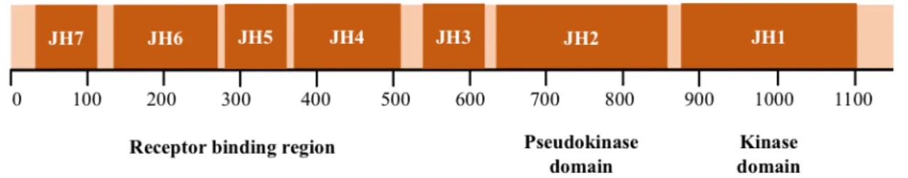
STATs are activated by a plethora of cytokines, growth factors and hormones that are able to control cell growth and differentiation, development and immune responses10. These extracellular ligands are necessary to stimulate membrane-bound receptor complexes, that subsequently allow STAT phosphorylation on a single tyrosine (Y) located around residue 700 and initiate the signaling pathway11. Cytokines, including IFNs, ILs, neurotrophic factors, colony-stimulating factors (CSFs) and hormones, bind to cytokine receptors allowing them to associate with tyrosine kinases (TKs), including JAKs, which in turn phosphorylate STATs. Some growth factor receptors, such as epidermal growth factor (EGF) and platelet-derived growth factor (PDGF) receptors, have intrinsic tyrosine kinase activity (RTKs) that can activate STAT directly or indirectly by means of JAKs and members of Src family12. Non-receptor tyrosine kinases (NRTKs), such as Src and Abelson leukemia protein (ABL), also can activate STATs constitutively in the absence of ligand-induced receptor signaling. G-protein coupled receptors can activate STAT1 and STAT3 in T-cells. Finally, activation of STATs can also be mediated by other proteins that serve as adaptors to bring JAKs within close proximity to activate STATs13. Figure 1 – The STAT family of proteins. A, Structural representation of the STAT1 dimer. (Chen et al.,
1998). B, Functional motifs of STAT. STATs share several conserved domains, including a N-terminal domain, a coiled-coil domain, a DNA-binding domain, a linker domain, a SH2 domain and a C-terminal transactivation domain.
In normal cells, STAT activity is regulated by a variety of mechanisms, but the most predominant are post-translational modifications (PTMs), such as phosphorylation, methylation and acetylation. Tyrosine phosphorylation at the SH2 domain is essential for their activation. However, STAT1, STAT3, and STAT5 are also phosphorylated at a serine (S) in the TAD domain for maximal transcriptional activity. Furthermore, arginine (R) and lysine (K) methylation and lysine acetylation have also been described to increase DNA binding activity and gene transcription14–16. Another regulatory mechanism occurs at the level of gene expression. It has been reported that in response to IL-6 induction, STAT3 induces stat3 mRNA in the liver17. This autoregulatory mechanism for stat3 gene activation was first thought to be part of a positive feedback, but it could also lead to the displacement of activated STAT3 from the DNA.
STAT activity can be negatively regulated as well. Overexpression of truncated (β) isoforms of STAT1, STAT3 and STAT5, lacking the TAD domain, behave as dominant-negative factors in certain circumstances. Cytokine-inducible SH2-containing protein (CIS) competes with STATs for the same docking site on the phosphorylated receptors whereas suppressors of cytokine signaling (SOCSs) can suppress JAK activity through direct interaction or by inhibiting the activity of phosphorylated receptors. Unlike SOCS, protein inhibitor of activated STAT (PIAS) is constitutively expressed, interacts directly with phosphorylated STATs and negatively regulates them in the nucleus in a ligand-dependent manner18. Other examples of negative regulators include the JAK-binding proteins (JABs) and the STAT-induced STAT inhibitors (SSIs). Phosphatases, such as TC45, SH2-containing phosphatases 1 and 2 (SH1 and SH2), and protein-tyrosine-phosphatase-1B (PTP1B) can dephosphorylate and inactivate STATs19,20. Also, like most proteins, intracellular levels of STATs can be regulated by protein degradation via the ubiquitin-proteosome pathway21.
1.4.
JAK/STAT signaling pathway
The best studied pathway for STAT activation is perhaps through JAKs, more commonly known as the JAK/STAT pathway. Upon binding of the ligand, JAKs phosphorylate key tyrosine residues located on the cytoplasmic tail of the receptor, providing docking sites for the STAT proteins. Non-phosphorylated STAT dimers bind to the phosphorylated receptor site through their SH2 domains, and are subsequently phosphorylated at the key SH2 tyrosine residue by JAKs. Phosphorylated STAT (P-STAT) dimers dissociate from the receptor and translocate to the nucleus where they bind to specific DNA response elements to facilitate gene transcription22. In the nucleus, there are mainly two types of STAT response elements: ISRE and GAS. ISREs activate a multimeric complex called interferon-stimulated gene factor 3 (ISGF3) that comprises STAT1α, STAT1β, STAT2 and p48/IRF-9 and seems to be restricted to IFN signaling. Its target genes include ISG15, ISG54, 6-16, and 2-5A synthetase. GAS elements bind to hetero- and homodimers of STATs and include the sis-inducible element (SIE) in the c-fos promoter, the acute
phase response element (APRE) in promoters of acute phase proteins, and the prolactin response element (PRE) in the β-casein promoter. They are responsible to activate genes such as c-fos (through STAT1 and STAT3), IRF-1, 6-16, 9-27, 2-5 A synthetase, Ly-6E (through STAT1α), α2-macroglobulin, fibrinogen, and α1-acid glycoprotein, cell cycle regulator Cyclin D1, anti-apoptotic Bcl-XL, and Mcl-1 (through STAT3), and β-casein, Bcl-XL, and Cyclin D1 (through
STAT5)23.
There are four mammalian JAKs critical for cytokine signaling: JAK1, JAK2, JAK3 and tyrosine kinase 2 (TYK2). They range in size from 1200 to 1300 amino acids and are nearly ubiquitously expressed, with the exception of JAK3 which is only expressed in hematopoietic, vascular smooth muscle and endothelial cells. JAKs consist of seven conserved JAK homology (JH) domains (Fig. 2). The C-terminal portion includes a distinctive kinase domain (JH1) and a pseudokinase domain (JH2), which has a kinase domain fold but lacks crucial residues for catalytic activity and for nucleotide binding. The N-terminal JH domains, JH3-JH7, constitute a FERM (four-point-one, ezrin, radixin, moesin) domain and mediate association with receptors22,24. Each JAK family member has different affinities for cytokine and growth factor receptors, but in general they can be activated by receptors from the IFN family (IFNα/β, IFNγ, IL-10, IL-19, IL-20, IL-22), the glycoprotein 130 (gp130) family (IL-6, IL-11, oncostatin M (OSM), leukemia inhibitor factor (LIF), cardiotrophin-1 (CT-1), granulocyte colony-stimulating factor, leptin, IL-12, IL-23), the γC family (IL-2, IL-4, IL-7, IL-9, IL15, IL-21) or the single chain family (erythropoietin, growth factor, prolactin, thrombopoietin)8,25,26.
1.5.
Roles of STATs in biological processes
STATs present a dual role: they act as cytoplasmic proteins for signal transduction and as nuclear transcription factors for gene transcription. Essentially, STATs can regulate the expression of genes that promote cell growth, survival, differentiation, development and inflammation27. Nonetheless, as mentioned above, different STATs have a variety of activation pathways that may happen in different tissues at different time periods, depending on extracellular cues and physiological requirements. For that reason, each one of them has specific functions. STAT1 is mainly implicated in innate and adaptive immunity and can act as a key intermediary to
down-Figure 2 – JAK structure. JAKs share seven regions of high homology, JH1-JH7. JH1 has been shown
to encode the kinase. JH2 represents a pseudokinase domain, which appears to regulate JH1 catalytic activity. JH3-JH7 are implicated in receptor association.
regulate cell proliferation, apoptosis and tumor suppression. STAT2, STAT4, and STAT6 play an important role in the maturation of naive CD4+ T cells and, therefore, in regulation of immune responses. STAT3 and STAT5a/b promote growth and differentiation of various tissues and prevent cell death28. These biological functions were studied through generation of gene targeted mice (Table 1)18,29.
Table 1 – Role of STAT proteins. IFN – interferon; Th1 – T helper 1; IL – interleukin; NK – natural killer;
Th2 – T helper 2
STAT proteins Phenotype of null mice
STAT1 Impaired responses to IFNs; impaired growth control; defective macrophages activity; high sensitivity to viral infections
STAT2 Impaired responses to interferons
STAT3 Embryonic lethality; multiple defects in adult tissues including impaired cell survival (both positive and negative) and impaired response to pathogens STAT4 Impaired Th1 differentiation owing to loss of IL-12 responsiveness
STAT5a Impaired mammary gland development and lactation owing to loss of prolactin responsiveness; partial defect in T cell growth
STAT5b Defect in NK cell development owing to loss of growth hormone responsiveness;
STAT6 Impaired Th2 differentiation owing to loss of IL-4 responsiveness
Although STAT functions in humans include mostly physiology and development, they can also regulate oncogenic signaling in many different tumor types. STAT3 and STAT5 are the STATs most often implicated in human cancer progression30. Approximately 70% of human solid and hematological tumors exhibit overexpression or constitutively activated STAT3 compared with normal cells31. In cancer cells, activation of STAT3 and STAT5 leads to increased cell proliferation, cell survival, angiogenesis, and immune system evasion32. Conversely, their inactivation/knockout sensitizes cancer cells to antitumoral treatments33–35.
Dysregulation of STAT activation can also lead to other complications. For example, malfunction of the JAK/STAT pathway has been reported to be associated with various cardiovascular diseases36, central nervous system inflammation and neurodegenerative diseases37.
1.6.
STAT3
STAT3 was independently discovered and studied by two research groups and described in 1994. Zhong et al. thought that if IFNα and IFNβ transcriptional responses used different STATs that shared some amino acids, it seemed likely that additional family members could also influence the activity of other STATs. As a result, they discovered STAT3 as a DNA-binding protein in response to EGF in hepatocytes38. Akira et al. purified and cloned STAT3 from mouse liver nuclear extracts and named it as acute phase response factor (APRF). Furthermore, they identified
STAT3 as a DNA-binding factor that selectively binds to IL-6-responsive element within the acute phase gene promoter17.
STAT3 is also activated by such diverse agents as growth factors, oncogenes, and IFNs, and plays an important role in cell growth, survival, differentiation of various tissue types, such as skin, liver, mammary gland, thymus and nervous system, inflammation and motility39. The classic pathway of STAT3 is known as the JAK/STAT3 pathway. Members of the IL-6 family of cytokines exert their action through their corresponding receptor complexed with the gp130 transmembrane protein, which in turn initiates intracellular signaling through JAK by phosphorylating STAT3 at tyrosine 705 (Y705)40. Y705 phosphorylation converts STAT3 from an inactive conformation to an active one. Originally, P-STAT3 was thought to be released from the receptor as a monomer, and then form homo- or heterodimers (e.g. with STAT1). However, recent data demonstrated that STAT3 resides largely in the cytoplasm of resting cells as unphosphorylated homodimers. These homodimers can be recruited to the receptor-associated JAK upon ligand binding. Y phosphorylation allows STAT3 dimers to be released from the receptor and reorient their conformation where the SH2 domains come closer with each other and create a stronger interaction41. Active STAT3 dimers are then recognized by importin α3, which binds the coiled-coil domain of STAT3 and mediates its nuclear import to activate transcription42. It is now recognized the existence of non-canonical STAT3 pathways, in which the functions of STAT3 are independent of Y705 phosphorylation or even nuclear translocation. Both active and inactive STAT3 dimers undergo nucleocytoplasmic shuttling and regulate gene transcription, although the target genes could be different. In addition, STAT3 was also discovered to be present in isolated mitochondria, suggesting that STAT3 exerts a direct impact on mitochondrial function43.
A critical role for STAT3 in malignant transformation was initially revealed through a oncoprotein, v-Src. Studies showed that STAT3 is constitutively activated in v-Src transformation44 suggesting that STAT3 may have key roles in oncogenesis. Aberrantly activated STAT3 is now known to initiate, contribute and sustain a variety of cancers, such as breast, ovaries, prostate, lung, pancreas, brain and hematologic malignancies14,45,46. STAT3 dysregulation, constitutive activation or mutations that disrupt the epigenetic control of endogenous regulators of STAT3 signaling have been reported to induce cell proliferation and resistance to apoptosis42,47. Constitutive activation of STAT3 can also be caused by loss of negative regulation, excessive stimulation, positive feedback loops and somatic mutations that confer a hyperactive property to STAT348. No STAT3 mutations are associated with malignant tumors, but several have been identified in benign inflammatory hepatocellular adenoma (IHCA), including L78R, D502Y, K658Y, E166Q and Y640F49. These mutations produce constitutively
activated STAT3 conformations that accumulate in the nucleus and enhance STAT3 transcriptional activity.
Although STAT3 hyperactivation is present in many cancers50, the role of STAT3 in oncogenesis is still unclear and is likely dependent on tumor type and cellular context. Given the fact that it is able to regulate both oncogenes and tumor suppressor genes, STAT3 has been reported to either promote or inhibit oncogenesis. STAT3 can directly regulate genes leading to multiple tumor promoting processes from cell survival, invasion, angiogenesis, and immune escape; and indirectly regulate other transcription factors that support tumor growth by promoting survival, angiogenesis, metastasis, and even pluripotency30. Numerous target genes and their cellular function have been identified for STAT3 (Table 2)51,52.
Table 2 - Gene Regulation by STAT3. c-Fos – cellular Fos; HIF – hypoxia inducible factor; Oct – octamer
transcription factor; Hsp – heat shock protein; IL – interleukin; COX – cyclooxygenase; MMP – matrix metalloproteinase; ICAM – intracellular adhesion molecular; NGAL – neutrophil gelatinase associated lipocalin; POMC – proopiomelanocortin; SAA – serum amyloid A; VEGF-A – vascular endothelial growth factor A; bFGF – basic fibroblast growth factor; HGF – hepatocyte growth factor; TNF – tumor necrosis factor; S1P – shingosine-1-phosphatase; R – receptor; MUC - mucin
Function STAT3-Regulated genes
Transcription Factors c-Fos, HIF-1α, c-Myc, Sox2, Nanog, Twist, Zeb1, p53, Oct-1
Apoptosis and Proliferation Bcl-2, Mcl-1, Bcl-xL, Survivin, Fas, Hsp70, Hsp90α/β, Cyclin-D1
Immune Suppression and Inflammation IL-10/23, TGF-β, COX-2
Metastasis MMP-1/2/3/9, Fascin, Vimentin, RhoU,
ICAM-1, NGAL, POMC, SAA1
Angiogenesis VEGF-A, bFGF, HGF
Cell Signaling AKT, PIM-1, TNF-R2, S1P-R1, MUC-1
The inhibition of STAT3 suppresses tumor growth and enhances the sensitivity to anticancer agents in a variety of tumors, thus suggesting STAT3 as a potential target for anticancer therapy32. There have been developed inhibitors that target steps of STAT3 activation and function. Inhibition of EGFR, TKR, JAK or SFK can inhibit phosphorylation/activation of STAT3 in various tumors. Inhibiting intermolecular interactions that involve STAT3 can affect STAT3 SH2 domain. Inhibition of nuclear import/export of STAT3 where the targets are importins α3, α5 and α7, importin β and exportin 1 and inhibition of STAT3-mediated transcription by targeting its DNA binding site are also processes being developed. Finally, application of natural products for unspecified targets is being considered as well53,54. During the past few years, many unexpected new roles of STAT3 in cancer and its underlying mechanisms have emerged55. These newly discovered pathways have been pointed to unique directions for the creation of new therapeutics for cancer trying to overcome its complexity.
Although STAT3 role in cancer remains a major topic in biomedical research, there has been evidence that STAT3 activation or suppression has a significant contribution in the progression or remission of a series of other human diseases. High levels of STAT3 are found in myocardial ischemia/reperfusion injury, rheumatoid arthritis, renal fibrosis, inflammatory lung diseases/pulmonary fibrosis, psoriasis, inflammatory bowel disease and atherosclerosis. Low levels of STAT3 are found in cardiomyopathy, liver fibrosis, obesity and diabetes type 256. STAT3 level and activity are found up- or down-regulated in Alzheimer’s disease (AD), and for that reason STAT3 role in this condition is still controversial57.
Regarding the central nervous system (CNS), high levels of STAT3 have been implicated in neural regeneration and glial reactivity58. Upon brain trauma, star-shaped glial cells called astrocytes undergo important morphological modifications including hyperplasia and hypertrophy. This process is called reactive astrogliosis and involves dramatic changes in gene expression, especially the ones that regulate the morphology, energy metabolism, function and proliferation of astrocytes. Within days after insult, a glial scar, a physical and functional wall, is formed around the injured brain tissue and in severe cases it can be permanent59,60. Due to the variety of responses that astrocytes can provide, reactive astrogliosis has been divided in three categories: (1) mild to moderate reactive astrogliosis, (2) severe diffuse reactive astrogliosis and (3) severe reactive astrogliosis with compact scar formation. Compact astroglial scars are comprised of newly proliferated astrocytes with densely overlapping processes that form borders to damaged tissue and inflammation61. This important step has been reported to be dependent on the activation of the JAK/STAT3 pathway62.
1.6.1. Post-translational modifications of STAT3
Proteins can be regulated by PTMs, including phosphorylation/dephosphorylation (on tyrosines, serines and threonines), methylation/demethylation (on arginines and lysines), acetylation/deacetylation (on lysines), isomerization, ubiquitination (on lysines), proteolytic cleavage and others. There are a number of PTMs that regulate STAT3 activity. Y705 phosphorylation is probably the most studied PTM and it is essential for STAT3 activation. When mutated to phenylalanine (STAT3-Y705F), this mutant could not be phosphorylated and showed a dominant-negative activity influencing the activation of Wild-type (WT) STAT363. Serine 727 (S727) is phosphorylated in different situations to facilitate gene expression64. Interestingly, constitutive S727 phosphorylation without Y705 phosphorylation was reported to be a hallmark of chronic lymphocytic leukemia65. Furthermore, phosphorylation at S727, but not at Y705, is necessary for the localization of STAT3 to the mitochondria of hepatocytes and myocardial cells66,67.
Several different PTMs of lysine residues have been reported to affect the STAT3 function as well. Lysine 685 (K685) is rapidly acetylated by p300 in response to OSM and IL-6 (as early as
15 and 20 min, respectively). It enhances tyrosine phosphorylation and is necessary for dimerization15,68. A significant increase of acetylation at K685 is detected in tumor tissues. CD44 is a transmembrane glycoprotein that has been recognized as a marker for tumor cells. When activated, it can bind to STAT3 and p300 in the nucleus and acetylate STAT3 at K685 leading to cell proliferation69. In addition, K685 acetylation can also lead to DNA methylation and silencing of tumor suppressor genes through recruitment of DNA methyltransferase 1 (DNMT1)70. K49 can either be acetylated or dimethylated. Acetylation at K49 is associated with positive regulation of gene expression and DNA binding. More specifically, IL-6-induced gene expression requires p300-mediated acetylation of K4971. It was later discovered that this residue could be also dimethylated by the histone methyltransferase enhancer of zeste homolog 2 (EZH2) and influence gene transcription in response to IL-672. K49 dimethylation is enhanced by exposure of cells to cytokines, but they only occur after Y705 phosphorylation. For example, EZH2 is bound preferentially to tyrosine-phosphorylated STAT3. K140 is another residue that can be dimethylated. Dimethylation is mediated by methyl transferase SET9 and takes place in the nucleus. Like K49, K140 dimethylation occurs in response to IL-6 stimulation, but only after Y705 and S727 phosphorylation. This PTM is required to maintain normal cellular responsiveness to cytokines by reducing the duration and extent of expression of the potent negative-regulator SOCS316.
1.6.2. Unphosphorylated STAT3
Initially, it was thought that STAT3 existed as a cytoplasmic monomer, dimerizing and translocating to the nucleus only upon tyrosine phosphorylation. P-STAT3 would dimerize by means of the SH2 domains of the STAT3 molecules, and only phosphorylated dimers could bind to DNA. This view still persists in the literature in spite of an increasing body of evidence against it. STAT3 proteins exist as stable homodimers prior to activation (currently known as latent/unstimulated/unphosphorylated STAT3, U-STAT3 dimers). Both the amino and/or carboxyl termini are important to stabilize homodimerization of these inactive dimers and may facilitate subsequent activation73. There are two possible unphosphorylated dimer orientations termed as “antiparallel” and “parallel”, based on whether the SH2 domain is on the opposite or the same end of the dimer. The antiparallel conformation was speculated as the predominant structure in the latent state prior to stimulation. However, FRET analysis showed that the C-terminal domains had close proximity in latent state, supporting a parallel orientation similar to activated STAT374. Dimers of unphosphorylated STAT3 are also able to enter the nucleus and bind to DNA, regulating the transcription of a different set of genes than phosphorylated STAT375.
1.6.2.1.
Nucleocytoplasmic shuttling
Latent STAT3 enters the nucleus independently of its phosphorylation76, unlike other STATs, such as STAT1 and STAT2, which accumulate in the nucleus only following their
phosphorylation77. The variety of behavior between different STAT proteins can be due to the involvement of distinct mechanisms that regulate their intracellular trafficking. Large proteins, like STATs, are only able to travel freely into and from the nucleus due to their nuclear-localization sequences (NLS) or nuclear-export sequences (NES). These signals can either result in protein modifications altering the protein conformation, or in association with another protein which includes importins and exportins for nuclear import and export, respectively78,79. In the canonical nuclear translocation, the P-STAT3 dimer is released from the receptor and translocates to the nucleus through importin-α3, which recognizes the coiled-coil domain and mediate its nucleocytoplasmic shuttling. However, recent studies showed that STAT3 can bind constitutively to importin-α3 and α6 and shuttling in and out of the nucleus independently of its phosphorylation80. In contrast, the phosphorylation of the nuclear localization signal of STAT1 is a prerequisite for its interaction with importin-α5 and subsequent nuclear import81,82. While some studies revealed that U-STAT3 could translocate to the nucleus through importins, others demonstrated that it was independent of the binding of NLS and NES and importins. U-STAT3 mutants lacking NLS and NES were generated, and they could shuttle between cytoplasm and nucleus indicating that the NLS and NES of STAT3 are not necessary for nucleocytoplasmic shuttling of U-STAT3. In addition, when the NTD was deleted from the monomeric form, it could shuttle faster than the WT-STAT3, indicating that dimerization of U-STAT3 is not necessary for nucleocytoplasmic shuttling83. Furthermore, this STAT3 monomer could not accumulate in the nucleus after stimulation with cytokines, indicating that the NTD is essential not only for latent dimerization but also for nucleocytoplasmic shuttling84. Although there are inactive monomers and dimers in the nucleus, the pre-association of STAT3 complexes occurs strongly within the cytoplasmic environment85. The constitutive presence of latent STAT3 in the nucleus is not static: there is a subcellular distribution of latent STAT3 with high cytoplasmic and low nuclear concentrations that represents a steady state resulting from continuous nuclear import and export86. In general, inactive monomers dimerize in the cytoplasm in a parallel orientation which is mostly stabilized by homotypic interactions between NTDs. The protomers are orientated in a form which structurally allows the separate SH2 domains to interact with each other resulting in additional stabilization74.
1.6.2.2.
Mitochondria
Mitochondria are double-membrane subcellular compartments that contain their own circular genome, i.e. mitochondrial DNA (mtDNA). They are best known as powerhouses that supply eukaryotes with cellular energy, producing the majority of ATP needed for cellular processes through oxidative phosphorylation. Nevertheless, mitochondria are also crucial for other biological processes, such as maintaining ion homeostasis, producing precursors of macromolecules (i.e. lipids, proteins and DNA), and regulating apoptosis by sequestering
potentially damaging byproducts including reactive oxygen species (ROS). Furthermore, recent evidence demonstrates that mitochondria play an active role in integrating signaling pathways and communicating with other organelles87. Notably, mitochondrial dysfunction not only affects ATP synthesis leading to a variety of mitochondrial diseases, but is also linked to cancer, metabolic disorders and neurodegenerative diseases, such as, Alzheimer’s, Parkinson’s and Huntington’s diseases88.
In 2009, it was discovered a small pool of STAT3 present in mitochondria (mitoSTAT3)66and its presence in different cells and tissues was confirmed by a number of follow-up studies. Currently, it is known that mitoSTAT3 is involved in cellular metabolism, cell development and death, cancer transformation, ischemia/reperfusion (I/R) heart injury, sperm motility, T cell immunity and others89. Although mitoSTAT3 is not necessary for the maintenance and formation of mitochondria, it enhances the activity of the complexes I, II and V of the electron transport chain. In cells with loss of STAT3, the activity of these complexes is reduced but can be restored by reconstituting these cells with a mitochondrially restricted form of STAT3. This fact awakened a huge interest in the scientific community, which confirmed these findings and additionally showed that STAT3 can also affect the activity of complexes III and IV of the electron transport chain90. In addition, Ras-mediated cellular transformation was shown to be dependent on mitoSTAT391.
Focusing on post-translational modifications, both Y705 and S727 phosphorylation of STAT3 have been found in mitochondria but only S727 phosphorylation is critical for mitoSTAT3 to enhance the activity of complexes I and II66. STAT3 Y705 phosphorylation or its transcriptional activity are not required for its role on the electron transport chain in several cell types.
To understand how mitoSTAT3 works it is important to know how STAT3 is transported to mitochondria. This field continues to be unclear but there has been some progress: the C-terminus of STAT3 has been shown to be required for mitochondrial transport89. Moreover, it has been observed that the gene associated with retinoid interferon-induced cell mortality 19 (GRIM-19), a component of mitochondrial complex I, can regulate STAT3 translocation into mitochondria when the S727 residue is phosphorylated. GRIM-19 seems to have an important role on other mitoSTAT3 functions. It enhances STAT3 integration in complex I, intensifying the production of ROS92; and it interacts directly with the NLS-domain of STAT3, impairing STAT3 nuclear localization and inhibiting its transcriptional activity93.
1.6.2.3.
Transcriptional activity of unphosphorylated STAT3
Surprisingly, recent data indicate that STAT3 transcriptional activity is partially independent of phosphorylation. Unphosphorylated STAT3 is also capable of binding to DNA and be a transcriptional activator and chromatin/genomic organizer by itself. It can bind to DNA-response
elements, such as GAS, as a monomer and as a dimer; and to AT-rich DNA sequence sites which are important for regulation of gene expression and/or chromatin organization due to their particular structure94. Phosphorylated STAT3 dimers can also drive the expression of the STAT3 gene itself, leading to a large ligand-dependent increase in the intracellular levels of U-STAT3, which persists for many days and drives the expression of a set of genes distinct from the set activated by P-STAT3 dimers75. For example, high levels of U-STAT3 can activate the expression of RANTES, an important mediator of acute and chronic inflammation, and some oncogenes including MET and MRAS95.
1.6.2.4.
Important residues for unphosphorylated STAT3 activity
Since phosphorylated STAT3 activates its own transcription, an increase in unphosphorylated STAT3 can be caused by an abnormal constitutive activation of STAT3, which can lead to tumorigenesis77. This is one of the reasons why current efforts are also focusing on unphosphorylated STAT3. Nowadays there is a need to understand deeply the contribution of specific residues to unphosphorylated STAT3 activity. Some of these residues have been identified. Two intermolecular disulfide bridges, C367-C542 and C418-C426, influence the structure and stability of U-STAT3 dimers. When these bonds are reduced, the dimeric form starts to dissociate. Mutations in these residues trigger structural changes in unphosphorylated STAT3 dimers and abolished its DNA-binding activity96. L78 and R609 residues are also relevant to unphosphorylated STAT3 dimerization. STAT3-L78R itself cannot dimerize without stimulation, thus supporting the significance of the NTD for latent dimer formation74. Concerning the R609Q mutation, it not only prevents interaction with pY motifs and the binding pocket in the SH2 domain, but also leads to a complete reorientation of this domain resulting in a monomer structurally similar to a dimer, which possibly disturbs any kind of interaction. This suggests that R609 is essential for dimer formation and for SH2 domain to stabilize latent dimers97.Two arginine residues within the DNA biding domain, R414 and R417, are important for tyrosine phosphorylation and modulation of nuclear accumulation of latent STAT3. Mutations of these residues to glutamine attenuated STAT3 phosphorylation, and impaired nuclear import/export and distribution of latent STAT3, leading to a higher nuclear accumulation98.
The residue K685 is known to be one important PTM of STAT3. It is found to be acetylated in U-STAT3 and is necessary for the expression of most U-STAT3-dependent genes. A global analysis revealed that more than 70% of the gene expression induced by a high level of unphosphorylated STAT3 was impaired by the K685R mutation99. Chronic activation of ATR1 (angiotensin II type-1 receptor) induces unregulated expression of the STAT3 gene, leading to nuclear accumulation of U-STAT3, which correlates with the progression of cardiac hypertrophy100. K685 acetylation in response to angiotensin II was demonstrated to be critical for
U-STAT3-driven gene expression in the context of heart failure, revealing an essential role of this residue in important pathological situations.
2. Objectives
STAT3 can act and induce transcriptional activity both as a stimulated or unstimulated dimer. Recent evidence indicates that unstimulated STAT3 is involved in human pathologies such as cancer. Mutations or post-translational modifications in specific residues (K49, L78, K140, R609, K685, Y705 and S727) can alter STAT3 signaling pathway. The main aim of this project was to determine the relative contribution of specific residues to the homodimerization and intracellular localization of unstimulated STAT3 homodimers.
3. Material and Methods
3.1.
The Venus-STAT3 bimolecular fluorescent complementation system
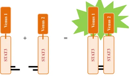
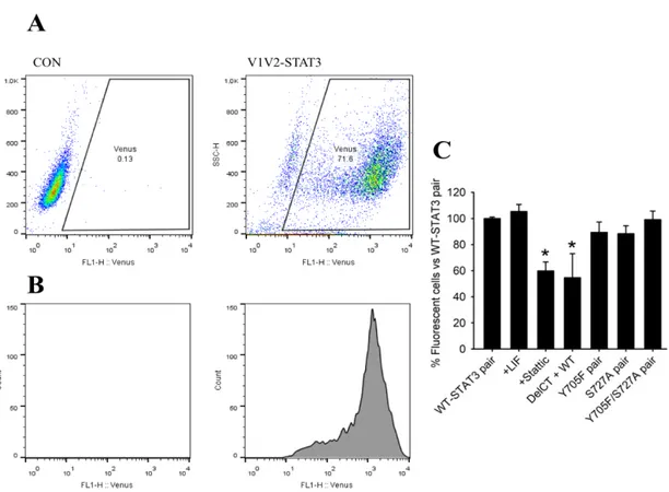
Bimolecular fluorescence complementation (BiFC) assays are systems used to study protein-protein interactions in living cells. A Venus-STAT3 BiFC system was designed and developed in our lab (Cell Structure and Dynamics, ITQB-NOVA, Oeiras, Portugal). In this assay, STAT3 proteins were fused to non-fluorescent halves of the Venus fluorescent reporter protein, a member of the green fluorescent protein (GFP) family (Fig. 3).
When the STAT3 proteins dimerize, the two halves of Venus get close enough to directly contact with each other and reassemble the functional reporter protein. Consequently, a signal is emitted and is proportional to the amount of STAT3 dimers formed. Fluorescence can then be analyzed qualitatively and quantitatively by conventional methods, such as flow cytometry and fluorescence microscopy.
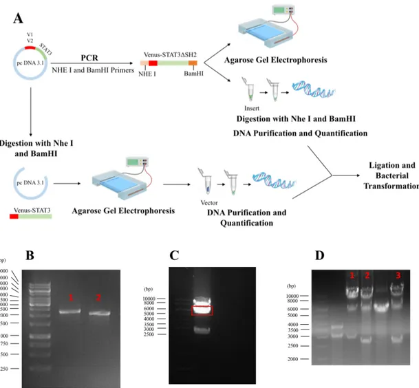
Figure 3 – A BiFC cellular model for the visualization of STAT3 dimers in living cells. Venus BiFC
fragments (Venus 1 and Venus 2) were fused to the N-terminal of the STAT3 sequence in two independent constructs. When two STAT3 molecules dimerize, the Venus halves interact directly with each other and the protein becomes functional and emits fluorescence.
3.2.
Generation of Venus-STAT3 BiFC constructs
In order to generate single and double STAT3 mutants, the original Venus-STAT3 BiFC system carrying Wild-type STAT3 or the Y705F mutant were used as a template for Polymerase chain reaction (PCR)-based site-directed mutagenesis, respectively. These sets of template constructs were already available at Dr. Herrera´s laboratory. In total, 10 different STAT3 mutant constructs were generated: five fused with Venus 1 (amino acids 1-157) and another five fused with Venus 2 (amino acids 158-238).
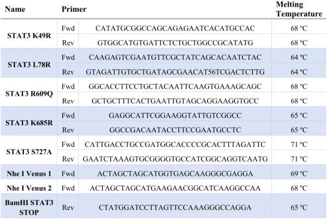
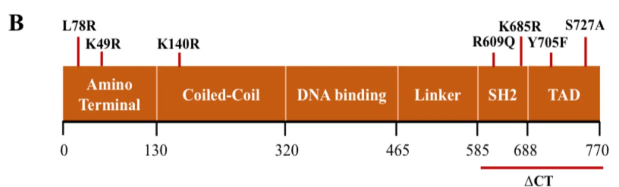
For the mutagenesis, five pairs of primers (forward and reverse, Table 3) were designed using PrimerX free software (http://www.bioinformatics.org/primerx/) with the aim of generating mutations by replacing a single residue by another without altering the fundamental amino acid structure of STAT3 (Fig. 4A). The original leucine (L) residue on position 78 and the lysine (K) residues on positions 49 and 685 were replaced by arginine (R); the arginine residue on position 609 by glutamine (Q);and the serine (S) residue on position 727 by alanine (A) (Fig. 4B).
Table 3 – Primers used for mutagenesis and PCR cloning. Fwd – forward; Rev – reverse.
PCR was carried out with Pfu Turbo DNA polymerase (2.5 U/μL), (Agilent; CA, USA), Venus-STAT3 BiFC constructs as DNA templates (10 ng) and the corresponding pair of primers (125 ng). The conditions used for the denaturation, annealing and extension were 30 s at 95 ºC, 16
Name Primer Melting Temperature
STAT3 K49R Fwd CATATGCGGCCAGCAGAGAATCACATGCCAC 68 ºC Rev GTGGCATGTGATTCTCTGCTGGCCGCATATG 68 ºC STAT3 L78R Fwd CAAGAGTCGAATGTTCGCTATCAGCACAATCTAC 64 ºC Rev GTAGATTGTGCTGATAGCGAACAT56TCGACTCTTG 64 ºC STAT3 R609Q Fwd GGCACCTTCCTGCTACAATTCAAGTGAAAGCAGC 68 ºC Rev GCTGCTTTCACTGAATTGTAGCAGGAAGGTGCC 68 ºC STAT3 K685R Fwd GAGGCATTCGGAAGGTATTGTCGGCC 65 ºC Rev GGCCGACAATACCTTCCGAATGCCTC 65 ºC
STAT3 S727A Fwd CATTGACCTGCCGATGGCACCCCGCACTTTAGATTC 71 ºC
Rev GAATCTAAAGTGCGGGGTGCCATCGGCAGGTCAATG 71 ºC
Nhe I Venus 1 Fwd ACTAGCTAGCATGGTGAGCAAGGGCGAGGA 69 ºC
Nhe I Venus 2 Fwd ACTAGCTAGCATGAAGAACGGCATCAAGGCCAA 68 ºC
BamHI STAT3
cycles of 30 s at 95 ºC, 1 min at 68 ºC and 9 min at 72 ºC. The cycles were adjusted according to the type of mutation desired and the annealing temperature was also adapted to the primers (Table 3). After PCR amplification, reactions were digested with DpnI restriction enzyme (10 U/μL) (Thermo Fisher Scientific; Waltham, MA, USA) for 1 h at 37 ºC. DpnI cleaves only methylated DNA, which is only present in the template DNA due to their previous amplification in bacteria. Thus, it digests the non-mutated templates and in principle leaves mutated constructs intact. Mutagenesis reactions were then transformed into thermocompetent Escherichia coli (NZYtech; Lisbon, Portugal). Briefly, the reaction mix was added to competent bacteria and placed on ice for 30 min. A heat-shock of 45 s at 42 ºC and 2 min in ice was given to the mix. Later, 300 μL of Luria Broth (LB) medium (1% w/v Tryptone; 0.5% w/v Yeast extract; 171 mM NaCl) without antibiotics were added and incubated in agitation at 120 rpm, 37 ºC for 1 h. Transformed bacteria were seeded on LB agar 1x (1% w/v Tryptone; 0.5% w/v Yeast extract; 171 mM NaCl; 1.5% w/v Agar) in petri dishes containing 100 μg/mL of ampicillin and grown overnight at 37 ºC. For DNA extraction and sequencing, colonies were collected and grown in 3 mL of LB medium containing 100 μg/mL of ampicillin at 37 ºC in agitation at 180 rpm overnight. The ZymoPure™ Mini Prep Kit (Zymo Research; CA, USA) allowed the DNA extraction and it was quantified on a Nanodrop 2000c from Thermo Fisher Scientific Inc. (Waltham, MA, USA) using H2O Mili-Q as blank. The success of mutagenesis was confirmed by sequencing (GATC, Germany).
Venus-STAT3 constructs without the SH2 and TAD domains (Venus-STAT3ΔCT) were obtained through cloning. Venus-STAT3 BiFC system carrying Wild-type STAT3 as template (Fig. 5A). This plasmid was submitted to cloning by amplifying the Venus-STAT3ΔCT fragment from the original pcDNA 3.3 TOPO vector and subcloned into the same vector. The primers used are shown in Table 3, and the restriction enzymes chosen to digest the Venus-STAT3ΔCT inserts without the C terminus were NheI and BamHI (10 U/μL) (Thermo Fisher Scientific; Waltham, CA, USA). PCRs were carried out using Phusion DNA polymerase (2 U/μL) (Thermo Fisher Scientific; Waltham, CA, USA), Venus-STAT3 BiFC constructs as DNA templates (10 ng), the corresponding pair of primers (100 ng) and DMSO (100%) (Thermo Fisher Scientific; Waltham, CA, USA). The conditions used for denaturation, annealing and extension were 30 s at 98 ºC, 30 cycles of 10 s at 98 ºC, 30 s at 67 ºC and 1 min at 72 ºC, and 10 min at 72 ºC. The extension time was calculated according to the operation rate of the polymerase (15-30 s/kb) and the size of Venus-STAT3ΔCT (approximately 2500 kb). The melting temperatures of primers were also taken into consideration for the annealing temperature. After the agarose gel electrophoresis confirmed that the primers worked (Fig. 5B), the inserts were purified using the NZYGelpure kit (NZYtech; Lisbon, Portugal).
Both the Venus-STAT3 BiFC system carrying Wild-type STAT3 and the PCR products were digested with NheI and BamHI restriction enzymes to remove the fragment containing Venus-STAT3 from the pcDNA 3.3 vector and generate compatible cohesive ends between them. Agarose gel electrophoresis was executed to confirm the efficiency of digestion and the insert was purified and quantified (Fig. 5C). DNA insert ligation into vector DNA was carried out using T4 DNA ligase (New England BioLabs; Ipswich, MA, USA) for 2 h at room temperature. The ligation products were then transformed into bacteria, and three colonies were selected for further growth, DNA extraction and quantification, as described above. Cloning was confirmed by digestion with NheI and BamHI and sequencing (Fig. 5D).
Figure 4 – Generation of the STAT3 constructs. A, Schematic representation of the generation of STAT3-L78R mutant construct. B, Localization of the STAT3 mutants on STAT3 BiFC constructs.
L78R, R609Q, Y705F-K49R, Y705F-K685R, Y705F-S727A mutations were generated in both V1- and V2-STAT3 BIFC constructs and the C-terminal was successfully removed from V2-V2-STAT3 BiFC construct. K49R, K140R, K685R, Y705F and S727A mutations were already made by mutagenesis.
3.3.
Cell Culture
HeLa human cervix adenocarcinoma cells (reference CRM-CLL-2) were maintained in 100 mm tissue culture dishes (VWR®; PA, USA) at 37 ºC in a humidified incubator with 5% CO2 in Dulbecco’s Modified Eagle’s Medium (DMEM, Biowest; Nuaillé, France) supplemented with 10 % fetal bovine serum (Biowest; Nuaillé, France), 2 mM glutamine (Lonza; Verviers, Belgium) and 1x Penicillin/Streptomycin solution (Gibco®; Carlsbag, CA, USA). Medium was changed every other day and cells where passed once a week by trypsinization (Trypsin 0.05% w/v, for 5 min at 37 ºC) (Corning®; Corning, NY, USA). For all experiments, cells were counted using a Neubauer Chamber from Assistent (Sonheim, Germany) and seeded in different types of dishes according to the type of assay. For flow cytometry experiments, cells were seeded at 8x10⁵
Figure 5 – Generation of Venus-STAT3ΔCT. A, Schematic representation of PCR cloning. B, Agarose
gel electrophoresis confirmed that (1) V1-STAT3ΔCT and (2) V2-STAT3ΔCT fragments were successfully amplified with NheI and BamHI primers. C, After digestion with NheI and BamHI, agarose gel electrophoresis showed three bands: the first being the Venus-STAT3 linearized plasmid (around 8000 kDa), the second (in red) being the pcDNA 3.3 vector (around 5000 kDa) and the third being the insert removed (V2-STAT3, around 2500 kDa). D, The reactions were digested with NheI and BamHI to confirm the ligation between pcDNA 3.3 vector and the Venus-STAT3ΔCT insert. (1) V1-STAT3ΔCT and (2)-(3) V2-STAT3ΔCT were the bands chosen for sequencing.
cells/well (6-well plates). For microscopy, cells were seeded at 8x10⁵ cells/dish in 35 mm
glass-bottom dishes. For western blotting, cells were seeded at 2x10⁶ cells/dish in 60 mm dishes.
3.3.1. Transfection
Cells were transfected with complementary pairs of Venus-STAT3 BiFC plasmids containing the gene sequence of Wild-type or mutant STAT3 using jetPRIME (Polyplus-transfection®; Illkirch, France) according to manufacturer’s instructions. Briefly, 24 h after cell seeding, the transfection mixture in a 1:3 ratio (1 µg of DNA: 3 µl of jetPRIME) was prepared and added to cells. Twenty-four hours after transfection, STAT3 dimerization was evaluated by flow cytometry, STAT3 expression and phosphorylation by Western Blot, and intracellular STAT3 localization by fluorescence microscopy. For some experiments, the STAT3 inhibitor Sttatic (Selleckchem, Houston, TX, USA) or the Leukemia inhibitory factor (LIF) (R&D systems, Minneapolis, MN, USA) were used as indicated in the corresponding figure legends.
3.4.
Fluorescence Microscopy
Living or fixed cells were examined in a custom-built Nikon Eclipse TE2000-S inverted fluorescence microscope equipped with a Hamamatsu Flash 2.8 sCMOS camera. For fixation, cells were washed with PBS and fixed and permeabilized in 100% ice-cold methanol, at -20 ºC, for 10 min. They were washed again and stored in PBS at 4 ºC.
Photos were taken using a 100x objective and handled with ImageJ free software. Cells were classified qualitatively in three categories according to the relative intensity and localization of the fluorescence signal (Fig. 6A, 6B, 6C): 1) predominantly in the cytoplasm, 2) predominantly in the nucleus, or 3) homogeneously distributed through nucleus and cytoplasm. These categories are mutually exclusive meaning their sum is 100% of cells. Furthermore, the percentage of cells with mitochondrial signal, where the fluorescent signal co-localizes with Mitotracker Red (Life Technologies; Eugene, OR, USA), or aggregates where the fluorescent signal did not co-localized with the dye (Fig. 6D), was also determined.
3.5.
Flow Cytometry
Cells were washed once with PBS and trypsinized (0.05% w/v) for 5 min at 37 ºC. They were collected in microcentrifuge tubes and centrifuged (300xg, 5 min at RT). The supernatant was discarded and the cell pellet resuspended in PBS. Fluorescence was measured using a FACSCalibur flow cytometer (Becton Dickinson, CA, USA) equipped with low-power air cooler 15 mW blue (488 nm) argon laser and a red (635 nm) diode laser (band-pass filter 530/30 nm). For each experiment group 10000 events were analyzed. Data were analyzed by means of FlowJo software (Tree Star Inc., OR, USA).
3.6.
Protein Analysis
3.6.1. Extraction of total protein
Cells were washed once with PBS and lysed using NP-40 lysis buffer (150 mM NaCl, 50 mM Tris-HCl pH 8.0, 1% NP-40) supplemented with tablets of protease and phosphatase inhibitors (Roche; Basel, Switzerland). Cells were scrapped directly from the plates into microcentrifuge tubes and incubated 10 min in ice. A W-450 D sonicator (Emerson, Danbury, USA) was used for 5 s at 10% of amplitude in order to disrupt cell membranes and release intracellular proteins. Cells were then centrifuged at 10,000 xg for 10 min at 4 ºC and the soluble protein fraction was collected Figure 6 – Representative fluorescence microscopy pictures of the qualitative classification in HeLa cells. A, Predominantly in the cytoplasm. B, Predominantly in the nucleus. C, Homogenously in the
cytoplasm and nucleus. D, Mitochondrial localization and aggregates with and without Mitotracker Red. Scale bar: 20 μm.