ISSN 0100-879X
BIOMEDICAL SCIENCES
AND
CLINICAL INVESTIGATION
www.bjournal.com.br
www.bjournal.com.br
Volume 44 (12) 1194-1298 December 2011
Institutional Sponsors
The Brazilian Journal of Medical and Biological Research is partially financed by
Faculdade de Medicina de Ribeirão Preto Campus
Ribeirão Preto
Ex plor e H igh - Pe r for m a n ce M S Or bit r a p Te ch n ology I n Pr ot e om ics & M e t a bolom ics
analit icaw eb.com .br S C I E N T I F I C
Braz J Med Biol Res, December 2011, Volume 44(12) 1222-1230
doi: 10.1590/S0100-879X2011007500143
Inhibition of STAT3 by RNA interference suppresses angiogenesis
in colorectal carcinoma
Inhibition of STAT3 by RNA interference
suppresses angiogenesis in
colorectal carcinoma
W.F. Qian
1, W.X. Guan
1, Y. Gao
1, J.F. Tan
1, Z.M. Qiao
1,
H. Huang
1and C.L. Xia
21Department of General Surgery, Suzhou Hospital Affiliated to Nanjing Medical University,
Suzhou, JS, China
2Department of Anatomy and Cytoneurobiology Laboratory, Medical College, Soochow University,
Suzhou Industrial Park, Suzhou, JS, China
Abstract
In order to investigate signal transduction and activation of transcription 3 (STAT3) signaling on angiogenesis in colorectal car -cinoma (CRC) after inhibiting STAT3 expression,we constructed the HT-29-shSTAT3 cell line by lentivirus-mediated RNAi. Cell
growth was assessed with MTT and the cell cycle distribution by flow cytometry. CRC nude mouse models were established and tumor growth was monitored periodically. On day 30, all mice were killed and tumor tissues were removed. Microvessel density (MVD) was determined according to CD34-positive staining. The expression of vascular endothelial growth factor A (VEGFA), matrix metalloproteinase-2 (MMP2) and basic fibroblast growth factor (FGF2) was monitored by quantitative real-time PCR and Western blot analysis. Knockdown of STAT3 expression significantly inhibited cell growth in HT-29 cells, with a significantly
higher proportion of cells at G0/G1 (P < 0.01). Consistently, in vivo data also demonstrated that tumor growth was significantly
inhibited in mice injected with HT-29-shSTAT3 cells. MVD was 9.80 ± 3.02 in the HT-29-shSTAT3 group, significantly less than that of the control group (P < 0.01). mRNA and protein levels of VEGFA and MMP2 in the HT-29-shSTAT3 group were significantly lower than in the control group (P < 0.05), but no significant difference was observed in the mRNA or protein level of FGF2 (P > 0.05). Taken together, these results demonstrate that STAT3 signaling is important to the growth of CRC and promotes angiogenesis by regulating VEGFA and MMP2 expression.
Key words: STAT3; RNA interference; Colorectal carcinoma, Angiogenesis; VEGFA; MMP2; FGF2
Introduction
Correspondence: W.F. Qian, Department of General Surgery, Suzhou Hospital Affiliated to Nanjing Medical University, 215002 Suzhou, China. E-mail: qianweifeng163@163.com
Received April 30, 2011. Accepted October 13, 2011. Available online October 28, 2011. Published November 28, 2011. Colorectal carcinoma (CRC) is the third most common
malignant neoplasm worldwide and the second leading
cause of cancer-related deaths (1). Despite recent advances
in diagnostic and therapeutic measures, the prognosis of colorectal cancer patients remains poor. Although
sig-nificant progress has been made in defining the molecular mechanisms of CRC development, the specific molecular regulation pathways involved in CRC progression are still largely unknown. In order to develop new therapeutic
strategies, it is critical to understand the mechanism of progression of CRC.
It is well known that all tumors undergo neovascular
-ization, or angiogenesis, in order to acquire nutrients for
continuing growth and metastatic spread. Not surprisingly,
angiogenesis is a complex multistage process and is regu-lated by the balance between angiogenic and angiostatic
factors (2). Recently, more studies have shown that signaling transduction and activation of transcription 3 (STAT3), a key
signaling transduction protein and an oncoprotein, appears
to play a central role in modulating angiogenesis in various
cancers including melanoma, breast cancer, gastric cancer,
and hepatocellular carcinoma (3-6).
The STAT3 protein was originally discovered as a latent
cytoplasmic transcription factor in response to extracel-lular signaling proteins, especially cytokines and growth
factors. Upon activation, STAT3 protein is phosphorylated
Downregulation STAT3 suppresses colorectal carcinoma 1223
activates transcription of the target genes of proliferation,
anti-apoptosis, or cell cycle progression by binding directly
to high-affinity DNA binding sites or by associating with other transcription factors. In addition to its diverse biologi
-cal functions involved in cell proliferation, differentiation, apoptosis, inflammation, and oncogenesis, accumulating evidence suggests that STAT3 also plays an important
role in angiogenesis in both physiological and
pathologi-cal situations (7-13). Several studies have reported that STAT3 directly regulates vascular endothelial growth factor A (VEGFA) expression to induce tumor angiogenesis in
breast cancer, gastric cancer and hepatocellular
carci-noma (4,5,14). In pancreatic cancer, STAT3-targeting RNA
interference (RNAi) inhibits angiogenesis by suppressing the expression of matrix metalloproteinase-2 (MMP2)
(15). In human melanoma brain metastases, activated
STAT3 contributes to tumor angiogenesis by regulating
the expression of basic fibroblast growth factor (FGF2) (3).
These results indicate that STAT3 signaling interferes with
the process of angiogenesis in various cancers through
different mechanisms.
Recent studies have shown that STAT3 was constitu
-tively activated in CRC cell lines (16). Suppression of STAT3 expression by small interfering RNA (siRNA) inhibited cell
invasion ability and induced anoikis in CRC cells (17). Clini
-cal data also showed that in colorectal adenocarcinoma, thephospho-Tyr-STAT3 (p-STAT3) level was significantly
correlated with the depth of tumor invasion, venous inva
-sion, lymph node metastasis, and increasing stages of the
Dukes’ classification (18,19). Taken together, these findings
demonstrate that the STAT3 pathway plays a critical role in the progression of CRC (20-22). However, it is unknown whether STAT3 signaling contributes to angiogenesis in CRC.
In the present study, we investigated the possible mecha -nism of STAT3 signaling and its effect on angiogenesis in CRC after inhibiting STAT3 expression with lentivirus-mediated short hairpin RNA (shRNA).
Material and Methods
Material
The HT-29 and 293T cell lines were purchased from the Institute of Biochemistry and Cell Biology, Shanghai Insti-tute for Biological Sciences, Chinese Academy of Science.
Fifteen syngeneic female BALB/c nude mice (6 weeks old) were provided by the Animal Center Laboratory of Soochow University and were housed under specific-pathogen-free
conditions. The use of animals in this study was approved by the Animal Ethics Committee of Soochow University and the protocol of animal treatment was approved by the Institutional Animal Care and Use Committee.
PRMI-1640 was purchased from Invitrogen (USA); fetal bovine serum was purchased from Hyclone (USA); trypsin was purchased from Sigma (USA); pRNAT-U6.2/Lenti
lentivirus vector and packaging system (pMD1g-pRRE, pRsv-REV and pMD2G) were purchased from Invitrogen;
Trizol reagent and lipofectamine 2000 were purchased from
Invitrogen; oligo(dT), RNase inhibitor, Taq Dnase, dNTP
mixture (10 mM), 10X PCR buffer and MgCl2 (25 mM) were purchased from Promega (China); M-MLV reverse
transcriptase and SYBR Green Real-Time PCR Master Mix kit were purchased from Toyobo (Japan); Nuclear-Cytosol
Extraction Kit was purchased from KeyGen Biotech (China); STAT3, p-STAT3, VEGFA, MMP2, FGF2, and GAPDH antibodies were purchased from Santa Cruz (USA); CD34
antibody, goat anti-rabbit IgG-horseradish peroxidase, and
antigen-removing solution of sodium citrate were purchased
from Boshide (China).
Construction of the recombinant lentivirus vector pRNAT-shSTAT3 and preparation of recombinant lentiviruses
According to a previous screening study (23), we
chose the site 2092-2112 of STAT3 (GeneBank
acces-sion No. NM_003150) as the target site of RNAi. The STAT3 hairpin oligos were synthesized (Invitrogen) and annealed. The sense was 5’-GATCCCGTCTTACCGCTG ATGTCCTTCTTTGATATCCGAGAAGGACATCAGCGG TAAGATTTTTTCCAAC-3’ and the anti-sense was 5’-GTT
GGAAAAAATCTTACCGCTGATGTCCTTCTCGGATATC AAAGAAGGACATCAGCGGTAAGACGGGATC-3’). A
non-targeting shRNA unrelated to human gene sequences was used as a negative control. The DNA fragments correspond
-ing to the target gene were amplified by PCR. The up (+)
position is a BamHI restriction site and the down (-) position is an XhoI restriction site. The PCR product and
pRNAT-U6.2/Lenti lentivirus vector driven by the U6 promoter and carrying the reporter gene, enhanced green fluorescence
protein (GFP), were mixed together and digested with
BamHI and XhoI. A T4 ligase reaction system was used to connect the appropriately digested oligonucleotide and
expression vector in recombinant clone ligation buffer. Competent DH5α cells were transformed with the ligated products and allowed to grow in culture. The correct positive clones were confirmed by sequencing analysis (Shanghai Invitrogen, Co., China).
The recombinant lentiviruses were produced by transient
transfection of 293T cells according to standard protocols
(24). Briefly, subconfluent 293T cells on 10-cm plates were cotransfected with 20 μg of a plasmid vector, 15 μg pMD1g-pRRE, 10 μg pRsv-REV, and 7.5 μg pMD2G by calcium phosphate precipitation. After 16 h the medium was changed, and recombinant lentiviruses were harvested
24 h later.
Transduction with recombinant lentiviruses and generation of stable cell lines
with either the recombinant STAT3-shRNA lentiviruses or the non-targeting shRNA recombinant lentiviruses in com -plete medium for 48 h (MOI 1:20). Cells were washed and transferred to complete medium for 48 h. After selection
by fluorescence-activated cell sorting using GFP as the
marker, the transfected cells were obtained and expanded
into stable cell lines. Cells were then harvested for the sub
-sequent experiments. The HT-29 cells transfected with the recombinant STAT3-shRNA lentivirus were named
HT-29-shSTAT3 cells, while cells transfected with the recombinant
non-targeting shRNA lentiviruses were named HT-29-GFP
cells. The parental HT-29 cells as well as HT-29-GFP cells
served as controls.
Quantitative real-time PCR
Total RNA was extracted from cells of each group (HT-29, HT-29-GFP and HT-29-shSTAT3) using Trizol reagent according to manufacturer instructions. Two micrograms
RNA was reverse transcribed in a 20-μL reaction solution containing 10 U M-MLV reverse transcriptase and 0.5 µg oligo(dT) primer. Quantitative real-time PCR, using SYBR
Green Real-Time PCR Master Mix kit, was performed according to the manufacturer protocol. The STAT3 gene
was amplified using specific oligonucleotide primers and
the housekeeping gene GAPDH was used as the internal
standard. The sequences of the STAT3 primers were sense 5’-GAGGACTGAGCATCGAGCA-3’, antisense 5’-CATGTGATCTGACACCCTGAA-3’, and the size of the products was 85 bp. The sequences of the GAPDH
primers were sense 5’-ACTCCTCCACCTTTGACGC-3’,
antisense 5’-CTCTTCCTCTTGTGCTCTTGC-3’, and the
size of the products was 180 bp. Thedata were quantified by the 2-DDCTmethod. All analyses were performed in triplicate.
Western blot analysis
Cell lysates were prepared with the Nuclear-Cytosol
Extraction Kit, proteins were partially separated by 10% SDS-PAGE and blotted onto a membrane. After blocking at 37°C in 5% milk in phosphate-buffered saline (PBS) contain
-ing 0.1% Tween 20 for 2 h, membranes were incubated with
primary antibody against STAT3,p-STAT3 and GAPDH at
4°C overnight. After extensive washing, immunocomplexes were detected with HRP-conjugated secondary antibodies
at room temperature. An enhanced chemiluminescence kit was used for detection. The
amount of GAPDH detected was used as a normalization control for protein loading.
Cell proliferation assay and cell cycle analysis
Cells were plated onto
96-well micro96-well plates at a density of 1000 cells/well. Cell
prolifera-tion was determined by the MTT assay to count cells at
570 nm absorbance (A570) on a multiwell plate reader.
For cell cycle analysis, cells were harvested and stained with propidium iodide to analyze DNA content by flow
cytometry.
In vivo experiments
Six-week-old syngeneic female BALB/c nude mice were
randomly divided into three groups: HT-29, HT-29-GFP, and HT-29-shSTAT3 (5 mice per group). Cells were suspended
in PBS and 1 x 10⁷ cells were injected subcutaneously into
the right back of the mice. Tumor size was calculated every 5 days using the simplified formula (L x W2 x 0.5). On day 30, all mice were killed and tumor tissue was removed for
further study.
Histopathological and immunohistochemical examination
Tissues were fixed in 10% neutral formalin, embedded
in paraffin and cut into 5-µm thick sections. Hematoxylin
and eosin (HE) staining was performed for histopathological
analysis. In addition, 5-µm thick sections of formalin-fixed,
paraffin-embedded specimens were stained with anti-CD34
antibodies (1:100) according to the manufacturer protocol
for immunohistopathological analysis. Microvessel density
(MVD) in the tumor samples was determined by the method
of Weidner (25). The immunostained sections were scanned at low magnification (40X) and the tumor area with the high
-est density of distinctly highlighted microvessels (“hot spot”)
was selected. MVD was then determined in the hot spot by
counting all vessels at 200X magnification, and the mean of the counts for three fields was calculated. Large vessels
with thick muscular walls were excluded from the counts,
and vessel lumens were not necessary for a structure to be defined as a vessel.
Quantitative real-time PCR and Western blot analysis
Tumor RNA of each of the 5 nude mice in each group was isolated and taken for quantitative real-time PCR of VEGFA, MMP2 and FGF2. The expression of the house -keeping gene GAPDH was used as an internal standard.
The real-time PCR primer sequences are shown in Table 1. Similarly, tumor tissue protein was exacted and subjected to Western blot analysis of VEGFA, MMP2 and FGF2 ac -cording to manufacturer instructions.
Table 1. Real-time PCR primer sequences.
Gene Sense Antisense Length (bp)
Downregulation STAT3 suppresses colorectal carcinoma 1225
Statistical analysis
The statistical significance of the means was calculated
by one-way ANOVA, followed by the
Student-Newman-Keuls test for multiple comparisons. P < 0.05 was required for statistical significance. All computations were carried out using the SPSS13.0 statistical program.
Results
Creation of stable cell lines and inhibition of the expression and activity of STAT3 by STAT3 shRNA in HT-29-shSTAT3 cells
The recombinant lentivirus vector was successfully constructed and confirmed by DNA sequencing. After the recombinant lentivirus vector was packaged in 293T cells, recombinant lentiviruses were obtained in the supernatant
at a titer of 2 x 10⁷ TU/mL. After being infected with the recombinant lentiviruses carrying the reporter gene GFP, the
transfected cells expressed GFP proteins. The proportion of
transfected HT-29 cells was analyzed under a fluorescence microscope. The results indicated that 85% HT-29 cells were transfected with the recombinant lentiviruses (Figure 1). Our data suggested that the lentivirus shRNA vector pRNAT-U6.2/Lenti was highly efficient in infecting HT-29 cells. After fluorescence-activated cell sorting, the HT-29-shSTAT3 line
and HT-29-GFP cell lines were obtained.
STAT3 mRNA expression in each cell group was analyzed by real-time PCR. The results demonstrated that endogenous STAT3 mRNA of the HT-29-shSTAT3 cells was markedly decreased compared to HT-29-GFP and HT-29 cells (Figure 2A). Consistently, as shown in Figure 2B, the expression of STAT3 protein in HT-29-shSTAT3 cells was down-regulated compared to HT-29-GFP and HT-29 cells.
To determine the levels of p-STAT3 protein, we performed
Western blot analysis using antibodies against p-STAT3. As shown in Figure 2B, p-STAT3 was also markedly
de-Figure 1. Micrographs of HT-29 cells infected with recombinant lentiviruses. After being in-fected with the recombinant lentiviruses carrying the reporter gene GFP, the transin-fected cells
expressed GFP proteins and could be observed under a fluorescence microscope. The results
indicated that 85% HT-29 cells were transfected with the recombinant lentiviruses. A and C,
creased in HT-29-shSTAT3 cells after STAT3 silencing by RNAi. This indicated that STAT3 shRNA reduced not only
the steady-state levels of total STAT3 protein, but also the levels of tyrosine-phosphorylated STAT3 protein in
HT-29-shSTAT3 cells.
STAT3 shRNA inhibited cell growth and induced
G0/G1 arrest
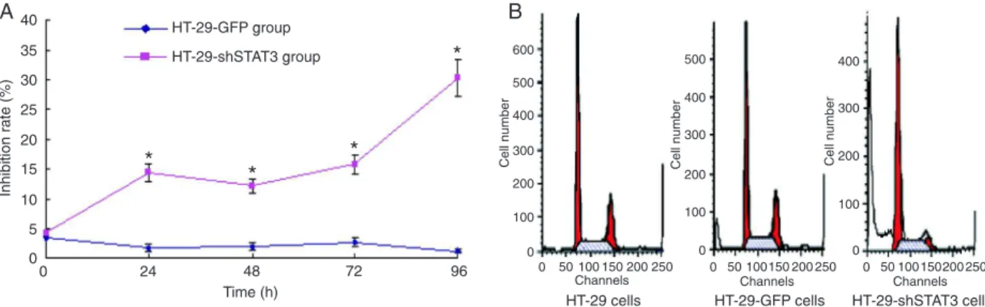
To determine whether STAT3 down-regulated by RNAi had an inhibitory effect on HT-29 cell growth, cell prolifera-tion rate was determined by MTT assay. As shown in Figure 3A, inhibition of STAT3 expression by RNAi suppressed the growth of HT-29-shSTAT3 cells. Cell cycle analysis (Figure 3B) showed that the proportion of G0/G1 of HT-29 cells and HT-29-GFP cells was 38.65 ± 2.33 and 38.48 ± 1.63%, respectively. The proportion of G0/G1 of HT-29-shSTAT3 cells was 68.73 ± 2.88%, significantly increased compared
to HT-29-GFP and HT-29 cells (P < 0.01).
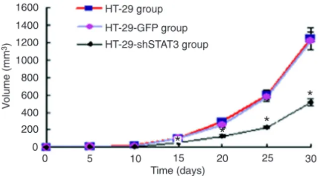
Therapeutic effect on tumor-bearing nude mice
In nude mice, HT-29 and HT-29-GFP cells rapidly formed
tumors after injection. However, tumors grew slowly in the
HT-29-shSTAT3 nude mouse group, with a smaller size than
those in the HT-29 and HT-29-GFP groups from day 15 after inoculation (P < 0.05). From day 25 after injection there was a very significant difference (P < 0.01). Although the average
tumor size in the HT-29-GFP group was smaller than that in the
HT-29 group, the difference was not significant (Figure 4).
Effect of STAT3 suppression on histopathology and immunohistochemistry
HE-stained sections were examined under a light mi
-croscope (Figure 5). In the HT-29 and HT-29-GFP groups there were many heterokaryocytes and blood vessels. The MVD of the two groups was 28.73 ± 5.11 and 27.60 ± 4.27, respectively (Figure 5). In contrast, in the HT-29-shSTAT3 group there were few blood vessels, with an MVD of 9.80
Figure 2.A, STAT3 mRNA expression in cells of each group. Group 1, HT-29 cells; group 2, HT-29-GFP cells; group 3, HT-29-shSTAT3
cells. Data are reported as means ± SEM (N = 3). *P < 0.01, significantly different from the other two groups (ANOVA). B, Expression and activation of STAT3 proteins in cells of each group. Lane 1 = HT-29 cells; lane 2 = HT-29-GFP cells; lane 3 = HT-29-shSTAT3
cells.
Figure 3. A, Growth inhibition curves of HT-29-shSTAT3 and HT-29-GFP cells. Data are reported as means ± SEM (N = 3). *P < 0.01, significantly different from the HT-29-GFP group (ANOVA). B, Cell cycle of each cell group. Cell cycle analysis showed that the proportion of G0/G1 of HT-29 cells and GFP cells was 38.65 ± 2.33 and 38.48 ± 1.63%, respectively. The percentage of
Downregulation STAT3 suppresses colorectal carcinoma 1227
± 3.02, significantly lower than that of the HT-29 or
HT-29-GFP group (P < 0.01).
Expression of VEGFA,MMP2 and FGF2 in tumors
The mRNA expression of VEGFA, MMP2and FGF2 in
tumors was analyzed by quantitative real-time PCR (Figure 6A). The protein level of the target genes was determined by Western blot analysis (Figure 6B). In the HT-29-shRNA group, the mRNA and protein levels of VEGFA and MMP2 were significantly lower compared to the 29 and HT-29-GFP groups (P < 0.05), but no significant change was observed in the mRNA or protein level of FGF2 among
these three groups (P > 0.05). Figure 4. HT-29 and HT-29-GFP groups (ANOVA).Change of tumor volumes in each group. *P < 0.05 vs
Figure 5. HE staining and CD34 immunohistochemical analysis. A, HE staining of the HT-29; B, HT-29-GFP; C, HT-29-shSTAT3 groups. In A and B, there were many heterokaryocytes and blood vessels. However, in C there were few blood
vessels. D, E, and F, CD34 immunohistochemical analysis in the three groups, respectively. In D and E, microvessel
Discussion
STAT family proteins were originally identified as key
components of cytokine signaling pathways that
regu-late gene expression (26). Among the 7 members of the
mammalian STAT family, STAT3 is the one most strongly
implicated in oncogenesis (27). Many studies have sug
-gested that persistently activated STAT3 participates in
oncogenesis through disregulation of genes encoding the antiapoptotic proteins bcl-xL, mcl-1, and bcl-2, as well as the proliferation-associated proteins cyclin D1 and c-Myc (28-30). In addition, STAT3 is able to regulate the innate and
adaptive immune responses during cancer development (31). Recently, the constitutively activated STAT3 has been reported to induce tumor angiogenesis in various cancers
including melanoma, breast cancer, gastric cancer, and
hepatocellular carcinoma (3-6). It is not known if STAT3
signaling contributes to angiogenesis in CRC.
In order to investigate the possible mechanism of
STAT3 signaling and its effect on angiogenesis in CRC
after inhibiting STAT3 expression, we first constructed the HT-29-shSTAT3 cell line successfully by lentivirus-mediated RNAi. The siRNA can be used effectively in vitro and in vivo
to suppress gene expression in eukaryotic cells (32), but the inhibitory effect of siRNAs on target gene expression
only lasts a few days. Recently, investigators have learned that a lentivirus vector can integrate stably into the chromo
-somes of their target cells, a likely requisite for long-term expression, and transduce nondividing cells. Thus, lentivirus vector-mediated siRNA can inhibit target gene expression for a long time. Therefore, a lentivirus siRNA vector is an attractive tool, which can inhibit gene expression with high efficiency and has become a new tool for human gene therapy. Our data demonstrate that the rate of lentivirus vector transfected to HT-29 cells was 85%. Quantitative
real-time PCR analysis and Western blot assay indicated
that the expression of STAT3 in HT-29 cells transfected
with STAT3 shRNA was significantly suppressed at both the mRNA and protein level. The present data show that
STAT3 shRNA inhibited not only STAT3 expression but also
the activity of STAT3 in HT-29 cells.
Accumulating evidence has demonstrated that STAT3
plays an important role in cell proliferation of some cancers (33). The mechanism by which STAT3 participates in cell proliferation is based on induction of cell cycle genes, such as cyclin D1, c-Myc, etc. Both cyclin D1 and c-Myc respond
to mitogenic signaling and are required for progression of
cells from the G1 to the S phase of the cell cycle. Here, in order to determine whether STAT3 regulated HT-29 cell proliferation, the cell growth rate and cell cycle of HT-29-shSTAT3 cells were analyzed. The results showed that down-regulation of STAT3 also inhibited the growth of HT-29-shSTAT3 cells and induced G0/G1 arrest of these cells. In addition, the present in vivo study showed that tumors in the HT-29-shSTAT3 group grew slowly, with a smaller size than in the HT-29 and HT-29-GFP groups. This clearly indicated that STAT3 signaling had an important effect on tumor growth in CRC-bearing nude mice.
For tumor growth, angiogenesis is essential and
impor-tant, a process that involves endothelial cell proliferation, selective degradation of the basement membrane and the
surrounding extracellular matrix, endothelial cell migration, and the formation of a tubular structure. Malignant tumors depend on capillary proliferation to maintain the oxygen and nutrient supply for their continuous growth.
Numer-ous lines of evidence have shown that angiogenesis, as quantitated according to MVD, which can reflect the status of angiogenesis in tumor tissue, plays a significant clinico
-pathologic role in cancer patients (34,35). In CRC, MVD is an important, highly significant and accurate prognostic factor (36,37). The MVD method, i.e., counting microvessels in hot spots, has been the preferred method (25).In order
Figure 6. A, VEGFA, MMP2 and FGF2 mRNA expression of each group. Data are reported as means ± SEM (N = 5). *P < 0.05, significantly different from the other two groups (ANOVA). B,
Expression of VEGFA, MMP2 and FGF2 proteins (an example of a mouse tumor sample from each
Downregulation STAT3 suppresses colorectal carcinoma 1229
to count microvessels, visualization of endothelial cells by antibodies specifically targeting them is essential. At pres -ent, CD34, CD31 and factor VIII are the antibodies most commonly used for this purpose. In the present study, the results of CD34 immunohistochemistry showed that MVD
was significantly lower in the HT-29-shSTAT3 groups than
in the other two groups, implying that STAT3 signaling has an important effect on angiogenesis in CRC.
Angiogenesis is a complex multistage process and is regulated by the balance between many stimulatory and
inhibitory factors. In CRC, studies have shown that some proangiogenic factors, such as VEGFA, MMP2 and FGF2,
play important roles in angiogenesis (37-39). In breast cancer, gastric cancer and hepatocellular carcinoma,
STAT3 signaling controls VEGFA expression (4,5,14).
In pancreatic cancer, STAT3 signaling regulates the
ex-pression of MMP2 (15), and in human melanoma brain metastases, activated Stat3 regulates the expression of FGF2 (3). These results demonstrate that constitutive
STAT3 signaling contributes to tumor angiogenesis through multiple mechanisms. In order to probe the possible mechanism of the STAT3 signaling pathway underlying angiogenesis in CRC, we determined the expressions of VEGFA, MMP2 and FGF2. The result of quantitative real-time PCR analysis and Western blot assay showed that the
expression of VEGFA and MMP2 was significantly
down-regulated in xenografts originating from HT-29-shSTAT3
cells. That is, down-regulated STAT3 signaling caused a
significantly decreased expression of VEGFA and MMP2 in CRC. However, it was surprising that there was no significant difference in FGF2 expression among these
three groups. On the basis of these data, we speculate
that STAT3 signaling activates and promotes angiogenesis by regulating VEGFA and MMP2 expression in CRC. It is highly possible that STAT3 activation is a common signal regulating VEGFA and MMP2 expression, and VEGFA and
MMP2 are the downstream target molecules of STAT3 signaling in CRC. In contrast, FGF2 may not participate in the STAT3 signaling pathway in CRC.
Our findings show that STAT3 signaling plays an im -portant role in HT-29 cell growth. STAT3 targeting shRNA can remarkably silence the expression of the gene STAT3 in HT-29 cells, resulting in inhibition of cell growth and cell cycle arrest at the G0/G1 phase. In nude mice, STAT3 sig-naling is important for the growth of CRC and can promote
angiogenesis by regulating VEGFA and MMP2 expression.
These studies suggest that targeting STAT3 signaling can be an important approach to the control of tumor growth and angiogenesis in CRC.
Acknowledgments
Research supported by the Suzhou Social Development
Program (#SS08033).
References
1. Jemal A, Murray T, Samuels A, Ghafoor A, Ward E, Thun MJ.
Cancer statistics, 2003. CA Cancer J Clin 2003; 53: 5-26. 2. Bergers G, Benjamin LE. Tumorigenesis and the angiogenic
switch. Nat Rev Cancer 2003; 3: 401-410.
3. Xie TX, Huang FJ, Aldape KD, Kang SH, Liu M,
Gershen-wald JE, et al. Activation of stat3 in human melanoma pro -motes brain metastasis. Cancer Res 2006; 66: 3188-3196. 4. Hsieh FC, Cheng G, Lin J. Evaluation of potential
Stat3-regulated genes in human breast cancer. Biochem Biophys Res Commun 2005; 335: 292-299.
5. Gong W, Wang L, Yao JC, Ajani JA, Wei D, Aldape KD, et al. Expression of activated signal transducer and activator of transcription 3 predicts expression of vascular endothelial
growth factor in and angiogenic phenotype of human gastric cancer. Clin Cancer Res 2005; 11: 1386-1393.
6. Wang XH, Liu BR, Qu B, Xing H, Gao SL, Yin JM, et al.
Silencing STAT3 may inhibit cell growth through regulating signaling pathway, telomerase, cell cycle, apoptosis and angiogenesis in hepatocellular carcinoma: potential uses for gene therapy. Neoplasma 2011; 58: 158-171.
7. Chen Z, Han ZC. STAT3: a critical transcription activator in
angiogenesis. Med Res Rev 2008; 28: 185-200.
8. Valdembri D, Serini G, Vacca A, Ribatti D, Bussolino F. In vivo activation of JAK2/STAT-3 pathway during angiogen -esis induced by GM-CSF. FASEB J 2002; 16: 225-227. 9. Osugi T, Oshima Y, Fujio Y, Funamoto M, Yamashita A, Ne
-goro S, et al. Cardiac-specific activation of signal transducer and activator of transcription 3 promotes vascular formation
in the heart. J Biol Chem 2002; 277: 6676-6681.
10. Niu G, Wright KL, Huang M, Song L, Haura E, Turkson J, et al. Constitutive Stat3 activity up-regulates VEGF expression
and tumor angiogenesis. Oncogene 2002; 21: 2000-2008. 11. Wei D, Le X, Zheng L, Wang L, Frey JA, Gao AC, et al. Stat3
activation regulates the expression of vascular endothelial
growth factor and human pancreatic cancer angiogenesis and metastasis. Oncogene 2003; 22: 319-329.
12. Haura EB, Turkson J, Jove R. Mechanisms of disease:
Insights into the emerging role of signal transducers and
activators of transcription in cancer. Nat Clin Pract Oncol 2005; 2: 315-324.
13. Shailubhai K, Dheer S, Picker D, Kaur G, Sausville EA, Ja -cob GS. Atiprimod is an inhibitor of cancer cell proliferation and angiogenesis. J Exp Ther Oncol 2004; 4: 267-279.
14. Li J, Piao YF, Jiang Z, Chen L, Sun HB. Silencing of signal
transducer and activator of transcription 3 expression by
RNA interference suppresses growth of human hepatocel-lular carcinoma in tumor-bearing nude mice. World J Gas-troenterol 2009; 15: 2602-2608.
1099-1106.
16. Lin Q, Lai R, Chirieac LR, Li C, Thomazy VA, Grammatika
-kis I, et al. Constitutive activation of JAK3/STAT3 in colon
carcinoma tumors and cell lines: inhibition of JAK3/STAT3 signaling induces apoptosis and cell cycle arrest of colon carcinoma cells. Am J Pathol 2005; 167: 969-980.
17. Fan Y, Zhang YL, Wu Y, Zhang W, Wang YH, Cheng ZM, et
al. Inhibition of signal transducer and activator of transcrip
-tion 3 expression by RNA interference suppresses invasion
through inducing anoikis in human colon cancer cells. World J Gastroenterol 2008; 14: 428-434.
18. Kusaba T, Nakayama T, Yamazumi K, Yakata Y, Yoshizaki
A, Nagayasu T, et al. Expression of p-STAT3 in human
colorectal adenocarcinoma and adenoma; correlation with clinicopathological factors. J Clin Pathol 2005; 58: 833-838.
19. Ma XT, Wang S, Ye YJ, Du RY, Cui ZR, Somsouk M.
Con-stitutive activation of Stat3 signaling pathway in human
colorectal carcinoma. World J Gastroenterol 2004; 10: 1569-1573.
20. Windham TC, Parikh NU, Siwak DR, Summy JM, McCon
-key DJ, Kraker AJ, et al. Src activation regulates anoikis in
human colon tumor cell lines. Oncogene 2002; 21: 7797-7807.
21. Laird AD, Li G, Moss KG, Blake RA, Broome MA,
Cher-rington JM, et al. Src family kinase activity is required for signal tranducer and activator of transcription 3 and focal adhesion kinase phosphorylation and vascular endothelial
growth factor signaling in vivo and for anchorage-dependent and -independent growth of human tumor cells. Mol Cancer Ther 2003; 2: 461-469.
22. Orlovsky K, Theodor L, Malovani H, Chowers Y, Nir U.
Gamma interferon down-regulates Fer and induces its
association with inactive Stat3 in colon carcinoma cells. Oncogene 2002; 21: 4997-5001.
23. Ruan JL, Gao Y, Guan WX, Qiao ZM, Qian WF. Construction
of STAT3 lentiviral vector. J Nantong Univ Med Sci 2010; 30:
83-85.
24. Zufferey R, Nagy D, Mandel RJ, Naldini L, Trono D. Multiply
attenuated lentiviral vector achieves efficient gene delivery in vivo. Nat Biotechnol 1997; 15: 871-875.
25. Weidner N. Intratumor microvessel density as a prognostic
factor in cancer. Am J Pathol 1995; 147: 9-19.
26. Murray PJ. The JAK-STAT signaling pathway: input and
output integration. J Immunol 2007; 178: 2623-2629. 27. Haura EB. SRC and STAT pathways. J Thorac Oncol 2006;
1: 403-405.
28. Mora LB, Buettner R, Seigne J, Diaz J, Ahmad N, Garcia
R, et al. Constitutive activation of Stat3 in human prostate
tumors and cell lines: direct inhibition of Stat3 signaling in-duces apoptosis of prostate cancer cells. Cancer Res 2002;
62: 6659-6666.
29. Bowman T, Broome MA, Sinibaldi D, Wharton W, Pledger
WJ, Sedivy JM, et al. Stat3-mediated Myc expression is required for Src transformation and PDGF-induced mitogen -esis. Proc Natl Acad Sci U S A 2001; 98: 7319-7324. 30. Masuda M, Suzui M, Yasumatu R, Nakashima T, Kuratomi Y,
Azuma K, et al. Constitutive activation of signal transducers and activators of transcription 3 correlates with cyclin D1 overexpression and may provide a novel prognostic marker in head and neck squamous cell carcinoma. Cancer Res 2002; 62: 3351-3355.
31. Wang T, Niu G, Kortylewski M, Burdelya L, Shain K, Zhang
S, et al. Regulation of the innate and adaptive immune re -sponses by Stat-3 signaling in tumor cells. Nat Med 2004;
10: 48-54.
32. Lewis DL, Hagstrom JE, Loomis AG, Wolff JA, Herweijer H. Efficient delivery of siRNA for inhibition of gene expression
in postnatal mice. Nat Genet 2002; 32: 107-108.
33. Aggarwal BB, Sethi G, Ahn KS, Sandur SK, Pandey MK, Kunnumakkara AB, et al. Targeting
signal-transducer-and-activator-of-transcription-3 for prevention and therapy of
cancer: modern target but ancient solution. Ann N Y Acad Sci 2006; 1091: 151-169.
34. Weidner N, Semple JP, Welch WR, Folkman J. Tumor
an-giogenesis and metastasis - correlation in invasive breast
carcinoma. N Engl J Med 1991; 324: 1-8.
35. Weidner N, Folkman J, Pozza F, Bevilacqua P, Allred EN, Moore DH, et al. Tumor angiogenesis: a new significant
and independent prognostic indicator in early-stage breast carcinoma. J Natl Cancer Inst 1992; 84: 1875-1887. 36. Gao J, Knutsen A, Arbman G, Carstensen J, Franlund B, Sun
XF. Clinical and biological significance of angiogenesis and
lymphangiogenesis in colorectal cancer. Dig Liver Dis 2009;
41: 116-122.
37. Des Guetz G, Uzzan B, Nicolas P, Cucherat M, Morere JF, Benamouzig R, et al. Microvessel density and VEGF
expression are prognostic factors in colorectal cancer. Meta-analysis of the literature. Br J Cancer 2006; 94:
1823-1832.
38. Kim TS, Kim YB. Correlation between expression of matrix metalloproteinase-2 (MMP-2), and matrix metalloprotei-nase-9 (MMP-9) and angiogenesis in colorectal adenocar-cinoma. J Korean Med Sci 1999; 14: 263-270.
39. Kos M, Dabrowski A. Tumour’s angiogenesis - the function