Temporal analysis of oxidative effects on the pulmonary inflammatory
response in mice exposed to cigarette smoke
Keila Karine Duarte Campos
a, Rafaela Gontijo Manso
b, Evandro Guedes Gonçalves
b,
Marcelo Eustáquio Silva
c, Wanderson Geraldo de Lima
d, Cristiane Alves Silva Menezes
e, Frank Silva
Bezerra
a,⇑aLaboratory of Metabolic Biochemistry (LBM), Department of Biological Sciences (DECBI), Center of Research in Biological Sciences (NUPEB), Federal University of Ouro Preto, Ouro Preto, MG, Brazil
bGraduating in Medicine, Federal University of Ouro Preto, Ouro Preto, MG, Brazil cLaboratory of Experimental Nutrition (LABNEX), School of Nutrition, Ouro Preto, MG, Brazil
dLaboratory of Immunopathology (LIMP), Department of Biological Sciences (DECBI), Institute of Exact and Biological Sciences (ICEB), Federal University of Ouro Preto, MG, Brazil eLaboratory of Immunoparasitology (LIP), Department of Biological Sciences, Center of Research in Biological Sciences, Federal University of Ouro Preto, MG, Brazil
a r t i c l e
i n f o
Article history:
Received 3 December 2012 Accepted 9 July 2013 Available online 18 July 2013
Keywords:
Cigarette smoke Flow cytometry Lung inflammation Oxidative stress Mice
a b s t r a c t
The most common factor related to the chronic obstructive pulmonary disease (COPD) development is the chronic smoking habit. Our study describes the temporal kinesis of pulmonary cellular influx through BALF analyses of mice acutely exposed to cigarette smoke (CS), the oxidative damage and antioxidative enzyme activities. Thirty-six mice (C57BL/6, 8 weeks old, male) were divided in 6 groups: the control group (CG), exposed to ambient air, and the other 30 mice were exposed to CS. Mice exposed to CS pre-sented, especially after the third day of exposure, different cellular subpopulations in BALF. The oxidative damage was significantly higher in CS exposed groups compared to CG. Our data showed that the eval-uated inflammatory cells, observed after three days of CS exposure, indicate that this time point could be relevant to studies focusing on these cellular subpopulation activities and confirm the oxidative stress even in a short term CS exposure.
Ó2013 Elsevier Inc. All rights reserved.
1. Introduction
Chronic obstructive pulmonary disease (COPD) is characterized by chronic airway inflammation, irreversible airflow limitation and emphysema[1,2]. The risk factors of COPD include indoor air pol-lution from biomass fuel, pulmonary tuberculosis, chronic asthma, socioeconomic status, genetic background and environmental fac-tors[3], however, the disease occurs predominantly in adult ciga-rette smokers[4]. Despite this, over 1 billion people continue to smoke and half of them are likely to develop a serious smoking-re-lated disease. Although the efforts to reduce smoking prevalence has to be brought into focus, understanding the processes that con-tribute to the inception and progression of smoking-related ill-nesses are of equal importance, given the highly addictive nature of cigarette smoke[5].
The lungs are an important way of exposure to environmental pathogens and antigens; nonspecific and specific defense mecha-nisms are involved in cleaning up these foreign substances from
the lungs. Protection against the foreign material reaching the lung alveoli involves innate and adaptive immune responses.
The innate defense system of the lung is provided by the epithe-lial barrier and the acute inflammatory response which follows tis-sue injury, including the recruitment and activation of neutrophils, eosinophils and macrophages[7]. Resident and inflammatory lung macrophages exhibit different origins and lifespans in lungs and have been identified as key regulators of pathological and repara-tive processes. Alveolar macrophages, which are considered tis-sue-resident macrophages, populate lung tissue during early embryogenesis and remain viable for prolonged periods with min-imal replenishment from bone marrow-derived monocytes. In con-trast, inflammatory macrophages originate from bone marrow-derived monocytes and have a shorter half-life[7]. Macrophages are activated by CS extract and secrete not only elastolytic en-zymes, but also many inflammatory chemokines (e.g., interleu-kin-8 and CXCR3-ligands), attracting neutrophils and cells from acquired immunity[6].
The adaptive immune response is dependent upon B- and T-lymphocytes (CD4+and CD8+), and has a longstanding memory for previous damage[6]. The acquired immunity involves specific immune responses that are elicited by antigens of various origins and that are executed primarily by T and B cells. Acute smoke
0008-8749/$ - see front matterÓ2013 Elsevier Inc. All rights reserved.
http://dx.doi.org/10.1016/j.cellimm.2013.07.002 ⇑ Corresponding author.
E-mail address:[email protected](F.S. Bezerra), [email protected](F.S. Bezerra).
Contents lists available atScienceDirect
Cellular Immunology
exposure resulted in significant increases in neutrophils and mononuclear cells within the lung[5], suggesting that all the dif-ferent inflammatory cells together are responsible for lung injury caused by cigarette smoke[6].
Oxidative stress has been implicated as a strong factor favoring the pathogenesis and progression of COPD [9]. Cigarette smoke (CS) is associated to the oxidative stress in several organs because it contains high concentrations of free radicals and reactive oxygen species (ROS)[10]. Oxidants present in cigarette smoke can stimu-late alveolar macrophages to produce ROS and to release media-tors, some of which attract neutrophils and other inflammatory cells into the lungs.[11]. Inin vitrostudies using alveolar macro-phages and bronchial epithelial cells, ROS have been shown to in-duce gene expression of inflammatory mediators, such as IL-1 and TNF alpha. The direct or indirect oxidant stress to the airway epi-thelium and alveolar macrophages may also generate cytokines, such as TNF alpha and IL-1beta, which in turn can activate airway epithelial cells to induce pro-inflammatory genes, such as TNF al-pha, IL-8, IL-1, iNOS, COX-2, ICAM-1, VCAM-1, IL-6, MMP-9, MIP-1alpha, GM-CSF, stress response genes and antioxidative enzymes (such as MnSOD and thioredoxin)[12].
Evidences support an imbalance between oxidant and antioxi-dant agents in the lungs and bloodstream of cigarette smokers and COPD patients [13]. Structural changes to essential compo-nents of the lung are caused by oxidative stress, contributing to irreversible damage of both parenchyma and airway walls [14]. These changes result in inflammatory cells influx followed by the increase of lipid peroxidation products, pro-inflammatory cyto-kines and altered antioxidative capability[15]. In order to mini-mize the oxidative damage, mammalian lungs present an integrated antioxidative enzymatic system[16,17]. The main com-ponents of this antioxidative enzymatic system are superoxide dis-mutase (SOD), catalase (CAT) and glutatione peroxidase (GPx). SOD is the primary enzymatic defense in the lungs against the damag-ing effects of O2 and H2O2by converting O2 into H2O2, which is a substrate for CAT and GPx[18]. The antioxidative enzymes consti-tute a critical mechanism to protect the lung parenchyma from damage caused by free radicals[19].
The present work aimed to evaluate, phenotypically, the tempo-ral cellular influx, the oxidative damage and antioxidative enzy-matic system activities in the lung of mice acutely exposed to cigarette smoke.
2. Methods
2.1. Animals
Male C57BL/6 mice, 8 weeks old (Laboratory of Experimental Nutrition, Department of Food – School of Nutrition, Federal Uni-versity of Ouro Preto) were housed under controlled conditions in standard laboratory cages. They were provided free access to water and food. All in vivo experimental protocols in animals at the Federal University of Ouro Preto were approved by the Ethics Committee.
2.2. Reagents
Coomassie blue, bovine serum albumin (BSA), thiobarbituric acid (TBA), trichloroacetic acid, tetramethoxypropane (TMP), adrenaline, glycine buffer, catalase, nicotinamide adenine dinucle-otide phosphate (NADPH), ethylenediaminetetraacetic acid (EDTA), hydrogen peroxide, phosphate buffered saline (PBS), glutathione reductase, NaHCO3, sodium azide, monoclonal antibodies (anti-CD4-PE, CD8-PerCP, GR1-FITC, F4/80 APC and CD11b PE).
2.3. Cigarette smoke exposure protocol
C57BL6 male mice (n= 36) were exposed to 6 commercial full-flavor filtered Virginia cigarettes (10 mg of tar, 0.9 mg of nicotine and 10 mg of carbon monoxide) per day for 5 days by using a smoking chamber previously described[20] [21]. The groups ex-posed to CS for 1, 2, 3, 4 or 5 days were called CSD1, CSD2, CSD3, CSD4, and CSD5, respectively. Briefly, each group of mice was placed in the inhalation chamber (40 cm long, 30 cm wide and 25 cm high), inside an exhaustion chapel. A cigarette was coupled to a plastic 60 mL syringe so that puffs could be drawn in and sub-sequently expelled into the exposure chamber. One liter of smoke from one cigarette was aspirated with this syringe (20 puffs of 50 mL) and the puff was immediately injected into the chamber. The 6 animals of each group were maintained in this smoke-air condition (3%) for 6 min, then the cover was removed from the inhalation chamber and by turning on the exhaust fan of the cham-ber of the chapel, the smoke was evacuated within 1 min. The mice were then immediately exposed to CS from a second cigarette for 6 min. The treatment was performed three times per day (morning, noon and afternoon), being two cigarettes per inhalation. The mice exposed to ambient air were used as the control group (CG;n= 6)
[22].
2.4. Bronchoalveolar lavage fluid (BALF), cell staining and flow cytometry
The animals were killed by cervical displacement. Airspaces were washed with buffered saline solution (0,5 mL) for three con-secutive times in the lung (final volume 1.2–1.5 mL). The fluid was withdrawn and stored on ice. Cells from BALF samples were counted using standard morphologic criteria and used to flow cytometry analyses.
BALF cells were incubated with the different antibody solutions for 30 min at 4°C, washed with phosphate-buffered saline (PBS, pH 7.2) and fixed in a formaldehyde-containing solution. The expres-sion of surface molecules was investigated combining differentially labeled CD4 and CD8 or F4/80, CD11b and anti-GR1. All the antibodies were from BD-Pharmingen, San Jose, CA. Fixed samples were maintained in the dark at 4°C until the acqui-sition of FACSCalibur (Becton–Dickinson). The data analyses were performed using the software FlowJo (Tree Star).
2.5. Processing and homogenized tissue
After performing BALF, the right ventricle was perfused with saline to remove blood from the lungs. The right lung was clamped so that just the left lung could be perfused with 4% buffered forma-lin (pH 7.2) at a pressure of 25 cm H2O for 2 min, via trachea. The left lung was removed and then immersed in a fixative solution for 48 h. Then, the material was processed as follows: bath with tap water for 30 min.
2.6. Analysis of oxidative damage and antioxidant enzyme activities
As an index of lipid peroxidation we used the formation of TBARS during an acid-heating reaction as previously described by Draper [23]. Briefly, the samples from lung homogenates were mixed with 1 mL of 10% trichloroacetic acid and 1 mL of 0.67% thiobarbituric acid; they were subsequently heated in a boiling water bath for 30 min. TBARS were determined by the absorbance at 532 nm and were expressed as malondialdehyde equivalents (nm/mg protein). Lung homogenates were used to determine SOD, CAT, and GPx activities. The SOD activity was assayed by mea-suring the inhibition of adrenaline auto-oxidation as absorbance at 480 nm[24]. CAT activity was measured by the rate of the decrease in H2O2at 240 nm[25]. GPx activity was measured by monitoring the oxidation of NADPH at 340 nm in the presence of H2O2[26]. The total protein content in each sample was determined by the method of Bradford[27].
2.7. Statistical analysis
The normal distribution of each variable was evaluated using the Kolmogorov–Smirnov test and were expressed as mean-s ± SEM. For comparimean-son among groupmean-s, one-way ANOVA followed by the Tukey post-test were performed (p< 0.05). The Kruskal– Wallis test followed by the Dunns post-test was used to analyze discrete data (p< 0.05). The InStat Graphpad software was used to perform the statistical analyses (GraphPad InStat version 5.00 for Windows 7, GraphPad Software, San Diego, CA, USA).
3. Results
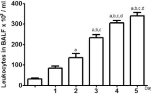
In order to verify the dynamic of the cellular influx during the five days of CS exposure, we determined the amount of leucocytes, presented in bronchoalveolar lavage fluid (BALF) of the analyzed groups (Fig. 1). A gradual increase of inflammatory cells in pulmon-ary tissue was observed from the second day until the last day of cigarette smoke exposure. The increscent quantity of cells on the second day (135 ± 21.56103/mL), third day (233 ± 13.82103/ mL), fourth day (305 ± 12.04103/mL) and fifth day (340 ± 15.92103/mL) of exposure were higher when compared to the cellular amount in control group (CG) (31.67 ± 4.01103/ mL) (Fig. 1). A pulmonary tissue analysis showed that the CG
ex-posed to ambient air presented normal-size air spaces and normal alveolar septa (Fig. 2). In the CS group, the alveolar spaces were similar to those in the control group, but leukocytes were more fre-quently observed in the alveoli since CS exposure leads to an influx of cells into the lung (Fig. 2).
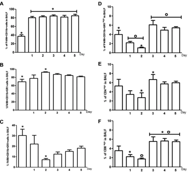
To better understand the cellular populations recruited to the lung during acute exposure to cigarette smoke, we evaluated, using flow cytometry, the cellular BALF content. We investigated the presence of four different cellular populations: CD4+ T lympho-cytes, CD8+T lymphocytes, neutrophils and macrophages (Fig. 3).
We observed that macrophages (F4/80+CD11b+cells) were the main cellular group present in BALF of CS exposed mice (CS1D 80.2%, CS2D 82.2%, CS3D 84.2%, CS4D 81.9% and CS5D 84.6%). Be-sides, macrophages were the first cellular population to increase in number, right after the first cigarette smoke exposure (Fig. 3A). Macrophages were persistent during all the period of cigarette smoke exposure (Fig. 3A). Alveolar macrophages (AMs) and other monocytes are the most important part of the innate immune re-sponse in the lungs [8]. We decided to classify, based on GR1 expression, the macrophages population observed by us as inflam-matory lung macrophages (F4/80+CD11b+GR1+) or resident macrophages (F4/80+CD11b+GR1 ). We observed that on the sec-ond day of CS exposure, the number of resident macrophages (F4/80+CD11b+GR1 ) was increased (92.73%) when compared to CG (69.63%) and the inflammatory lung macrophage numbers (7.25%) (F4/80+CD11b+GR1+) were dramatically reduced compared to CG (30.37%) (Fig. 3B and C). In relation to neutrophils (GR1 high-CD11b+F4/80 ), cigarette smoke exposure lead to a reduction of this cell population, on the second day of exposure (CS2D 1.1%) when compared to control group (CG 3.9%) (Fig. 3D). We observed that neutrophil population became prominent and persistent after the third day of cigarette smoke exposure (CS3D 6.1%, CS4D 4.9% and CS5D 5.4%) when compared to the first (2.2%) and second days (CS2D 1.1%) of CS exposure (Fig. 3D). Concerning to lymphocyte subpopulations, an increase in CD4+cells on the third day of expo-sure (CS3D 6.6%) was observed when compared to the percentage of these cells on the second day (CS2D 2.7%) (Fig. 3E). The increase in CD4+cells was persistent after the third day of cigarette smoke exposure (Fig. 3E). Similarly, CD8+T lymphocytes were recruited to the lung mainly after the third day of cigarette smoke exposure (CS3D 5.5%) when compared to the first (CS1D 2.2%) or second days of exposure (CS2D 1.7%) (Fig. 3F). CD8+cells were persistent in BALF after the third day of exposure (Fig. 3F).
Considering that macrophages were the most predominant cel-lular type on BALF during the five days of acute CS exposure (CS1D 80.2%, CS2D 82.2%, CS3D 84.2%, CS4D 81.2% and CS5D 84.6%) (Fig. 3A), we decided to compare the percentages of combined lym-phocyte subpopulations (CD4+and CD8+cells) and neutrophils in each point of cigarette smoke exposure, trying to identify the sec-ond most predominant cellular population during acute exposure (Fig. 4). We observed no differences between percentages of lym-phocyte (CG 8.8%) and neutrophil (CG 5.5%) populations in the CG (Fig. 4A). However, from the first until the last day of acute cig-arette smoke exposure, lymphocyte population increased (CS1D 5.7%, CS2D 4.0%, CS3D 13.0%, CS4D 11.0% and CS5D 11.0%) when compared to the neutrophil population (CS1D 2.2%, CSD2 1.1%, CS3D 6.1%, CS4D 4.9% and CS5D 5.4%) (Fig. 4B–F).
Macrophages and neutrophils present on pulmonary site con-tribute to an oxidant–antioxidant imbalance[28]. As we observed an influx of those cells into the lung through BALF analyses, we decided to determine if the acute exposure to cigarette smoke associated with cellular influx could lead to an oxidant-antioxidant imbalance. For this, we used the formation of TBARS as an index of lipid peroxidation and measured the activities of SOD, CAT, and GPx from lung homogenates. Data are presented inTable 1. Fig. 1.Analysis of cellular influx in BALF of control and cigarette smoke exposed
groups. The letter (a) represents a significant difference between the fifth days of exposure to CS (CS5D), fourth day of exposure to CS (CS4D), third day of exposure to CS (CS3D), second day of exposure to CS (CS2D) compared to the Control group (CG). (b) Represents a significant difference between the fifth days of exposure to CS (CS5D), fourth day of exposure to CS (CS4D), third day of exposure to CS (CS3D) compared to the first day of exposure to CS (CS1D). (c) Difference between the fifth days of exposure to CS (CS5D), fourth day of exposure to CS (CS4D), third day of exposure to CS (CS3D) compared to the second day of exposure to CS (CS2D). (d) Difference between the fifth days of exposure to CS (CS5D), fourth day of exposure to CS (CS4D) compared to the third day of exposure to CS (CS3D). Data were expressed as mean ± SEM (n = 6) and were analyzed by one-way ANOVA followed by Tukey post-test (p< 0.05).
The TBARS formation was evaluated to obtain an index of the oxidative damage resulted from exposure to CS. As shown in Ta-ble 1, the CS groups, from the second day of exposure, presented an elevated TBARS content in lung homogenates: CS2D (1.05 ± 0.03 U/mL/mg prot), CS3D (1.03 ± 0.02 U/mL/mg prot), CS4D (1.02 ± 0.03 U/mL/mg prot) and CS5D (1.20 ± 0,01 U/mL/ mg prot) when compared to CG (0.87 ± 0.02 U/mL/mg prot). SOD and CAT are the most important antioxidative enzymes responsible for the oxidative balance in the lungs and they are generally regu-lated by oxidative stress. SOD activity was decreased in lung homogenates in the groups exposed to CS during 3 days, CS3D (53.8 ± 13.8 U/mg prot) or 5 days CS5D (57.6 ± 2.7 U/mg prot) when compared to the SOD activity from CG (119.7 ± 4.8 U/ mg prot).
On the other hand, the CAT activity was higher in exposed ani-mals CS3D (27.1 ± 6.7 U/mg prot) and CS4D (22.3 ± 5.5 U/mg prot) when compared to CG (12.3 ± 4.8 U/mg prot). The CAT activity was not significantly different to CS1D (13.7 ± 2.5 U/mg prot), CS2D (12.9 ± 2.0 U/mg prot) and CS5D (15.1 ± 4.3 U/mg prot) when compared to CG. GPx, an antioxidative enzyme that reduces H2O2 to H2O by oxidizing glutathione, was also measured. Similarly to CAT results, the GPx activity was higher in CS exposed groups CS1D (213 ± 60.0 mM/min/mg prot 1) and CS2D (232 ± 22.4 mM/ min/mg prot 1) when compared to CG (112 ± 6.9 mM/min/ mg prot 1). The values of the GPx activity to CS3D (181.8 ± 37.8 mM/min/mg prot 1), CS4D (160.5 ± 19.4 mM/min/ mg prot 1) and CS5D (191.4 ± 9.4 mM/min/mg prot 1) were not significantly different when compared to CG. Taken together, these data confirm the establishment of antioxidant mechanisms in the model of acute CS exposure.
4. Discussion
In this study, we performed a phenotypic characterization of the pulmonary cellular influx of animals acutely exposed (during 5 days) to cigarette smoke as well as the oxidative damage of this exposure reflected by lipid peroxidation and the activities of the antioxidative enzymes SOD, GPx and CAT in that context.
Cigarette smoke (CS) has been implicated as the main risk factor for the development of COPD[29,30]. CS components can cause an inflammatory response upon inhalation and this exposure is con-sidered to be the starting point for the pathogenesis in COPD
[31]. CS is a mix of carcinogenic compounds, toxins, solid reactives,
oxidants and free radicals[32,33] which could initiate, promote and/or amplify oxidative damage[30]. They could also alter immu-nological functions that affect both humoral and cell-mediated im-mune responses such as elevated white blood cell count, increased numbers of circulating lymphocytes and an abnormal T-cell profile
[34].
Multiple studies indicate that repeated exposure to cigarette smoke may induce prolonged airway inflammation associated with cellular infiltration of macrophages and neutrophils[35]. To evalu-ate if a short term CS exposure could generevalu-ate a cellular influx from peripheral blood to the pulmonary tissue, we determined the amount of cells present in the bronchoalveolar lavage in animals acutely exposed (CSD1, CSD2, CSD3, CSD4 and CSD5) or not ex-posed (CG) to CS. We observed that CS exposure lead to a signifi-cant increase of inflammatory cells in the pulmonary tissue of animals after two days of CS exposure as compared to non-exposed animals. Our findings are corroborating previous studies of Bezerra and co-workers[30]that reported that macrophages were numer-ous in the lungs of CS groups and that polymorphonuclear cells were present in this tissue as well. Besides, a well-documented ef-fect of cigarette smoking in humans is leukocytosis (an increased number of blood leukocytes). However, the function of these cells is greatly reduced[8].
smoke than the inflammatory lung macrophages population since the last population presented a decrease in their number on the second day of exposure to CS. It is unlikely that the de-crease of inflammatory lung macrophages on the second day re-sults from ongoing CS exposure apoptosis, but rather suggests macrophage emigration from the lung parenchyma to alveolar space. A similar result was reported by Landsman and co-work-ers[37]. In that study, one day following DTx treatment, inflam-matory lung and resident macrophage numbers were reduced. The decrease in resident macrophage number was followed by their rapid reconstitution, and 2 days after the treatment, their amount almost reached initial levels. Inflammatory lung macro-phages, in contrast, continued to decline, reaching their lowest value on the fourth day[37].
Neutrophils are key effector cells in COPD, the presence of air-way neutrophils and the level of IL-8 and Neutrophil elastase (NE) are related to the severity of airflow obstruction. CS can influ-ence the accumulation of airway neutrophils, which is associated with the production of innate immune mediators and with an in-crease in airflow obstruction [31]. Besides macrophage popula-tions, we identified neutrophils in BALF of mice acutely exposed
to CS. This cellular population presented an increase in number starting from the third day of exposure when compared to the non-exposed group. However, on the second day of exposure, we observed a decrease in this population when compared to the con-trol group. D’Hulst and co-workers[6], reported the development of a progressive neutrophilia in BALF of animals exposed to CS dur-ing 24 weeks. Our data corroborate a previous study usdur-ing daily exposure of 4 or 8 cigarettes during 7 consecutive days, in which neutrophil recruitment was observed on the fourth day of sure, indicating that the cellular influx is dependent on the expo-sure time and the applied cigarette amount[36]. The observation of the neutrophil decrease on the second day of exposure might be explained by the presence of toxic substances present in the cig-arette smoke, such as superoxide, in contact with lung paren-chyma. On the other hand, the pulmonary influx of neutrophils observed after the third day of CS exposure may be due to the necessity of a previous macrophage activation followed by the re-lease of proinflammatory mediators in lung epithelial fluid, which would then amplify the inflammatory cascade by the activation of epithelial cells as well as the recruitment of neutrophils to the air-ways[38].
Fig. 3.Percentage of macrophages, neutrophils and lymphocytes in BALF of control and cigarette smoke exposed groups. Percentage of macrophage total population (F4/ 80+CD11b+cells) in BALF of mice exposed or not to cigarette smoke,n= 6 (A). Percentage of resident macrophages (F4/80+CD11b+GR1 ) in BALF of exposed or not to cigarette
smoke,n= 6 (B). Percentage of inflammatory lung macrophages numbers (F4/80+CD11b+GR1+) in BALF exposed or not to cigarette smoke,n= 6 (C). Percentage of neutrophils
(F4/80 CD11b+GR1high) in BALF of mice exposed or not to cigarette smoke,n= 6 (D). Percentage of CD4+T lymphocytes (CD4High) in BALF of mice exposed or not to cigarette
smoke,n= 6 (E). Percentage of CD8+T lymphocytes (CD8High) in BALF of mice exposed or not to cigarette smoke,n= 6 (F). Data were expressed as mean ± SEM (n= 6) and were
analyzed by one-way ANOVA followed by Tukey post-test (p< 0.05).
We observed that T lymphocytes, CD4+and CD8+, are present in BALF of acutely CS exposed mice and an increase of recruited T cells was noticed after the third day of CS exposure, similarly
to what was observed to neutrophils. Inflammatory cells, espe-cially macrophages and lymphocytes, have been directly associ-ated to subsequent development of COPD in experimental Fig. 4.Comparison between the percentage of T lymphocytes and neutrophils in BALF of control and cigarette smoke exposed groups. Percentage of lymphocytes (CD4highand
CD8high) and neutrophils (F4/80 CD11b+GR1high) in BALF of mice not exposed to cigarette smoke,n= 6 (A). Percentage of lymphocytes (CD4highand CD8high) and neutrophils (F4/
80 CD11b+GR1high) in BALF of mice after a day of exposure to cigarette smoke,n= 6 (B). Percentage of lymphocytes (CD4highand CD8high) and neutrophils (F4/80 CD11b+GR1high)
in BALF of mice after two days of exposure to cigarette smoke,n= 6 (C). Percentage of lymphocytes (CD4highand CD8high) and neutrophils (F4/80-CD11b+GR1high) in BALF of mice
after three days of exposure to cigarette smoke,n= 6 (D). Percentage of lymphocytes (CD4highand CD8high) and neutrophils (F4/80-CD11b+GR1high) in BALF of mice after four days
of exposure to cigarette smoke,n= 6 (E). Percentage of lymphocytes (CD4highand CD8high) and neutrophils (F4/80-CD11b+GR1high) in BALF of mice after five days of exposure to
cigarette smoke,n= 6 (F).Data were expressed as mean ± SEM (n= 6) and were analyzed by one-way ANOVA followed by Tukey post-test (p< 0.05).
Table 1
Biochemical assessment of the lung tissue of animals CG and CS.
CG CS 1D CS 2D CS 3D CS 4D CS 5D p⁄
TBARS (U/mL/mg prot) 0.87 ± 0.02a 0.93 ± 0.01a 1.05 ± 0.03b 1.03 ± 0.02b 1.02 ± 0.03b 1.20 ± 0.01c <0.0001
SOD (U/mg prot) 119.7 ± 4.86a 61.20 ± 7.81 75.20 ± 13.04a 53.80 ± 13.80b 69.80 ± 12.67a 57.60 ± 2.76b 0.0098
CAT (U/mg prot) 12.3 ± 4.8a 13.7 ± 2.5a,b 12.9 ± 2.0a,c 27.1 ± 6.7d 22.3 ± 5.5b, c,d 15.06 ± 4.3a,d 0.0001
GPx (mM/min/mg prot -1)10 5 112 ± 6.9a 213.3 ± 60.0b,e 232 ± 22.4b,c 181.8 ± 37.8a,b,c 160.5 ± 19.7a,e 191.4 ± 9.7a,b,c <0.0001
Different letters indicate statistical differences when comparing the data represented in a same row.
models[39,40]. The recruitment of lymphocytes is possibly a re-sult of the pulmonary aggression by the toxic compounds of CS. T lymphocytes, especially CD4+, contribute to the recruitment of other cellular types such as macrophages and neutrophils [41]. There was no difference between the number of lymphocytes or neutrophils in BALF of non-exposed mice. However, from the first until the last day of CS exposure, the percentage of lymphocytes recruited to pulmonary site was higher than the percentage of re-cruited neutrophils. The lower percentage of neutrophils in our model is consistent with the data of other groups, since the inflammation observed after acute exposure to CS becomes en-riched with neutrophils after one week of CS exposure [42]. On the other hand, chronic exposure is characterized by a BALF con-sisting of neutrophils, macrophages and lymphocytes after one month of CS exposure as previously reported by us and others
[42]. Macrophages and neutrophils recruited to the lung in re-sponse to the aggression of CS compounds are endogenous gener-ators of oxidants and can contribute to oxidative stress that is an imbalance between the production of oxidants and the body’s ability to detoxify the reactive intermediates or repair the result-ing damage.
Cigarette smoke exposes the lung to extreme levels of oxidative stress [43]. Products of oxidative stress can activate signaling mechanisms to enhance the inflammation of upper air pathways
[38]. Polyunsaturated fats and fat acids in cell membranes are important target to free radical attack, resulting in lipid peroxida-tion, a process that in its end can generate peroxides and aldehydes
[17]. The exposure to CS is associated with increased level of mal-ondialdehyde (MDA), a marker of oxidative stress that can be mea-sured in body fluids by the TBARS method[23]. In our work, we showed increased levels of MDA in the homogenized lung in ani-mals exposed to CS from day 2 until day 5, CS2D, CS3D, CS4D and CS5D, when compared to control group. Our data are corrobo-rating another work in which was observed that a short-term cig-arette smoke exposure is associated with acute lung inflammation and oxidative damage[44].
ROS are inherent by-products of activated leukocytes and con-tribute to the inflammatory response and parenchyma destruction. Additionally, the direct increase in the oxidative burden, produced by the release of oxygen radicals from inflammatory neutrophils and macrophages, has relevance to the oxidant-antioxidant imbal-ance committed thereby to the establishment of an oxidative stress
[45]. Inflammatory mediators and redox markers were assayed in lung homogenates by Valenca and co-workers in 2012, whereas leukocyte numbers were quantified in BALF. They suggest a tempo-ral response between parenchyma and BALF.
In this research, SOD, CAT and GPx activities were evaluated to understand their contributions to the redox imbalance during the time course of CS exposure. SOD is the main enzymatic lung de-fense against the deleterious effects of O2 acting through the con-version of O2into H2O2, a substrate to CAT and GPx. It was the first time that one work shows a decrease in the SOD activity on the third and fifth day of animal exposure to the cigarette smoke when compared to CG. Interestingly, it was observed a decrease in SOD activity in CS exposed animals and an increased activity of CAT in groups exposed to CS during 3 and 4 days, CS3D and CS4D, when compared to control group. GPx is another key enzyme for the maintenance of redox equilibrium. The enhanced GPx activity has been shown in lung of rats exposed to CS during 21 days
[38]. Nevertheless, COPD patients present low levels of GPx activity
[46]. In our work, the activity of GPx was enhanced in animals ex-posed to CS during 1 and 2 days, CS1D and CS2D, when compared to control group and, during 3 and 4 days of CS exposure, CS3D and CS4D. It is possible that in our short term CS exposure, GPx and CAT assume the protective role at the beginning and at the end of the CS exposure, respectively.
Taken together, our data show that it is possible to observe a cellular influx into the pulmonary site even in acute CS exposure model. This cellular influx is characterized by a massive influx of macrophages into the lungs, detected in the BALF, accompanied by a discrete increase in lymphocyte and neutrophil numbers. Be-sides, our short term CS exposure was enough to show an oxidative damage in lung parenchyma evidenced by the enhancement of GPx and CAT activities in different stages of CS treatment. These results validate an acute CS model in which is possible to obtain cellular and biochemical data from short term CS exposure. Studies con-cerning the function of those cells in the acute CS exposure will be necessary to determine the role of those cellular populations in this short term exposure context.
Acknowledgments
This work was supported by CAPES (Coordenação de Aper-feiçoamento de Pessoal de Nível Superior) and UFOP (Universidade Federal de Ouro Preto). We are grateful to the laboratories of the Research Center in Biological Sciences (NUPEB) from the Federal University of Ouro Preto for their technical support and Dra. Lis Ribeiro do Vale Antonelli who kindly donated the antibodies for flow cytometry assays.
References
[1]M. Decramer, W. Janssens, M. Miravitlles, Chronic obstructive pulmonary disease, Lancet 379 (9823) (2012) 1341–1351.
[2]M. Roth, Pathogenesis of COPD. Part III. Inflammation in COPD, Int. J. Tuberc. Lung Dis. 12 (4) (2008).
[3]A.M. Wood, R.A. Stockley, The genetics of chronic obstructive pulmonary disease, Respir. Res. 7 (2006) 130.
[4]M.D. Eisner, J. Balmes, P.P. Katz, L. Trupin, E.H. Yelin, P.D. Blanc, Lifetime environmental tobacco smoke exposure and the risk of chronic obstructive pulmonary disease, Environ. Health 4 (1) (2005) 7.
[5]F.M. Botelho, G.J. Gaschler, S. Kianpour, C.C.J. Zavitz, N.J. Trimble, J.K. Nikota, C.M.T. Bauer, M.R. Stampfli, Innate immune processes are sufficient for driving cigarette smoke-induced inflammation in mice, Am. J. Respir. Cell Mol. Biol. 42 (4) (2010) 394–403.
[6]A.I. D’Hulst, K.Y. Vermaelen, G.G. Brusselle, G.F. Joos, R.A. Pauwels, Time course of cigarette smoke-induced pulmonary inflammation in mice, Eur. Respir. J. 26 (2) (2005) 204–213.
[7]A. Misharin, L. Morales-Nebreda, G.M. Mutlu, G.R. Budinger, H. Perlman, Flow cytometric analysis of the macrophage and dendritic cell subsets in the mouse lung, Am. J. Respir. Cell Mol. Biol. (2013).
[8]M. Sopori, Effects of cigarette smoke on the immune system, Nat. Rev. Immunol. 2 (5) (2002) 372–377.
[9]C.A. Owen, Proteinases and oxidants as targets in the treatment of chronic obstructive pulmonary disease, Proc. Am. Thorac. Soc. 2 (4) (2005) 373–385. discussion 394–375.
[10] C.R. Rueff-Barroso, E.T. Lima Trajano, J.N. Alves, R.O. Paiva, M. Lanzetti, K.M. Pereira Pires, F.S. Bezerra, R.A. Pinho, S.S. Valenca, L.C. Porto, Organ-related cigarette smoke-induced oxidative stress is strain-dependent, Med. Sci. Monit. 16 (7) (2010) BR218–BR226.
[11]I.T. Lee, C.M. Yang, Role of NADPH oxidase/ROS in pro-inflammatory mediators-induced airway and pulmonary diseases, Biochem. Pharmacol. (2012).
[12]I. Rahman, W. MacNee, Oxidative stress and regulation of glutathione in lung inflammation, Eur. Respir. J. 16 (3) (2000) 534–554.
[13]S. Loukides, P. Bakakos, K. Kostikas, Oxidative stress in patients with COPD, Curr. Drug Targets 12 (4) (2011) 469–477.
[14]V.P. Balasubramanian, B. Varkey, Chronic obstructive pulmonary disease: effects beyond the lungs, Curr. Opin. Pulmon. Med. 12 (2) (2006) 106–112. [15]E.M. Drost, K.M. Skwarski, J. Sauleda, N. Soler, J. Roca, A. Agusti, W. MacNee,
Oxidative stress and airway inflammation in severe exacerbations of COPD, Thorax 60 (4) (2005) 293–300.
[16]S.A.A. Comhair, S.C. Erzurum, Antioxidant responses to oxidant-mediated lung diseases, Am. J. Physiol. Lung Cell. Mol. Physiol. 283 (2) (2002) L246–L255. [17]H. Van der Vaart, D.S. Postma, W. Timens, N.H.T. Ten Hacken, Acute effects of
cigarette smoke on inflammation and oxidative stress: a review, Thorax 59 (8) (2004) 713–721.
[18]S.S. Valenca, F.S. Bezerra, A.A. Lopes, B. Romana-Souza, M.C. Marinho Cavalcante, A.B. Lima, V.L. Goncalves Koatz, L.C. Porto, Oxidative stress in mouse plasma and lungs induced by cigarette smoke and lipopolysaccharide, Environ. Res. 108 (2) (2008) 199–204.
[19]R.E. Oberley-Deegan, E.A. Regan, V.L. Kinnula, J.D. Crapo, Extracellular superoxide dismutase and risk of COPD, Copd-J. Chron. Obstruct. Pulmon. Dis. 6 (4) (2009) 307–312.
[20]K.M. Pereira Pires, F.S. Bezerra, M.N. Machado, W.A. Zin, L.C. Porto, S.S. Valenca,
N-(2-mercaptopropionyl)-glycine but not Allopurinol prevented cigarette smoke-induced alveolar enlargement in mouse, Respir. Physiol. Neurobiol. 175 (3) (2011) 322–330.
[21]M.A. Santos-Silva, A.C. Nagato, E.T. Lima Trajano, J.N. Alves, A.C. Balthar Bandeira, L.C. Porto, F.S. Bezerra, The oxidative response of mouse hearts is modulated by genetic background, Arq. Bras. Cardiol. 100 (2) (2013) 157–163. [22]S.S. Valenca, K. Da Hora, P. Castro, V.G. Moraes, L. Carvalho, L. Porto, Emphysema and metalloelastase expression in mouse lung induced by cigarette smoke, Toxicol. Pathol. 32 (3) (2004) 351–356.
[23]H.H. Draper, E.J. Squires, H. Mahmoodi, J. Wu, S. Agarwal, M. Hadley, A comparative evaluation of thiobarbituric acid methods for the determination of malondialdehyde in biological materials, Free Radic. Biol. Med. 15 (4) (1993) 353–363.
[24]J.V. Bannister, L. Calabrese, Assays for superoxide dismutase, Methods Biochem. Anal. 32 (1987) 279–312.
[25]H. Aebi, Catalase in vitro, Methods Enzymol. 105 (1984) 121–126.
[26]L. Flohé, W.A. Günzler, Assays of glutathione peroxidase, Methods Enzymol. 105 (1984) 114–121.
[27]M.M. Bradford, A rapid and sensitive method for the quantitation of microgram quantities of protein utilizing the principle of protein-dye binding, Anal. Biochem. 72 (1976) 248–254.
[28]J.L. Lin, P.S. Thomas, Current perspectives of oxidative stress and its measurement in chronic obstructive pulmonary disease, COPD 7 (4) (2010) 291–306.
[29]P.J.C. Biselli FDTQSL, H.T. Moriya, D.H.R.F. Rivero, A.C. Toledo, P.H.N. Saldiva, T. Mauad, M.A. Martins, Short-term exposure of mice to cigarette smoke and/or residual oil fly ash produces proximal airspace enlargements and airway epithelium remodeling, Braz. J. Med. Biol. Res. 44 (2011) 9.
[30]F.S. Bezerra, S.S. Valenca, M. Lanzetti, W.A. Pimenta, P. Castro, V.L. Goncalves Koatz, L.C. Porto, Alpha-tocopherol and ascorbic acid supplementation reduced acute lung inflammatory response by cigarette smoke in mouse, Nutrition 22 (11–12) (2006) 1192–1201.
[31]K.J. Baines, J.L. Simpson, P.G. Gibson, Innate immune responses are increased in chronic obstructive pulmonary disease, PLoS ONE 6 (3) (2011).
[32]J.K. Nikota, M.R. Stämpfli, Cigarette smoke-induced inflammation and respiratory host defense: Insights from animal models, Pulm. Pharmacol. Ther. 25 (4) (2012) 257–262.
[33]M.R. Stampfli, G.P. Anderson, How cigarette smoke skews immune responses to promote infection, lung disease and cancer, Nature Reviews/Immunology, vol. 9, SCIENCE E, SOCIETY, 2009, p. 8.
[34]Z. Jubri, A.A. Latif, A.G.M. Top, W.Z.W. Ngah, Perturbation of cellular immune functions in cigarette smokers and protection by palm oil vitamin E supplementation, Nutr. J. (2013) 12.
[35]P. Castro, A. Legora-Machado, L. Cardilo-Reis, S. Valenca, L.C. Porto, C. Walker, C. Zuany-Amorim, V.L.G. Koatz, Inhibition of interleukin-1 beta reduces mouse lung inflammation induced by exposure to cigarette smoke, Eur. J. Pharmacol. 498 (1–3) (2004) 279–286.
[36]P. Castro, A.L. Machado, L.C. Reis, S. Valenca, Inhibition of interleukin-1h reduces mouse lung inflammation induced by exposure to cigarette smoke, Eur. J. Pharmacol. 498 (2004) 7.
[37]L. Landsman, S. Jung, Lung macrophages serve as obligatory intermediate between blood monocytes and alveolar macrophages, J. Immunol. 179 (6) (2007) 3488–3494.
[38]I. Rahman, Antioxidant therapies in COPD, Int. J. Chron. Obstruct. Pulmon. Dis. 1 (1) (2006) 15–29.
[39]T. Maeno, A.M. Houghton, P.A. Quintero, S. Grumelli, C.A. Owen, S.D. Shapiro, CD8(+) T cells are required for inflammation and destruction in cigarette smoke-induced emphysema in mice, J. Immunol. 178 (12) (2007) 8090– 8096.
[40]T. Bandoh, H. Mitani, M. Niihashi, Y. Kusumi, M. Kimura, J. Ishikawa, T. Totsuka, I. Sakurai, S. Hayashi, Fluvastatin suppresses atherosclerotic progression, mediated through its inhibitory effect on endothelial dysfunction, lipid peroxidation, and macrophage deposition, J. Cardiovasc. Pharmacol. 35 (1) (2000) 136–144.
[41]S. Hodge, G. Matthews, V. Mukaro, J. Ahern, A. Shivam, G. Hodge, M. Holmes, H. Jersmann, P.N. Reynolds, Cigarette smoke-induced changes to alveolar macrophage phenotype and function are improved by treatment with procysteine, Am. J. Respir. Cell Mol. Biol. 44 (5) (2011) 673–681.
[42]W.-Y.H. Wan, A. Morris, G. Kinnear, W. Pearce, J. Mok, D. Wyss, C.S. Stevenson, Pharmacological characterisation of anti-inflammatory compounds in acute and chronic mouse models of cigarette smoke-induced inflammation, Respir. Res. (2010) 11.
[43]S.S. Valença, W.A. Pimenta, C.R. Rueff-Barroso, T.S. Ferreira, A.C. Resende, R.S. Moura, L.C. Porto, Involvement of nitric oxide in acute lung inflammation induced by cigarette smoke in the mouse, Nitric Oxide 20 (3) (2009) 175–181.
[44]M. Lanzetti, F.S. Bezerra, B. Romana-Souza, A.C. Brando-Lima, V.L. Goncalves Koatz, L.C. Porto, S.S. Valenca, Mate tea reduced acute lung inflammation in mice exposed to cigarette smoke, Nutrition 24 (4) (2008) 375–381. [45]M. Lanzetti, C.A. da Costa, R.T. Nesi, M.V. Barroso, V. Martins, T. Victoni, V.
Lagente, K.M. Pereira Pires, P.M. Rodrigues e Silva, A.C. Resende, et al., Oxidative stress and nitrosative stress are involved in different stages of proteolytic pulmonary emphysema, Free Radical Biol. Med. 53 (11) (2012) 1993–2001.