Article
J. Braz. Chem. Soc., Vol. 22, No. 3, 546-551, 2011. Printed in Brazil - ©2011 Sociedade Brasileira de Química 0103 - 5053 $6.00+0.00
A
*e-mail: [email protected]
Simple Route for the Synthesis of Copper Hydroxy Salts
Jhon Mauricio Aguirre,a,d Adamo Gutiérrezb,d and Oscar Giraldo*,c,d
aDepartamento de Ingeniería Química, bDepartamento de Ingeniería Eléctrica,
Electrónica y Computación, cDepartamento de Física y Química and dLaboratorio de Materiales Nanoestructurados y Funcionales,
Universidad Nacional de Colombia, Carrera 27 No. 64-60, Manizales, Colombia
No presente trabalho é apresentada uma nova e rápida rota de síntese em escala de laboratório de hidroxinitrato de cobre(II), Cu2(OH)3NO3, mediante reação de hidróxido de magnésio com solução
aquosa de nitrato de cobre(II). O material foi caracterizado por difratometria de raios X (estrutura monoclínica), absorção atômica, espectroscopia vibracional (infravermelho com transformada de Fourier e Raman), análise térmica e microscopia eletrônica de varredura com EDX.
The current work introduces a new and rapid synthetic route for the in-laboratory preparation of copper hydroxy nitrate, through the reaction of magnesium hydroxide and copper nitrate aqueous solution. The material with the formula Cu2(OH)3NO3 and monoclinic phase was characterized by
X-ray diffraction, atomic absorption, Fourier transform infrared and Raman spectroscopy, scanning electron microscopy with EDX, and thermal analysis.
Keywords: hydroxy salts, anionic clay, copper hydroxynitrate, gerhardtite
Introduction
Two-dimensional solids have gained increasing interest in recent years given to possible applications in many ields, particularly exploring its capacity to intercalate ionic or neutral species in the interlayer region. There are several classes of materials with layered structures, that are characterized by weak interactions in the interlayer region.1 One of them corresponds to
negatively charged aluminum silicates, titanates and birnessite-type manganese oxides. A second group corresponds to neutral lamellar materials like graphite, MoO3-type oxides, and some coordination compounds. A third group corresponds to positively charged layered materials like hydrotalcite and some hydroxy salts of transition metal ions.2-4
The hydroxy salts (HS), also known as basic metallic salts,1,2 encompass a class of layered materials classiied
within the family of anionic clays, and they resemble layered double hydroxides (LDH) or hydrotalcite-like materials.3,4 Their structure are similar to the mineral
form of brucite or magnesium hydroxide [Mg(OH)2],5
where the OH− anions are hexagonally packed and the
magnesium(II) ions occupy octahedral sites, forming extended sheets.6,7
The copper(II) hydroxynitrate, Cu2(OH)3NO3, crystallizes
in an orthorhombic phase with cell parameters a = 6.087 Å, b = 13.813 Å, and c = 5.597 Å. It is naturally found in the mineral called Gerhardtite, but a stable monoclinic phase is obtained by conventional synthetic routes.8-10
The structure of Cu2(OH)3NO3 can be imagined as being obtained by replacement of 25% of the OH− anions
in the cooper hydroxide sheets by NO3− anions directly
coordinated to copper(II) cations through one of its oxygen atoms, as shown in Figure 1. The stacking of such a sheets in the z direction lead to the formation of a neutral three-dimensional structure.4,11 However, the nitrate ions can be
displaced by others negative charged molecules.
Metallic hydroxy salts with the composition M2(OH)3X (M = Cu, Co; X = organic anion)5,8 have been extensively
reported in this work a new simple method for preparation of copper hydroxynitrate, an interesting member of this class of layered materials.
Experimental
Synthesis
Copper hydroxynitrate, Cu2(OH)3NO3, was synthesized
by the reaction of a dispersion of analytical grade magnesium hydroxide, (Mg(OH)2), from BDH Chemicals
Ltd. Poole England, with a solution of analytical grade copper nitrate trihydrate, (Cu(NO3)2·3H2O, from Merck®).
Magnesium hydroxide (1.01 g) was dispersed in 100 mL (0.17 mol L-1) of distilled and deionized water (DDW) and
a copper nitrate solution, prepared by dissolving 10.08 g of salt in 100 mL of DDW (0.42 mol L-1), such that the
Cu/Mg molar ratio was larger than 2.
Copper nitrate was kept in excess to promote the complete exchange of magnesium(II) for copper(II) cations, avoiding the possible formation of copper hydroxide (Cu(OH)2), copper oxide (CuO), or other non-desirable
phases. The reaction was conducted at room temperature and ambient pressure (23.4 ºC, 585 mm Hg), for 20 min, with constant magnetic stirring at 1200 rpm. The pH of the initial reaction mixture was about 5.0, but decreased to 4.3 at the end of the reaction, due to the consumption of hydroxide. The resulting precipitate was separated through vacuum iltration with Schleicher & Schuell ilter paper with 2 µm pore size.
The solid was washed several times with DDW to remove excess of salts or other soluble impurities. Then, the aqua-marine blue solid was dried at room temperature and relative humidity of 65 ± 10%. The washing solution was saved for atomic absorption analyses and the aqua-marine blue precipitate was stored for further characterization.
Characterization techniques
The metal content in the samples were determined by atomic absorption spectrometry (AAS; Perkin-Elmer 3110). The concentration of the solution was adjusted to the equipment’s optimal detection readout limits before measurements.
Powder X-ray diffraction analysis were carried out using a Rigaku Minilex II equipment with Bragg Brentano geometry, equipped with NaI detector and Cu Kα radiation source (λ = 0.1540562 nm), operating at 30 kV and 15 mA. The X-ray diffraction patterns (XRD) were collected in the 2θ interval of 3 to 70º at a scanning speed of 2º per min, in
normal environmental conditions.
The Fourier transform infrared spectra were obtained in a Nicolet 380 spectrophotometer, equipped with a DTGS detector. The samples were prepared by diluting 10 mg of the pulverized solid samples with KBr and pressing them to form a pellet. Registries were obtained in the 4000 to 400 cm-1 interval with a 4.0 cm-1 resolution.
The Raman spectra were obtained in a HR-800 spectrophotometer (Horiba-JobinYvon), equipped with a 473 nm excitation laser, in the 400 to 3800 cm-1 range.
The morphology and the composition of the material were examined by SEM/EDX, using a Jeol JSM 5910LV scanning electron microscope equipped with energy-dispersive X-ray spectrometry detector.
Thermal analyses (TGA and DTG) were conducted in a high-resolution TA Instruments Q-500 T0 thermogravimetric
analyzer. The samples were heated in nitrogen atmosphere at a speed of 10 º min-1, from room temperature to 800 ºC.
Results and Discussion
Generally, hydroxy salts are synthesized by one of the following methods: the pyrolysis of a metallic salt;4
the reaction of a metallic oxide with the corresponding aqueous solution of a salt of the desired metal;3-5 the slow
precipitation of HS with sodium hydroxide from a metal salt solution;4,8,11 the co-precipitation procedure employed
for LDH synthesis, introduced by Newman and Jones.4
Additionally, the reaction of a metal oxide (MeO) with zinc acetate in an aqueous media, was reported by Morioka
et al.2 Some of these procedures demand strict control of
experimental variables, especially pH, temperature and concentration of the reactants to avoid the formation of sub-products like Cu(OH)2 and CuO.8
The chemical composition and physical characteristics like surface area, pore size, particle size, and structure of the inal product are dependent on the reactants concentration, temperature, pressure and reaction time.3,4,8,22 Frequently,
Figure 1. Schematic representation of the structure of copper
the product of synthesis must be submitted to subsequent steps of aging and cleansing, in order to eliminate most of the impurities that could alter the morphology and properties, and consequently the inal use of the material.
As noted, the classical dispersion method employed by Meyn et al.3 and Bruschini and Hudson,5 using two
compounds of the same metal, generates an HS material with a high degree of purity and crystallinity, but has the disadvantage of needing above 24 h of reaction time. Newman and Jones,4 Henrist et al.8 and Pereira et al.22 reacted copper
nitrate with sodium hydroxide using precipitation, dispersion, and co-precipitation methods, respectively, in order to produce a copper HS material. They obtained good results using an elaborated method, by employing the salt at boiling temperature, vacuum conditions and long aging periods, in order to improve the degree of crystallinity of the material. Rajamathi and Kamath,23 reported the synthesis of HS using
the hydrolysis of urea for long periods of times (1.25 to 7.5 h). The complexity of all these procedures explains the dificulty in reproducing the synthesis of HS materials, which implies carefully monitoring and controlling all conditions at each of the steps in a routine way to obtain good quality materials.
All the previously mentioned works validates the importance of a simple procedure for the preparation of copper hydroxynitrate, like the one reported here based on the reaction of magnesium hydroxide with copper nitrate as described in equation 1. High purity materials were obtained with minimal workout, short reaction times, and under normal environmental conditions.
4(Cu(NO3)2)(aq) + 3(Mg(OH)2) →
2(Cu2(OH)3(NO3))(s) + 3(Mg(NO3)2)(aq) (1)
The atomic absorption technique was used to evaluate the content of Mg and Cu, both, in the hydroxy salt and in the wash waters of recently prepared materials. The possible presence of small amounts of magnesium was revealed by this technique. In fact, the copper content in the solid samples was more than 400 times larger than the magnesium content (Cu/Mg molar ratio = 413). A contrasting situation was found for the supernatant solution in equilibrium with the solid material. In this case, the Cu/Mg molar ratio was determined to be 1.35, conirming the presence of a signiicant excess of copper nitrate.
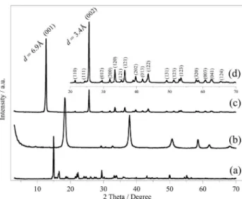
The crystal structure of the copper(II) hydroxynitrate was determined by X-ray diffraction, as shown in Figure 2, where its diffraction pattern is compared with that of copper nitrate and magnesium hydroxide (Figure 2a-2b), respectively. The expanded view in the 2θ range of 20 to 70º is shown in Figure 2d.
The high degree of crystallinity is evidenced by the presence of a series of sharp and well-deined (00l)
relections at periodic intervals, indicating the presence of a layered structure. The crystallographic phase was identified as belonging to the monoclinic P21 spatial
group using the JCPDS-ICDD 75-1779 ile, with cell parameters a = 5.605 Å, b = 6.087 Å, and c = 6.929 Å, and
β = 94.480º, at 25 ºC. The X-ray diffraction pattern, the Miller indexes for the main crystallographic planes, and the interlayer distances (d(001) = 6.9 Å and d(002) = 3.4 Å) for
the copper hydroxynitrate are presented in Figure 2. The sharp relections are in agreement with those reported in previous works.3,4,17,24
Clearly, a material with a crystalline phase distinct from that of copper nitrate and magnesium hydroxide precursors, with no peaks corresponding to CuO and/or Cu(OH)2, were
obtained. In fact, all evidences are consistent with copper hydroxynitrate, with the formula Cu2(OH)3NO3.
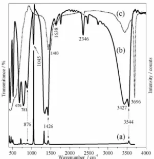
The FT-IR spectra of the magnesium hydroxide precursor and the prepared hydroxy salt are shown in Figure 3. Considering the structure of cooper hydroxynitrate some characteristic vibrational modes are expected in the IR spectrum. In the speciic case of the metal-oxygen bond, strong peaks are expected between 150 and 450 cm-1.
Medium intensity vibrational modes associated to the Cu–O–H bond should appear in the interval from 845 to 900 cm-1.27 Strong peaks associated with the NO
3− ion
vibrational modes are also expected between 1340 and 1380 cm-1. Finally, the presence of water should lead to
the appearance of a very strong peak around 1640 cm-1.
The IR spectrum of the magnesium hydroxide precursor is shown in Figure 3c. The strong bands centered at 3696 cm-1 and 3440 cm-1 were assigned to the stretching
Figure 2. X-ray diffraction (XRD) patterns of (a) copper nitrate;
modes of OH− groups, present in the layered structure and in
the water molecules present in the interlamellar space.11 The
peaks observed in the low frequency region were attributed to vibrational modes involving the Mg-OH bonds. 6
The peaks located at 1426 and 1483 cm-1 are consistent
with those found by Frost and Kloprogge,6 which show the
presence of magnesite (MgCO3), an impurity produced most
likely through contamination with CO2 from the environment.
The IR spectrum of copper hydroxynitrate prepared according to the method reported here (Figure 3b) exhibited few vibrational peaks. The very sharp peak at 3544 cm-1
and the broad one at 3427 cm-1 can be assigned to structural
OH− groups and to water molecules in the interlamellar
space, respectively. Notice that a similar sharp peak was found in Mg(OH)2 but shifted to higher wavenumbers.
(3696 cm-1) relecting the inluence of the M2+–OH bond.
The bandwidth is related to the degree of hydrogen bonding among neighboring OH groups.
There are two peaks in this region. The narrow band at 3544 cm-1 corresponds to free OH groups, while the one
centered at 3427 cm-1 indicates the presence of hydrogen
bonded OH groups.11,22 The peaks at 1045 (ν
1), 800 (ν2),
1343, 1375 and 1426 cm-1 (ν
3) can be assigned to vibrational
modes of nitrate (NO3–) ions,25 whose interaction with
cooper hydroxide layers is evidenced by the appearance of symmetric and asymmetric stretching modes of NO3– at
1426 and 1343 cm-1. The band at 1045 cm-1 corresponds
to the N–O stretching vibration of a monodentate O–NO group, which is active in both, FT-IR and Raman spectra (see Figure 4), in good agreement with the literature.22,26
The shoulder observed at 1375 cm-1is also assigned to free
nitrate ions in the interlamellar space.
The frequency of the vibrational modes attributed to Cu–O–H bonds are depend on the degree of hydrogen bonding and were found at 876, 781 and 676 cm-1, in good
agreementwith those reported by Henrist et al.22 (876, 784,
and 673 cm-1).
The synthesized copper hydroxynitrate exhibited some other vibrational peaks that were active in both infrared and Raman spectra (Figure 3c). The peak at 430 cm-1 was
assigned to a ν(Cu–O) vibrational mode, while the peak at 876 cm-1 was assigned to Cu–O–H vibrations.22 Another
peak at 1426 cm-1 was attributed to vibrational modes of the
nitrate group. A third peak at 1638 cm-1 has been assigned
to a bending mode of the hydroxyl group of water.6
Several studies have been conducted on the morphology of layered double hydroxides and other types of clays; however there are few reports on hydroxy salts.28 The SEM
images of copper hydroxynitrate are shown in Figure 4. The SEM images of the material show well crystallized particles with plate-like morphology (Figure 4) and average size of 1-2 µm.
Only copper was found in the hydroxy salt by EDX analyses (Figure 4c) confirming a highly efficient substitution of magnesium ions by copper ions in the synthesis. The oxygen/copper molar ratio was found to be 2.9 by EDX, very close to the theoretical value of 3.0. This result is quite satisfactory keeping in mind the limitations of the technique.
The thermogravimetric analysis (TGA) curves and the differential thermogravimetric (DTG) curve for the copper hydroxynitrate in nitrogen atmosphere are illustrated in Figure 5. There are two events leading to a total weight loss of 38%. The irst one in the interval from 20 ºC to 150 ºC, corresponding to a 15% weight loss, was attributed to the removal of weakly bond water molecules. The
Figure 3. Raman (a) and FT-IR (b) spectrum of copper hydroxynitrate
and (c) FT-IR of magnesium hydroxide.
Figure 4. Scanning electron microscopy (SEM) images of copper
second event with weight loss of 23%, occurring in the 150 ºC to 400 ºC interval, can be assigned to the thermal decomposition and destruction of the layered structure through dehydroxylation reactions and transformation of nitrate ions (present in the interlayer space) to nitrogen oxides, as reported in the literature for hydroxy salt materials.5,29-31 The most commonly accepted mechanism
for the thermal decomposition of copper hydroxynitrate is described by equation 2, which does not include the decomposition of nitric acid.22
Cu2(OH)3(NO3) → 2(CuO)(s) + HNO3(g) + H2O(g) (2)
The thermal behavior of the material is shown in Figure 5. The irst step (until up to 150 ºC) was assigned to the loss of water molecules adsorbed in the surface and in the interlayer space. De-hydroxylation and decomposition of the anion in the interlayer space should take place in the second step, leading to the destruction of the layered structure. The temperature of the second thermal decomposition process (Figure 5) was found to be 246 °C which is lower than for magnesium hydroxide (377 °C), evidencing its lower thermal stability.
Conclusions
A new and simple preparation method of copper hydroxynitrate (Cu2(OH)3NO3) by the reaction of
magnesium hydroxide and copper nitrate aqueous solution was described. The method is fast and does not require any variation of temperature or pressure, being carried out in mild conditions. The product showed a high degree of crystallinity despite of the fact that the material was not subject to long aging periods, as reported in other works. Our method can be extended to the preparation of other analogous layered hydroxide
materials based on transition metal ions such as nickel and cobalt. Studies in this direction are on the way in our laboratory.
Acknowledgments
The authors thank the Direction of Research (DIME) at National University of Colombia, Medellin Campus, for the economic support for this project (QUIPU code: 20101007735), and the Chemistry, Advanced Microscopy, Magnetism and Advanced Materials and Optics laboratories at this university for the analyses of Atomic Absorption, SEM/EDX, Raman, and TGA-DTG, respectively, and the laboratory of Analytic Chemistry at Quindío University for FT-IR analysis. Finally the authors thank Susan Diane Galvis for the helpful discussions of the manuscript.
References
1. Bera, P.; Rajamathi, M.; Hegde, M. S.; Kamath, P. V.; Bull. Mater. Sci.2000, 23, 141.
2. Morioka, H.; Tagaka, H.; Karasu, M.; Kadokawa, J.; Chiba, K.;
Inorg. Chem. 1999, 38, 4211.
3. Meyn, M.; Beneke, K.; Lagaly, G.; Inorg. Chem. 1993, 32, 1209.
4. Newman, S. P.; Jones, W.; J. Solid State Chem. 1999, 148, 26. 5. Bruschini, C. S.; Hudson, M. J.; Access in Nanoporous
Materials; Pinnavaia, T. J.; Thorpe, M. F., eds.; Plenum Press: New York, 1995, p. 161.
6. Frost, R. L.; Kloprogge, J. T.; Spectrochim. Acta, Part A 1999, 55, 2195.
7. Rajamathi, M.; Thomas, G.; Kamath P.; Proc. Indian Acad. Sci. (Chem. Sci.) 2001, 113, 671.
8. Pereira, D. C.; de Faria, D. L. A.; Constantino, V. R. L.; J. Braz. Chem. Soc. 2006, 17, 1651.
9. Hawthorne, F. C.; Schindler, M.; Can. Mineral. 2000, 38, 751. 10. Frost, R. L.; Erickson, K. L.; Weier, M. L.; Leverett, P.;
Williams, P. A.; Spectrochim. Acta, Part A 2005, 61, 607. 11. Rajamathi, J. T.; Britto, S.; Rajamathi, M.; J. Chem. Sci. 2005,
117, 629.
12. Fujita, W.; Awaga, K.; J. Am. Chem. Soc. 1997, 119, 4563.
13. Abdelouhab, S.; François, M.; Elkaim, E.; Rabu, P.; Solid State Sci. 2005, 7, 227.
14. Fujita, W.; Awaga, K.; Yokoyama, T.; Appl. Clay Sci.
1999, 15, 281.
15. Taibi, M.; Ammar, S.; Jouini, N.; Fiévet, F.; Molinié, P.; Drillon, M.; J. Mater. Chem. 2002, 12, 3238.
16. Kurmoo, M.; Kumagai, H.; Hughes, S. M.; Kepert, C. J.; Inorg. Chem. 2003, 42, 6709.
17. Park, S. H.; Lee, C. E.; J. Phys. Chem. B 2005, 109, 1118. 18. Arizaga, G. G. C. A.; Satyanarayana, K. G.; Wypych, F.; Solid
State Ionics2007, 178, 1143.
Figure 5. Thermogravimetric analysis curves, (a) DTG and (b) TGA, of
19. Rabu, P.; Angelov, S.; Legoll, P.; Belaiche, M.; Drillon, M.;
Inorg. Chem. 1993, 32, 2463.
20. Fujita, W.; Awaga, K.; Inorg. Chem. 1996, 35, 1915.
21. Laget, V.; Hornick, C.; Rabu, P.; Drillon, M.; Ziessel, R.; Coord. Chem. Rev. 1998, 178, 1533.
22. Henrist, C.; Traina, K.; Hubert, C.; Toussaint, G.; Rulmont, A.; Cloots, R.; J. Cryst. Growth 2003, 254, 176.
23. Rajamathi, M.; Kamath, P. V.; Int. J. Inorg. Mater. 2001, 3, 901.
24. Biswick, T.; Jones, W.; Pacula, A.; Serwicka, E.; J. Solid State Chem. 2006, 179, 49.
25. Nakamoto, K.; Infrared and Raman Spectra of Inorganic and Coordination Compounds, 4th ed., John Wiley & Sons: New York, 1986.
26. Arizaga, G. G. C.; Mangrich, A. S.; Ferreira, J. E.; Wypych, F.;
J. Colloid Interface Sci. 2008,320, 168.
27. Secco, E.; Worth, G.; Can. J. Chem.1987, 65, 2504.
28. Morioka, H.; Tagaya, H.; Kadokawa, J. I.; Chiba, K.; J. Mater. Sci. Lett. 1999, 18, 995.
29. Kandare, E.; Hossenlopp, J. M.; Inorg. Chem. 2006, 45, 3766.
30. Rojas, R.; Barriga, C.; Ulibarri, M. A.; Malet, P.; Rives, V.;
J. Mater. Chem. 2002, 12, 1071.
31. Rojas, R.; Ulibarri, M. A.; Barriga, C.; Rives, V.; Microporous Mesoporous Mater. 2008, 112, 262.
Submitted: December 3, 2009