Photoelastic analysis of stress generated by wires when
conventional and self-ligating brackets are used: A pilot study
Guilherme Caiado Sobral1, Mário Vedovello Filho2, Viviane Veroni Degan2, Milton Santamaria Jr3
Objective: By means of a photoelastic model, this study analyzed the stress caused on conventional and self-ligating brackets with expanded arch wires. Method: Standard brackets were adhered to artificial teeth and a photoelastic model was prepared using the Interlandi 19/12 diagram as base. Successive activations were made with 0.014-in and 0.018-in rounded cross section Nickel-Titanium wires (NiTi) and 0.019 x 0.025-in rectangular stainless steel wires all of which made on 22/14 Interlandi diagram. The model was observed on a plane polariscope — in a dark field microscope config-uration — and photographed at each exchange of wire. Then, they were replaced by self-ligating brackets and the process was repeated. Analysis was qualitative and observed stress location and pattern on both models analyzed. Conclusions:
Results identified greater stress on the region of the apex of premolars in both analyzed models. Upon comparing the stress between models, a greater amount of stress was found in the model with conventional brackets in all of its wires. Therefore, the present pilot study revealed that alignment of wires in self-ligating brackets produced lower stress in peri-odontal tissues in expansive mechanics.
Keywords:Orthodontic brackets. Dental arch. Corrective orthodontics.
How to cite this article: Sobral GC, Vedovello Filho M, Degan VV, Santama-ria Jr M. Photoelastic analysis of stress generated by wires when conventional and self-ligating brackets are used: A pilot study. Dental Press J Orthod. 2014 Sept-Oct;19(5):74-8. DOI: http://dx.doi.org/10.1590/2176-9451.19.5.074-078.oar
» Patients displayed in this article previously approved the use of their facial and in-traoral photographs.
Contact address: Milton Santamaria Jr
Av. Maximiliano Baruto, 500 ‒ Jd. Universitário Araras ‒ São Paulo/SP — Brazil. CEP: 13607-339 ‒ E-mail: [email protected]
1 MSc in Orthodontics, School of Dentistry — University of Araras
(UNIARARAS).
2 Professor, Department of Orthodontics, UNIARARAS. 3 Professor, Postgraduate program in Orthodontics,
UNIARARAS.
» The authors report no commercial, proprietary or financial interest in the products or companies described in this article.
Submitted em: May 12, 2013 - Revised and accepted: October 03, 2013
DOI: http://dx.doi.org/10.1590/2176-9451.19.5.074-078.oar
Objetivo: o presente estudo analisou, por meio de um modelo fotoelástico, a distribuição das tensões geradas em bra-quetes convencionais e autoligáveis quando ativados com arcos expandidos. Métodos: braquetes convencionais foram colados em dentes artificiais e, em seguida, foi confeccionado o modelo fotoelástico, utilizando como base o diagrama 19/12, de Interlandi. Foram feitas trocas sucessivas com fios de liga de níquel-titânio (NiTi) de secção circular 0,014" e 0,018" e de liga de aço de secção retangular 0,019" x 0,025", todos no diagrama 22/14 de Interlandi. A cada troca de fio, o modelo foi observado em polariscópio plano, na configuração de campo escuro, e fotografado. Foi feita a substituição por braquetes autoligáveis e repetido o experimento. A análise foi qualitativa, observando o local e o padrão da tensão das franjas nos dois modelos analisados. Conclusões: os resultados identificaram uma maior padrão de tensões das franjas na região do ápice de pré-molares em ambos os modelos analisados. Ao se comparar as tensões entre os modelos, observou--se uma maior quantidade de tensão nas franjas no modelo com braquetes convencionais em todos os fios utilizados no experimento. Portanto, o presente estudo mostrou que o alinhamento dos fios nos braquetes autoligáveis produz forças mais suaves nos tecidos periodontais nas mecânicas expansionistas.
INTRODUCTION
Nowadays, orthodontists have many techniques and methods available for treatment planning. There is a great variety of brackets, with different prescrip-tions and forms that allow the orthodontist to indi-vidualize each case according to patient’s needs.1
These needs make the scientiic community en-deavor to innovate in orthodontic appliances. Innova-tion, in turn, leads to better control of dental move-ment, given that one of the greatest challenges faced by the orthodontist is to come up with mechanical solu-tions to stimulate biological reacsolu-tions of the periodon-tium without compromising treatment outcomes.2
Correct management of orthodontic forces de-pends on a series of factors, including friction gener-ated between wires and brackets. In orthodontic slid-ing mechanics, friction poses clinical diiculties to the orthodontist. High levels of friction could decrease bracket eiciency, thereby reducing the speed of dental movement and hindering anchorage control.3
The concern of producing less friction, i.e., lower attrition between wires and brackets, contributed to the development of self-ligating brackets in which the tooth moves with the wires serving as a guide, since it does not involve the use of elastic ligatures which sig-niicantly increase friction between wires and the slot.4 The difference between conventional and self-ligating brackets system is the absence of elastic or metallic ligatures in the latter. In other words, brackets have a closing system that leaves the wire free inside the slot.5
One of the purposes of orthodontic mechanics is gaining space in the arch before alignment of crowd-ed teeth. Including badly-positioncrowd-ed teeth in the wire without previous space gain leads to unwanted displace-ments of adjacent teeth.6 On the other hand, according to Damon,5 lower friction treatment provides trans-versal adaptation that prevents potential side-efects of alignment, thereby providing treatment of crowded teeth without previous mechanic space gain.
In addition to treating crowding cases, this trans-versal adaptation might be used in favor of the ortho-dontist. For instance, in cases aiming at transversal expansion of one or both arches, Maltagliati6 showed that treatment with self-ligating brackets signiicantly increased the transversal dimensions. This unique be-havior of the self-ligating system in comparison to the
conventional one seems to derive from lower friction associated with heat activated nickel-titanium wires of small diameter acting as adjuvant in treatment results.6
One of the methods used to study the way forces manifest on bodies is by means of photoelasticity. The principle of photoelasticity is based on the fact that most materials turn birefringent (separation of light into two rays with different velocity and refrac-tion indexes) when subjected to mechanical stress.7,8
Birefringence is manifested by colored fringes in areas of induced stress. Orthodontic material repro-duces resilience of the periodontium.9 Monochro-matic tones are used for analysis of force quantity, while colored fringes provide more information on stress direction and distribution.10
By means of photoelasticity, the present study analyzed the stress caused on conventional and self-ligating brackets when combined with nickel-titanium wires.
MATERIAL AND METHODS
Photoelastic model
Only one photoelastic model was made. Initially, with conventional brackets (Kirium, Abzil Indústria e Comércio Ltda, São José do Rio Preto, Brazil) which were aterwards replaced by self-ligating brackets (Por-tia, Abzil Indústria e Comércio Ltda, São José do Rio Preto, Brazil) bonded with cyanoacrylate (Superbonder glue Loctite, Barueri, SP) to lower artiicial teeth (B2-306, Kilgore-Nissin, Kilgore International, USA).
The photoelastic model of the lower arch was manufactured on a wax roller based on Interlandi 19/12 diagram.11 The wax was cut so as to have a con-stant thickness throughout the model. A mold was made with the wax pressed on a muffle furnace.
The wax was thus removed with hot water, de-tergent and Remox (Vipi, Pirassununga, Brazil). The epoxy flexible photoelastic resin (Polipox, Ind. e Com. Ltda, São Paulo, Brazil) was handled in ac-cordance with the manufacturer’s specifications and placed on the space created by the wax until teeth roots were completely submerged. After 72 hours, the model was removed from the mold.
Plane polariscope characteristics
Photo-Figure 1 - Photoelastic model without residual tension.
elastic model and the analyzer (Keyko). The camera (SX120 IS, Canon Inc., Tokyo, Japan) was mounted on a tripod and positioned in front of the analyzer. The photoelastic model was placed on a rotating plat-form with measurement markings to ensure consis-tency in placing the model.
Prior to applying stress, the model was observed and photographed in frontal view, profile view (both left and right) and occlusal view. The objective was to assess absence of residual stress on the material and the initial conditions of the photoelastic resin (Fig 1).
Mechanical trial
In conventional brackets with elastic ligatures, 0.014-in and 0.018-in rounded cross section Nickel-Titanium (NiTi) wires and 0.019 x 0.025-in rectan-gular stainless steel wires were successively placed in the 22/14 Interlandi diagram. The photoelastic model was made on the bases of dimensions corresponding to 19/12 Interlandi diagram. Thus, it aimed at exert-ing expansive forces durexert-ing wire changes.
After each exchange of wire, the photoelastic model was photographed and analyzed for fringe standards. Self-ligating brackets were analyzed by the same means.
Photographs were taken based on the same criteria for both groups so as to avoid potential interference from other variables. All polariscope components re-mained within the same distance. Angling with the
camera lenses and the photoelastic model also re-mained the same throughout the experiment.
In order to ensure that the model would be posi-tioned in the exact same place after archwire place-ment, markings from the rotating platform were used. Photographs were taken at the same location under the same lighting conditions in the room.
Qualitative assessment of photoelastic model
In photograph analysis, the value of fringes de-pends on the type of material used, its width, length of light wave impacting and temperature of the mod-el.10 Therefore, this study assessed — by qualitative means — stress distribution on photoelastic models.12
Qualitative assessment was carried out by assess-ing stress pattern on the model, expressed by differ-ent fringe colors on the root surface of premolars and marked in scores, as follows:
Results evolve from lack of stress (-) to a small whitish halo (+), a bigger white halo (++), followed by a yellow (+++), violet or magenta halo (++++) and a cyan or light blue halo(+++++). Assessment was con-ducted in the apical region and middle third of lower premolar roots.
Results are presented in tables according to groups, either with conventional or passive self-ligating brackets. Moreover, results were determined via descriptive statistics by categorizing the scores ac-cording to the colors of fringes.
RESULTS
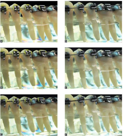
Figure 2 illustrates photographs of premolars and mo-lars under activation with conventional and self-ligating brackets. Figures A, B and C show the photoelastic mod-el with conventional brackets associated to 0.014-in and 0.018-in NiTi wires as well as 0.019 x 0.025-in stainless steel wires respectively. Figures D, E and F show the pho-toelastic model with self-ligating brackets activated with 0.014-in and 0.018-in NiTi wires and 0.019 x 0.025-in stainless steel wires, respectively.
Figure 2 - A, B, C) Conventional brackets;
D, E,F) Self-ligating brackets.
steel wires (Figs 2A, B and C; Table 1). In the mid-dle third, stress was only found in activations with conventional brackets, as yellow fringes were found in the three activations (Figs 2A, B and C; Table 2). The same was not found in self-ligating brackets (Figs 2D, E and F; Table 2).
DISCUSSION
This study reveals that both self-ligating and con-ventional brackets produce photoelastic stress under
conditions of alignment with expansive forces, since dia-gramming of wires was larger than the size of the pho-toelastic model dental arch. It also found that stress con-centrated in the apical region of premolars in both mod-els, but with greater stress concentration in conventional brackets models.
Furthermore, lower periodontal forces, seen in the photoelastic model, allow more physiological expansive treatment. Pandis13 conducted a study comparing the intercanine and intermolar distance ater treatment with
Table 1 - Qualitative analysis of the apical region of premolar roots expressed in scores according to the colors of the fringes on the photoelastic model.
Table 2 - Qualitative analysis of the middle third of premolar roots expressed in scores according to the colors of the fringes on the photoelastic model.
WIRE SELF-LIGATING
BRACKETS
CONVENTIONAL
BRACKETS
0.014-in NiTi ++++ +++
0.018-in NiTi +++++ +++
0.019 x 0.021-in
Stainless steel +++++ +++
WIRE SELF-LIGATING
BRACKETS
CONVENTIONAL
BRACKETS
0.014-in NiTi +++
-0.018-in NiTi +++
-0.019 x 0.021-in
Stainless steel +++
-A
B
C
D
E
conventional and passive self-ligating brackets. In the self-ligating group, intermolar distance was greater. However, buccal tipping of lower incisors was the same in both groups.
The system of self-ligating brackets can increase buccal tipping of incisors and the transverse dimen-sion of the maxilla and the mandible.14 However, in patients with muscular balance, buccal tipping of in-cisors might be desired and better controlled, thereby not changing patient’s facial profile.
Table 1 compares the stress observed in the apical region of premolar roots subjected to conventional and self-ligating systems. Greater concentration of photoelastic stress is observed in conventional brackets.
Lower friction between the wires and brack-ets,4,6 associated with resilient heat-activated nickel-titanium wires5 produce lower periodontal stress, as seen in the photoelastic study mode, thereby favor-ing expansive mechanics in crowdfavor-ing resolution. This treatment modality is indicated, for example, to patients with mainly horizontal growth, presence of muscular balance and some freedom for incisors to tip forward.
In this study, no heat-activated nickel-titanium wires were used. Additionally, all study models tested were free of crowding. Moreover, the self-ligating brackets used herein were passive. In active self-ligating brackets, the more the diameter of wires in-crease, the greater the friction which can be higher than conventional brackets.15
Therefore, according to the present results and the growing development of self-ligating brackets sys-tems, it is reasonable to assert that much has to evalu-ate with regards to stress produced by the use of self-ligating brackets compared to conventional brackets in expansive mechanics.
CONCLUSION
Based on the results of the current study, it is suggested that both bracket systems produced stress when activated. However, conventional brackets produced greater stress in comparison to passive self-ligating brackets. Therefore, the present pi-lot study reveals that self-ligating brackets produce softer forces in periodontal tissues in alignment ex-pansive mechanics.
ACKNOWLEDGMENTS
Special thanks is dedicated to the Department of Dental Material at the School of Orthodontics – State University of Campinas/Piracicaba (UNICAMP), es-pecially to Professor Dr. Américo Bortolazzo Correr.
1. Brito Júnior VS, Ursi WJS. O aparelho pré-ajustado: sua evolução e suas
prescrições. Rev Dental Press Ortod Ortop Facial. 2006;11(3):104-56.
2. Picchioni MS. Análise comparativa dos níveis de atrito em braquetes e
autoligados [dissertação]. São Bernardo do Campo (SP): Universidade Metodista de São Paulo; 2007.
3. Frank C. A comparative study of frictional resistances between orthodontic
bracket and arch wire. Am J Orthod. 1980;78(6):593-609.
4. Voudouris JC. Interactive edgewise mechanisms: form and function comparison
with conventional edgewise brackets. Am J Orthod Dentofacial Orthop. 1997;111(2):119-40.
5. Damon DH. The Damon low-friction bracket: a biologically compatible
straight-wire system. J Clin Orthod. 1998;32(11):670-80.
6. Maltagliati L. Sistema autoligado: quebrando paradigmas. Ortodontia SPO.
2009;42(5):360-1.
7. Rocha JET, Fuziy A, Tukasan PC, Oliveira RCG. Fotoelasticidade: aplicabilidade na
mecânica ortodôntica. Braz Oral Res. 2006;20(Spec issue1):81.
8. Vuolo JH. Polarização da luz e displays TN. São Paulo: IFUSP; 1998. p. 1-16.
9. Rossato C. Estudo fotoelástico das áreas de pressão, produzidas no periodonto,
por forças ortodônticas, na distalização do canino pelos métodos convencionais e com “Power arm”. [dissertação]. Bauru (SP): Unicersidade de São Paulo; 1982. 10. Glickman I, Roeber FW, Brion M, Pameijer JH. Photoelastic analysis of internal
stresses in the periodontium created by occlusal forces. J Periodontol. 1970;41(1):30-5.
11. Interlandi S. Diagrama de contorneamento ortodôntico para a técnica do arco
contínuo (Straight Wire). Ortodontia. 2002;35(4):91-105.
12. Dobranszki A. Estudo fotoelástico do controle vertical com arco de dupla chave na técnica Straight wire. Rev Dental Press Ortod Ortop Facial. 2009;14(4):123-8. 13. Pandis N. Self ligating vs conventional brackets in the treatment of mandibular
crowding: a prospective clinical trial of treatment duration and dental efects. Am J Orthod Dentofacial Orthop. 2007;132(2):208-15.
14. Kochenborger R. Avaliação das alterações dentárias e do peril facial obtidas no tratamento ortodôntico com braquetes autoligáveis [dissertação]. São Bernardo do Campo (SP): Universidade Metodista de São Paulo; 2009.
15. Lorenz M. Active and passive self-ligation: a myth? Angle Orthod. 2011;81(2):312-8.