CLINICAL SCIENCE
I Portuguese Hospital – Salvador/BA, Brasil.
II Department of Gastroenterology, Faculdade de Medicina da Universidade
de São Paulo - São Paulo/SP, Brazil.
III Laboratory of Immunology, Heart Institute, Faculdade de Medicina da
Universidade de São Paulo - São Paulo/SP, Brazil.
IV Federal University of Minas Gerais - Minas Gerais/MG, Brazil. V Instituto Israelita de Ensino e Pesquisa Albert Einstein - São Paulo/SP,
Brazil.
Email: [email protected] Tel.: 55 71 3203.3457
Received for publication on May 25, 2009 Accepted for publication on June 01, 2009
ANALYSIS OF HFE AND NON-HFE GENE MUTATIONS
IN BRAZILIAN PATIENTS WITH HEMOCHROMATOSIS
Paulo Lisboa Bittencourt,I,II Maria Lúcia Carnevale Marin,III Cláudia Alves
Couto,IV Eduardo Luiz Rachid Cançado, I Flair José Carrilho,I Anna Carla
GoldbergV
doi: 10.1590/S1807-59322009000900003
Bittencourt PL, Marin MLC, Couto CA, Cançado ELR,Carrilho FJ, Goldberg AC.Analysis of hfe and non-hfe gene mutations in brazilian patients with hemochromatosis. Clinics. 2009;64(9):837-41.
BACKGROUND: Approximately one-half of Brazilian patients with hereditary hemochromatosis (HH) are neither homozygous for the C282Y mutation nor compound heterozygous for the H63D and C282Y mutations that are associated with HH in Caucasians.
Other mutations have been described in the HFE gene as well as in genes involved in iron metabolism, such as transferrin receptor
2 (TfR2) and ferroportin 1 (SCL40A1).
AIMS: To evaluate the role of HFE, TfR2 and SCL40A1 mutations in Brazilian subjects with HH.
PATIENTS AND METHODS: Nineteen male subjects (median age 42 [range: 20-72] years) with HH were evaluated using the Haemochromatosis StripAssay A®. This assay is capable of detecting twelve HFE mutations, which are V53M, V59M, H63D, H63H, S65C, Q127H, P160delC, E168Q, E168X, W169X, C282Y and Q283, four TfR2 mutations, which are E60X, M172K, Y250X, AVAQ594-597del, and two SCL40A1 mutations, which are N144H and V162del.
RESULTS: In our cohort, nine (47%) patients were homozygous for the C282Y mutation, two (11%) were heterozygous for the H63D mutation, and one each (5%) was either heterozygous for C282Y or compound heterozygous for C282Y and H63D. No other mutations in the HFE, TfR2 or SCL40A1 genes were observed in the studied patients.
CONCLUSIONS: One-third of Brazilian subjects with the classical phenotype of HH do not carry HFE or other mutations that are currently associated with the disease in Caucasians. This observation suggests a role for other yet unknown mutations in the aforementioned genes or in other genes involved in iron homeostasis in the pathogenesis of HH in Brazil.
KEYWORDS: Hereditary hemochromatosis; Iron overload; HFE mutations; Gene mutations; Brazil.
INTRODUCTION
Hereditary hemochromatosis (HH) is a recessive inherited disorder of iron metabolism that is characterized by systemic iron overload as a result of enhanced intestinal absorption.1,2 The disease usually affects males in the fourth
to fifth decade of life and may lead to cirrhosis, diabetes mellitus, panhypopituitarism, cardiomyopathy, arthritis and skin hyperpigmentation.1,2 In European and North-American
Caucasians, HH has been linked to two missense mutations in the HFE gene, which maps to the 4 kb telomeric HLA-A locus on the short arm of chromosome 6.1-3 Furthermore,
80% to 100% of Caucasian patients of Northern European descent with HH were shown to be homozygous for the
C282Y mutation, whereas 4% to 6% were reported to be compound heterozygotes for the C282Y and H63D
mutations.4
The frequencies of these HFE mutations are lower in Italian and Brazilian patients with HH,5-7 and approximately
addition, none of these mutations were observed in African Americans with HH or in patients with African iron overload.8,9
Other mutations in the HFE gene, particularly S65C, have been implicated in HH and are generally found in a compound heterozygous state with either C282Y or H63D. 3,10-12 Furthermore, mutations have been found in genes that have
been recently implicated in iron homeostasis, including the transferrin receptor 2 (TfR2) gene on chromosome 7, the hepcidin antimicrobial peptide (HAMP) gene on chromosome 19, the hemojuvelin (HJV) gene on chromosome 1 and the ferroportin 1 (Fpn1, also known as SCL40A1) gene on chromosome 2. The identification of causative mutations for HH in these genes has improved our understanding of iron homeostasis as well as suggested a genetic basis for a new classification of iron overload disorders.1,3,13
HH has different phenotypic presentations. Types 1 and 3 HH are characterized by the classical HH phenotype, which includes parenchymal iron overload and the presence of HFE and TfR2 mutations, respectively. Type 2 HH is associated with the previously described phenotype of juvenile hemochromatosis and mutations in the HAMP and HJV genes, whereas type 4 HH is associated with heterogeneous clinical and laboratory findings of iron overload that are more pronounced in the reticuloendothelial system.13
Mutations in the TfR2 gene, particularly the Y250X
variant, and several different mutations in the SCL40A1 gene have been reported in Caucasian, African and Asian patients with non-HFE linked iron overload.13-18 The
prevalence of these recently described gene mutations in Brazilian patients with HH has not been assessed, and the purpose of the present study is to determine the frequency of non-classical HFE as well as TfR2 and SCL40A1 mutations in Brazilian subjects with the classical phenotype of HH.
PATIENTS AND METHODS
Subjects: Sixteen unrelated male patients from the metropolitan area of São Paulo, Brazil and three other subjects from Salvador, Bahia were studied. All had a diagnosis of HH based upon several criteria: 1) the absence of secondary causes of iron overload, including chronic hemolytic anemia, thalassemia major, sideroblastic and spur cell anemia, parenteral or dietary iron overload, alcohol abuse and chronic liver disease due to either hepatitis C or non-alcoholic steatohepatitis; 2) transferrin saturation greater than 50% and ferritin levels greater than 400 g/ L; and 3) grade III or IV siderosis by Perls stain and no other evidence of liver disease upon liver biopsy 2. Fifteen patients had been
enrolled in a previous study and were already typed for the HLA-A C282Y and H63D mutations.6 All patients gave
informed consent for participation in this study.
The median age of the patients was 42 (range: 20-72) years. The clinical, laboratory and histological features of the patients are summarized in table 1.
DNA extraction: Genomic DNA was extracted from peripheral blood leukocytes using the DTAB/CTAB technique.19
Determination of HFE and non-HFE mutations: DNA samples from all patients, with the exception of those who were previously confirmed as homozygous for the C282Y
mutation,6 were tested with a reverse transcriptase-based
assay, the Haemochromatosis StripAssay A® (Vienna Lab, Labordiagnostika, GmbH, Vienna, Austria), according to the manufacturer’s instructions. This assay can detect twelve mutations in the HFE gene, which are V53M, V59M, H63D, H63H, S65C, Q127H, P160delC, E168Q, E168X, W169X, C282Y and Q283P, four mutations in the TfR2 gene, which are E60X, M172K, Y250X and AVAQ594-597del, and two mutations in the SCL40A1gene, which are N144H and V162del.
Statistical analysis: Clinical and laboratory data are shown in the text and tables as median and range. Differences in the frequencies of clinical and laboratory parameters were compared using the Fisher exact probability test when appropriate. A p value < 0.05 was considered to be significant.
RESULTS
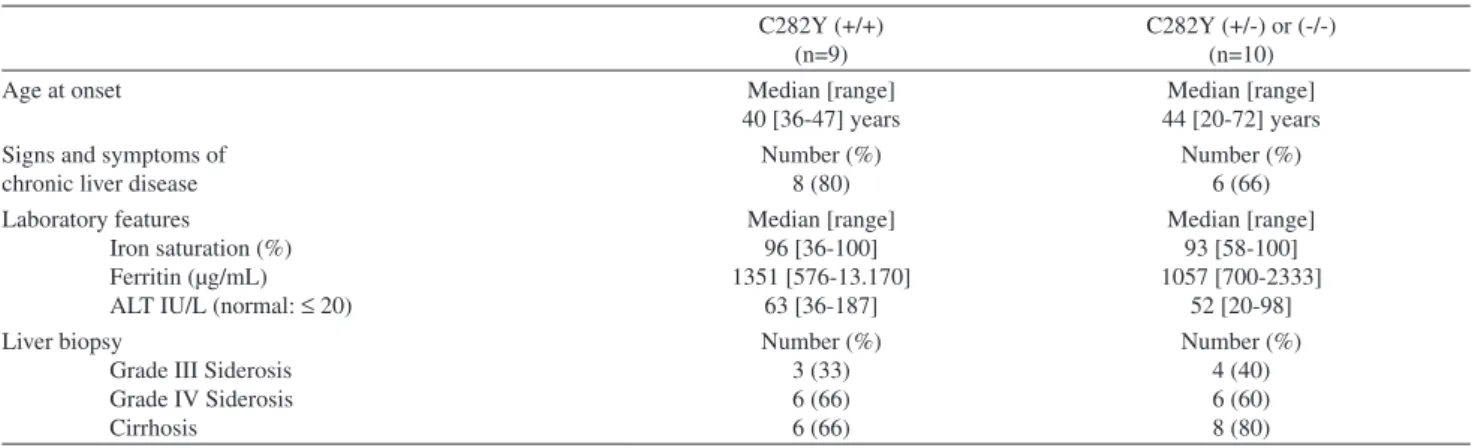
The C282Y and H63D mutation status for each patient in our cohort is shown in Table 2.
Table 1 - Clinical and laboratory features of Brazilian pa-tients with HH
Clinical and laboratory features
Age at onset Median [range]
42 [20-72 ] years Signs and Symptoms
Chronic liver disease Diabetes Impotence Skin hyperpigmentation Panhypopituitarism Cardiac Insufficiency Arthritis Number (%) 14 (74) 4 (21) 3 (19) 1 (15) 1 (15) 1 (15) 1 (15) Laboratory features
Iron saturation (%) Ferritin (µg/mL) ALT IU/L (normal: ≤ 20) Bilirubin mg/dL (normal ≤ 1.1) Albumin g/dL (normal 3.5-5.0)
Median [range] 93 [55-100] 1102 [563-13.170] 57 [20-187] 1.5 [0.6-7.2] 4.3 [2.7-4.9] Liver biopsy
Grade III Siderosis Grade IV Siderosis Cirrhosis
Table 2 - Clinical and laboratory features of Brazilian patients with hemochromatosis with and without homozygosity for C282Y mutation
C282Y (+/+) (n=9)
C282Y (+/-) or (-/-) (n=10)
Age at onset Median [range]
40 [36-47] years
Median [range] 44 [20-72] years Signs and symptoms of
chronic liver disease
Number (%) 8 (80)
Number (%) 6 (66) Laboratory features
Iron saturation (%) Ferritin (µg/mL) ALT IU/L (normal: ≤ 20)
Median [range] 96 [36-100] 1351 [576-13.170] 63 [36-187] Median [range] 93 [58-100] 1057 [700-2333] 52 [20-98] Liver biopsy
Grade III Siderosis Grade IV Siderosis Cirrhosis Number (%) 3 (33) 6 (66) 6 (66) Number (%) 4 (40) 6 (60) 8 (80) Numbers in parentheses are percentages
Nine (47%) patients were homozygous for the C282Y
mutation. In addition, two (11%) patients were heterozygous for H63D, and one each (5%) was either heterozygous for
C282Y or compound heterozygous for C282Y and H63D. None of the other ten HFE mutations and none of the TfR2 or SCL40A1 mutations that were included in the assay were found in these patients.
A comparison of the clinical and laboratory profiles of patients with and without homozygosity for the C282Y
mutation revealed that the former had higher ferritin levels and a lower age of onset for the disease when compared to their counterparts who were non-homozygous for C282Y. However, this difference was not significant (Table 2).
DISCUSSION
Mutations in the HFE gene other than the C282Y
substitution are uncommon in subjects with HH.1,3,10,13 In
fact, the majority of patients who are not homozygous for the C282Y mutation are usually found to be compound heterozygotes for the C282Y and H63D mutations. Rare
HFE mutations, such as S65C, E168X, W169X and Q238P,
have been found in HH subjects who are heterozygous either for C282Y or H63D.1,3,10-12,20,21 The occurrence of the
S65C mutation, as well as homozygosity for H63D,11,12,22,23
has recently been associated with a milder form of iron overload disease. In the present study, no other mutation in the HFE gene, apart from C282Y and H63D, was identified in subjects with HH. Similar results were also reported by Cancado et al.,7 who were unable to find subjects with the
S65C mutation in their cohort of Brazilian patients with iron overload. These findings are also in accordance with several other reports from Europe and North America,1,3 in which
the S65C or another HFE mutation was found without the additional presence of either the C282Y and H63D mutations
in only a few patients with HH or African iron overload. Non-HFE linked HH has been particularly associated with mutations in the ferroportin 1, transferrin receptor 2 and hepcidin genes.13 Hepcidin is a pleiotropic protein that plays
a major role in cellular iron trafficking. Mutations in the
HAMP gene have been associated with type 2 HH or juvenile iron overload, which is characterized by severe iron loading with end-stage organ damage in the second to third decade of life.1,3,13 Given that our patients do not share clinical features
that are compatible with type 2 HH, HAMP gene variants were not assessed in the present study.
On the other hand, our search for several common mutations in the TfR2 and SCL40A1 genes also yielded negative results. TfR2 variants have been implicated in HH type 3 HH, which exhibits a phenotype that is indistinguishable from classical disease phenotype of type 1 HH.13 Unexpectedly, none of our patients with HH carried
any one of the four most common TfR2 gene mutations,
E60X, M172K, Y250X and AVAQ594-597del, that had been previously associated with non-HFE HH in Italy, Portugal and Japan.13-17 In addition, two mutations in the SCL40A1
gene, which have been correlated with type 4 HH in Europe, North America and Australia,13 were not encountered in our
cohort of Brazilian patients with HH. This is not surprising due to the stringent criteria we employed in this study, selecting only patients with parenchymal iron overload and grade III or IV hepatic siderosis. Type 4 HH is mainly associated with iron loading in reticuloendothelial cells, especially macrophages,13 and none of our patients had
histopathological features that were consistent with type 4 HH upon liver biopsy.
two-thirds of Brazilian subjects with either iron overload or HH were previously reported to be either homozygous for the C282Y mutation or compound heterozygous for the C282Y and H63D mutations.6,7,24 Moreover, the frequency
of both of these HFE mutations was reported to be lower in Brazilians when compared to healthy subjects from Europe and North America.25,26
The absence of mutations in the TfR2 and SCL40A1 genes in subjects with non-HFE HH has also been reported in other of European and North American patient cohorts, particularly in African-Americans and in patients with African iron overload.27-30 This observation suggests a
possible role for other mutations in the known HH genes or even for other undiscovered genes that are also involved in iron homeostasis. In this regard, mutations in the iron responsive element of the ferritin heavy chain polypeptide 1 gene (FTH1) have been recently reported in association with an autosomal dominant form of HH in Japan.17
Due to the ethnic background of the Brazilian population, other strategies, including direct sequencing or denaturing HPLC screening of candidate genes, may be more valuable in order to identify novel variants in the steadily increasing group of candidate genes for HH.
As previously shown in our first report,6 patients
homozygous for the C282Y mutation tended to be younger and to have a more severe iron overload when compared to their counterparts who were non-homozygous for C282Y. Even after the inclusion of four additional subjects from the present cohort of HH patients, the difference is still not significant. Interestingly, C282Y homozygotes have been shown to have more pronounced iron overload when compared to heterozygotes or to patients without the C282Y mutation in other populations.31
In summary, only 52% of Brazilian HH patients who were analyzed in this study were either homozygous for
C282Y (n=9) or compound heterozygous for the C282Y and
H63D mutations (n=1). None of these patients had another HFE variant or any mutation in the TfR2 and SCL40A1
genes.
ACKNOWLEDGMENTS
This study was in part supported by the São Paulo Research Foundation (FAPESB), grants number 0553/05 and 01/09850-0 and Alves de Queiroz Family Fund for Research.
REFERENCES
1. Siah CW, Ombiga J, Adams LA, Trinder D, Olynyk JK. Normal iron metabolism and the pathophysiology of iron overload disorders. Clin Biochem Rev. 2006;27:5-16.
2. Tavill AS. American Association for the Study of Liver Diseases; American College of Gastroenterology; American Gastroenterological Association. Diagnosis and management of hemochromatosis. Hepatology. 2001;33:1321-8.
3. Fleming RE, Britton RS, Waheed A, Sly WS, Bacon BR. Pathogenesis of hereditary hemochromatosis. Clin Liver Dis. 2004;8:755-3, vii. 4. Bacon BR, Powell LW, Adams PC, Kresina TF, Hoofnagle JH. Molecular
medicine and hemochromatosis: at the crossroads. Gastroenterology. 1999;116:193-207.
5. Camaschella C, Fargion S, Sampietro M, Roetto A, Bosio S, Garozzo G, et al. Inherited HFE unrelated hemochromatosis in Italian families. Hepatology 1999;29:1563-4.
6. Bittencourt PL, Palácios SA, Couto CA, Cançado EL, Carrilho FJ, Laudanna AA, et al. Analysis of HLA-A antigens and C282Y and H63D mutations of the HFE gene in Brazilian patients with hemochromatosis. Braz J Med Biol Res. 2002;35:329-35.
7. Cançado RD, Guglielmi AC, Vergueiro CS, Rolim EG, Figueiredo MS, Chiattone CS. Analysis of HFE gene mutations and HLA-A alleles in Brazilian patients with iron overload. Sao Paulo Med J. 2006;124:55-60. 8. Monaghan KG, Rybicki BA, Shurafa M & Feldman GL. Mutation analysis of the HFE gene associated with hereditary hemochromatosis in African Americans. American Journal of Hematology. 1998;58:213-7.
9. McNamara L, MacPhail AP, Gordeuk VR, Hasstedt SJ & Rouault T. Is there a link between African iron overload and the described mutations of the hereditary haemochromatosis gene? British Journal of Haematology. 1998;102:1176-8.
10. Pointon JJ, Wallace D, Merryweather-Clarke AT, Robson KJ. Uncommon mutations and polymorphisms in the hemochromatosis: genetics and beyond. Genet Test. 2000;4:151-61.
11. Holmström P, Marmur J, Eggertsen G, Gåfvels M, Stål P. Mild iron overload in patients carrying the HFE S65C gene mutation: a retrospective study in patients with suspected iron overload and healthy controls. Gut. 2002;51:723-30.
12. Mura C, Raguenes O, Ferec C. HFE mutation analysis in 711 hemochromatosis probands: evidence for S65C implication in mild form of hemochromatosis. Blood. 1999;93:2502-5.
13. Pietrangelo A. Non-HFE hemochromatosis. Hepatology. 2004;39:21-9. 14. Camaschella C, Roetto A, Cali A, De Gobbi M, Garozzo G, Carella M,
et al. The gene TfR2 is mutated in a new type of haemochromatosis mapping to 7q22. Nat Genet. 2000;25:14-15.
15. Mattman A, Huntsman D, Lockitch G, Langlois S, Buskard N, Ralston D, et al. Transferrin receptor 2 TfR2 and HFE mutational analysis in non-C282Y iron overload: identification of a novel TfR2 mutation. Blood. 2002;100:1075-7.
17. Hayashi H, Wakusawa S, Motonishi S, Miyamoto K, Okada H, Inagaki Y, et al. Genetic background of primary iron overload syndromes in Japan. Intern Med. 2006;45:1107-11.
18. Gordeuk VR, Caleffi A, Corradini E, Ferrara F, Jones RA, Castro O, et al. Iron overload in Africans and African-Americans and a common mutation in SCL40A1 ferroportin gene. Blood cells Mol Dis. 2003;31:299-304.
19. Gustincich S, Mamfioletti G, Delsal G, Schneider C & Carnici P. A fast method for high-quality genomic DNA extraction from whole blood. BioTechniques 1991;11:298-302.
20. Salvioni A, Mariani R, Oberkanins C, Moritz A, Mauri V, Pelucchi S, et al. Prevalence of C282Y and E168X HFE mutations in an Italian population of Northern European ancestry. Haematologica. 2003;88:250-53.
21. Le Gac G, Dupradeau FY, Mura C, Jacolot S, Scotet V, Esnault G, et al. Phenotypic expression of the C282Y/Q283P compound heterozygosity in HFE and molecular modeling of the Q283P mutation effect. Blood Cells Mol Dis 2003;30:231-7.
22. Aguilar-Martinez P, Bismuth M, Picot MC, Thelcide C, Pageaux GP, Blanc F, et al. Variable phenotypic presentation of iron overload in H63D homozygotes: are genetic modifiers the cause ? Gut. 2001;48:836-42. 23. de Diego C, Opazo S, Murga MJ, Martinez-Castro P. H63D homozygotes with hyperferritinaemia: Is this genotype, the primary cause of iron overload? Eur J Haematol. 2007; 78:66-71.
24. Bittencourt PL. Assessment of HFE mutations in patients with iron overload. São Paulo Med J. 2007;125:65-7.
25. Agostinho MF, Arruda VR, Basseres DS, Bordin S, Soares MC, Menezes RC, et al. Mutation analysis of the HFE gene in Brazilian populations. Blood Cells Mol Dis. 1999;25:3242-7.
26. Pereira AC, Mota GF & Krieger JE. Hemochromatosis gene variants in three different ethnic populations: effects of admixture for screening programs. Human Biology. 2003,73:145-51.
27. Barton EH, Barton JC, Hollowell WW, Acton RT. Transferrin receptor-2 TfR2 mutation Y250X in Alabama Caucasian and African American subjects with and without primary iron overload. Blood Cells Mol Dis. 2001;27:279-84.
28. Aguilar-Martinez P, Esculie-coste C, Bismuth M, Giansily-Blaizot M, Larrey D, Schved JF. Transferrin receptor-2 gene and non-C282Y homozygous patients with hemochromatosis. Blood Cells Mol Dis 2001;27:290-5.
29. McNamara L, Gordeuk VR, Macphail AP. Ferrportin Q284H mutations in African families with dietary iron overload. J Gastroenterol Hepatol 2005;20:1855-8.
30. Barton JC, Acton RT, Lee PL, West C. SLC40A1 Q248H allele frequencies and Q248H-associated risk of non-HFE iron overload in persons of sub-Saharan African descent. Blood Cells Mol Dis. 2007;39:206-11.