Hereditary hemochromatosis: study of laboratory
alterations related to polymorphism
Hemocromatose hereditária: estudo de alterações laboratoriais relacionadas
com polimorfismos
Andressa C. Castilhos1; Bianca T. Canci2; Márcia K. Alves2; Karen Olívia B. Goulart1, 2
1. Centro Universitário da Serra Gaúcha (FSG), Rio Grande do Sul, Brazil. 2. Faculdade Nossa Senhora de Fátima, Rio Grande do Sul, Brazil.
First submission on 27/02/17; last submission on 19/06/17; accepted for publication on 23/06/17; published on 20/08/17
ABSTRACT
Introduction: Hereditary hemochromatosis (HH) is an inherited condition associated with excessive iron storage. Its strong presence is underestimated because patients are asymptomatic. In the city of Caxias do Sul (RS), due to the higher frequency of the disease, it is necessary to perform a study that relates the HH genotypes to laboratory analyses. Objective: The objective of this study was to evaluate
the association of the C282Y, H63D and S65C polymorphisms and laboratory markers in the study cases. Methods: The study was carried
out based on the analysis of medical records from 283 patients assisted from January 2010 to January 2013, older than 18 years of age, who performed the genotyping protocol for HH and laboratory exams. The analysis of markers between genotype and laboratory exams was performed by the SPSS20.0 software. Results: The most frequent genetic alterations were in H63D/WT 84 (31%) and C282Y/WT 17
(6.2%). The most compatible genotype for HH was C282Y/C282Y, even though it did not show signiicant results. The elevated serum ferritin level was slightly higher (p < 0.05) in the study groups, and all of them were above the reference value. We observed in C282Y/C282Y and H63D/C282Y genotypes an insigniicant increase in mean corpuscular volume (MCV) and in mean corpuscular hemoglobin (MCH).
Conclusion: According to the study, it was possible to observe a signiicant elevated serum ferritin level in all HH genotypes.
Key words: hemochromatosis; genetic polymorphism; genotype.
INTRODUCTION
Hereditary hemochromatosis (HH) is a genetic, autosomal recessive disorder characterized by the overload of iron in tissues(1, 2).
The HH gene (HFE) is located on the short arm of chromosome 6,
a major histocompatibility complex (MHC)(3). The protein expressed
by this gene is related to the control of intestinal iron absorption: interacts with the transferrin receptor (TfR) detecting its degree of saturation, thus providing signaling to the enterocyte whether there is a greater or lesser need for intestinal iron uptake(4).
The HH classiication is performed according to the genetic alteration found and the gene associated with iron
overload(5). Classical HH is associated with HFE gene mutations
(homozygosis for C282Y or compound heterozygosis for C282Y/H63D)(6). Mutations in this gene, for example, have an
inadequate mechanism of hepcidin production (systemic iron-regulatory hormone) in response to iron overload(4, 6).
Three allelic variants of the HFE gene were associated with
HH: C282Y, signiicantly associated with HH; H63D and S65C(7). In
the irst case, there is a single mutation at position 282, in which cysteine is replaced by tyrosine; in the second, the mutation was identiied at position 63, where histidine is replaced by aspartate; in the third, the mutation results from the substitution of cysteine for serine at position 65(5).
The frequency of C282Y and H63D mutants is still the subject of studies in the Brazilian population, but it is known that they are present in 2/3 of the Brazilian patients with HH, probably suggesting other mutations in the HFE gene or that other genes
may be involved in the regulation of iron metabolism(2). A recent
study showed a high prevalence of C282Y and H63D mutations in
HFE gene in patients from São Paulo and the southern region of
Brazil, in which 46.67% of the individuals had at least one of the
HH mutations(8). In Brazil there are few studies on the genotypes
mentioned, but the existing ones show patterns different from
those of the northern European studies, where the mutations are less frequent(2, 3, 7-9).
The southern region of the country, with Caucasian origin, should present different frequencies for the C282Y mutation when compared to the population of the Northeast region, for example, of predominantly African origin(10). Therefore, the present study
aims at clarifying the possible link between HH-associated polymorphisms and laboratory tests, identifying the HFE gene
mutations in the study population, relating them to the disease, as well as evaluating the association between C282Y, H63D and S65C polymorphisms and different biochemical and hematological markers in patients attended at a laboratory in Caxias do Sul, Rio Grande do Sul, Brazil.
METHODS
We studied the medical records of 283 patients who had their blood tests collected in a laboratory in the city of Caxias do Sul (RS), from January 2010 to January 2013. Patients who performed the genotyping for HH-associated genes, among other conventional laboratory tests, were selected, thus indicating that these patients were under speciic investigation for this pathology.
The inclusion criteria for the study were: to present a molecular biology test for HH, to be older than 18 years of age and not having positive serologic markers for hepatitis B and C. The sample was selected by the annual report iled in the laboratory information system. Patients with negative HH genotyping were considered as controls. From these 283 patients, 11 were excluded from the results due to the presence of hepatitis B and C markers reactive to the diseases, which could lead to a false result in the relationship between HH and biochemical tests.
The analysis of the reports was performed through the system used by the data grantor. Results of the following tests were collected:
• molecular biology – polymerase chain reaction (PCR) for detection of mutations(11, 12) H63D, C282Y and S65C;
• biochemical tests – lactate dehydrogenase (LDH), iron (Ir), gamma-glutamyl transferase (GGT), glucose (GLU), aspartate aminotransferase (AST) and alanine aminotransferase (ALT) performed by Cobas Integra 400 plus device, that uses absorption photometry technique;
• immune exams – hepatitis B surface antigen (HbsAg), antibodies to hepatitis C virus (HCV) and ferritin (FER) performed by Cobas 6000 device, that uses electrochemiluminescence
method. Transferrin saturation index (TSI) was performed by the supporting laboratory using the immunoturbidimetry method;
• hematimetric exams: red blood cells (RBC), hemoglobin (Hb), hematocrit (Hct), mean corpuscular volume (MCV), mean corpuscular hemoglobin concentration (MCHC) and total leukocytes (LEU) performed by low cytometry method in the Sysmex-XE 2100 device.
After collection, patients with complete laboratory data were selected and the information was stored in a database constructed by the researcher. Statistical analysis was performed in the SPSS 20.0 program. The frequency of genotypes present in the population and the average of the biochemical parameters were evaluated and the Chi-square test was used to verify the signiicance of the relationship between the biochemical and hematological markers and the genetic mutations. Values of p < 0.05 were considered signiicant.
The present study follows the ethical requirements for research and was approved by the Research Ethics Committee of the Associação Cultural e Cientíica Virvi Ramos, on 18 August 2015, under the number of opinion 1.181.585.
RESULTS
From the 272 records analyzed, 19.1% (n = 52) were female
patients and 80.9% (n = 220), were male patients. The mean age was 55.74 years, with minimum age of 23 years and a maximum of 85 years.
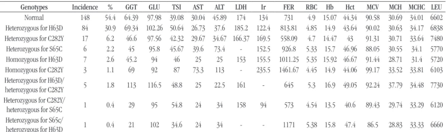
The Table shows the frequency of genotypes in the study
records, and the highest percentage is 54.4% (n = 148) of normal
patients, that is, without the presence of genotype mutations for HH, followed by H63D/WT 31% (n = 84) and C282Y/WT 6.2% (n = 17) genotype. Only medical records with complete laboratory
data were analyzed, and FER levels could be signiicantly increased
(p < 0.05) throughout the study population – all with values above
300 ng/ml [reference values (RV): men (M): 12-300 ng/ml and women (W): 12-150 ng/ml] –, even in patients without mutation in the HFE gene.
The hematimetric indexes were evaluated in all study groups, and we observed an increase in MCV levels in the C282Y/C282Y mutation carriers (mean: 99.17 l/RV: 82-98 l) and the mean corpuscular hemoglobin (MCH) in patients with H63D/C282Y genotype (mean: 37.79 pg/RV: 28-33 pg), but these data were not statistically signiicant. The other hematimetric parameters were within the range of normality.
In the Figure, the data demonstrate the elevated FER levels
according to the study groups.
advantage over men regarding the overload of iron, since they have the monthly loss of this element through menstruation and also in the gestational period. Although women beneit from the physiological loss of iron through menstruation, both men and women are affected by the disease, the ratio is 4 to 10:1(1).
Most cases of HH that have northern European origin are associated with HFE(13). Caxias do Sul is a city that oficially
began with European colonization, with Italian immigrants and people from other European countries. Even so, HH not associated with HFE, although rare, is found in several parts of the world,
regardless of race.
The hematimetric indexes found here are expected and, because of the FER levels in these patients, they are unlikely to have iron-deiciency anemia, as well as the increased MCV and MCH values in the C282Y/C282Y and H63D/C282Y mutation carriers, as also demonstrated by other biochemical markers.
Patients without mutation for the study genotypes were the majority (54.4%), but even in those patients that did not have HH mutations, FER values were elevated, with mean value of 731 ng/ml. Elevated FER may be linked to several factors, such as liver diseases, acute and chronic infection, B12 and folic acid deiciency, and excessive alcohol consumption(14, 15). Thus, the
HFE gene mutation seems to be a necessary condition, but not a requirement for the development of the disease, evidencing the theory of Beutler (2003)(16).
Cançado et al. (2007)(9) found a frequency of theHFE gene
mutation in patients with iron overload of 76%, higher than the frequency found in the present study. The most prevalent mutation TABLE − Genotypes present in the population and mean of the analytical parameters
Genotypes Incidence % GGT GLU TSI AST ALT LDH Ir FER RBC Hb Hct MCV MCH MCHC LEU
Normal 148 54.4 64.39 97.98 39.08 30.04 45.89 174 134 731 4.9 15.07 44.34 90.58 30.69 34.01 6602
Heterozygous for H63D 84 30.9 69.34 102.26 50.64 26.73 37.6 185.2 122.4 813.81 4.85 14.9 43.64 90.02 30.63 34.17 6838
Heterozygous for C282Y 17 6.2 46.6 97.56 42.32 29.67 34.67 166.37 169.5 558.09 4.7 14.47 43 91.31 30.71 33.64 7480
Heterozygous for S65C 6 2.2 45 95.8 45.67 39.6 73.4 - 152.5 926.8 5.33 15.7 46.96 88.05 30.55 34.1 5770
Homozygous for H63D 7 2.6 45.2 94 46 25 25 153 155.5 1011.25 5.35 15.92 46.67 91.44 28.71 31.4 5720
Homozygous for C282Y 3 1.1 69 92 87 73.3 113 - 235.5 1461.67 4.45 14.9 44.06 99.17 33.52 33.81 6103
Heterozygous for H63D/
heterozygous for C282Y 5 1.8 113 116.5 48.8 25 22.5 161 - 645 5.3 16.9 49.05 92.24 37.79 34.48 7730
Heterozygous for C282Y/
heterozygous for S65C 1 0.4 29 95 54.8 24 34 158 94 573 4.54 13.5 40.6 89.43 29.74 33.29 6120
Heterozygous for S65c/
heterozygous for H63D 1 0.4 21 102 34.6 24 34 - - 1171 5.38 15.8 47.4 86.5 28.83 33.33 6660
Reference values: GGT: M 10-50 U/l, W 7-32 U/l; GLU: 70-99 mg/dl; TSI: 20%-45%; AST: < 40 Ul/l; ALT: < 40 Ul/l; LDH: 100-190 E4:V18; Ir: 30-160 micrograms/dl; FER: M 12-300 ng/ml, W 12-150 ng/ml; RBC: M 5.3 ± 3 g/dl, W 13.6 ± 2.5 g/dl; Hb:12-15 g/dl; Hct: M 47 ± 7, W 42 ± 6; MCV: M/W 82-98 Fl; MCH: M/W 31-36 g/dl; MCHC: 30-35 g/dl; LEU: 5,000-10,000.
GGT: gamma-glutamyl transferase; GLU: glucose; TSI: transferrin saturation index; AST: aspartate aminotransferase; ALT: alanine aminotransferase; LDH: lactate dehydrogenase; Ir: iron; FER: ferritin; RBC: red blood cells; Hb: hemoglobin; Hct: hematocrit; MCV: mean corpuscular volume; MCH: mean corpuscular hemoglobin; MCHC: mean corpuscular hemoglobin concentration; LEU: total leukocytes; M: man; W: woman.
S65C/H63D (
n = 1) ± 0
FIGURE − FER mean values per genotype studied
FER: ferritin. 1600 1400 1200 1000 800 600 400 200 0
731 813.81 558.09
926.8 1011.25 1461.67
645 573 1171
Normal (
n = 148) ± 308
H63D/WT (
n = 84) ± 437
C282Y/WT (
n = 17) ± 335
S65C/WT (
n = 6) ± 307
H63D/H63D (
n = 7) ± 503
C282Y/C282Y (
n = 3) ± 475
Mean FER (p < 0.05)
H63D/C282Y (
n = 5) ± 318
C282Y/S65C (
n = 1) ± 0
DISCUSSION
in the present study was H63D/WT (30.9%), corroborating other Brazilian studies that also showed a higher frequency of this
mutation(17, 18). These patients are at low risk of developing the
disease; they have the heterozygous gene and may present an increase in FER and iron levels, as observed in this study. The other markers such as AST and ALT, which indicate the level of iron toxicity caused by HH, are not altered.
Patients with C282Y/C282Y genotype, admitted in studies as the most critical for the development of the disease, presented biochemical markers values altered, however with data not statistically signiicant. The presence of other genotypes should be considered for HH if iron overload is also diagnosed. In patients who presented these mutations, there was increase of serum iron (not compatible with HH), but they may develop the disease in the future according to their living habits.
C282Y mutation of HFE gene presents a wide variation, which
will depend fundamentally on the study population and the criteria used in the selection of each sample and in the diagnosis of iron overload. The frequency of mutation in patients with histological diagnosis of iron overload in the studies of Altes et al. (2003)(19)
and Brandhagen et al. (2000)(20) was 16% and 85%, respectively.
In the present study, it was the genotype that presented higher concentrations of serum FER and iron, besides saturation of the major transferrin, values compatible with the diagnosis for HH. These results conirm the correlation between C282Y/C282Y genotype and increased risk of iron overload(9).
When the selection of the sample occurs by the histological diagnosis of iron overload, the frequency of patients with C282Y/ C282Y genotype is higher when the sample is assessed on the biochemical parameters. The estimated percentage of C282Y/C282Y patients who will develop laboratory evidence of iron overload ranges from 40% to 70%(16, 21), thus evidencing the patient’s predisposition to
develop HH when the genetic mutation is present(16).
Among studies performed in Brazil, Bittencourt et al. (2002)(3)
showed thatC282Y and H63D mutations are present in about two thirds of Brazilian individuals with HH, in the sample we observed 53% of patients with C282Y/C282Y genotype and 13.5% with C282Y/ WT or H63D/WT. On the other hand, Bueno et al. (2006)(7) reported
that62% of patients with HH diagnosis had abnormal HFE, 37.5% homozygous for C282Y, 12.5% compound heterozygous (C282Y/ H63D) and 12.5% heterozygous for C282Y/WT mutation. The results differ from those found in the present study, thus showing that there is also a wide variation in the frequency of mutations among the samples evaluated in the country.
Individuals homozygous for H63D mutation (H63D/ H63D) and heterozygotes (C282Y/H63D) have a low phenotypic expression rate(11, 22). The S65C mutation implies disease usually
when compound with C282Y (C282Y/S65C)(6, 22), and, likewise
the mutations above mentioned, has a low rate of phenotypic expression, which corroborates the results found in the present study.
It was not possible to evaluate the association between biochemical, immunological and hematimetric markers with all genotypes, since the alterations were not statistically signiicant, most likely because the stage of the disease has not yet led to these laboratory alterations. On the other hand, it is clear the presence of genetic mutations in the study population, and early investigation should be performed in any and all evidence of iron overload in these patients to prevent the disease itself from occurring, often leading to irreversible damage to the body.
CONCLUSION
Data from the present study suggest that elevated FER values are associated with HH genotypes, indicating the importance of analyzing this marker in asymptomatic patients. It was also observed that the gene indicated as the most severe for the involvement of the disease, C282Y/C282Y, had a low frequency in the study population.
The correlation of the genotypes with the other markers was not statistically signiicant. Probably the laboratory changes are not yet noticeable due to the present stage of the disease. It is possible to observe a signiicant amount of HH-related genotypes in the research sample for the disease, thus showing a high frequency of this pathology, and the need of laboratory follow-up of these patients, as well as that of their relatives, for the purpose of preventing consequences of iron overload that may be triggered by external factors.
ACKNOWLEDGEMENTS
REFERENCES
1. Souza AFM, Carvalho-Filho RJ, Chebli JF. Hemocromatose hereditária: relato de caso e revisão da literatura. Arq Gastroenterol. 2001; 38(3): 194-202.
2. Bonini-Domingos CR. Aumento de ferro, hemocromatose hereditária e defeitos no gene HFE: o que conhecemos na população brasileira? Rev Bras Hematol Hemoter. 2007; 29(4): 341-2.
3. Bittencourt PL, Palácios SA, Couto CA, Cançado EL, Carrilho FJ, Laudanna AA. Analysis of HLA-A antigens and C282Y and H63D mutations of the HFE gene in Brazilian patients with hemochromatosis. Braz J Med Biol Res. 2002; 35: 329-35. PubMed PMID: 11887210.
4. Grotto HZW. Metabolismo do ferro: uma revisão sobre os principais mecanismos envolvidos em sua homeostase. Rev Bras Hematol Hemoter. 2008; 30(8): 390-7.
5. Santos PCJL, Cançado LD, Terada CT, Guerra-Shinohara EM. Alterações moleculares associadas à hemocromatose hereditária. Rev Bras Hematol Hemoter. 2009; 31(3): 192-202.
6. Cançado RD, Chiattone CS. Visão atual da hemocromatose hereditária. Rev Bras Hematol Hemoter. 2009; 32(6): 469-75.
7. Bueno S, Duch CR, Figueiredo MS. Mutations in the HFE gene (C282Y, H63D, S65C) in a Brazilian population. Rev Bras Hematol Hemoter. 2006; 28(4): 293-5.
8. Herkenhoff ME, Pitlovanciv AK, Remualdo VR. Prevalência das mutações C282Y e H63D no gene HFE em pacientes de São Paulo e do Sul do Brasil. J Bras Patol Med Lab. 2016; 52(1): 21-4.
9. Cançado RD, Guglielmi ACO, Vergueiro CSV, Rolim EG, Figueiredo MS, Chiattone CS. Estudo das mutações C282Y, H63D e S65C do gene HFE em doentes brasileiros com sobrecarga de ferro. Rev Bras Hematol Hemoter. 2007; 29(4): 351-60.
RESUMO
Introdução: A hemocromatose hereditária (HH) é uma doença hereditária associada ao armazenamento em excesso de ferro. Sua
forte presença é subestimada por ser assintomática nos pacientes. Na cidade de Caxias do Sul (RS), por conta da maior frequência da doença, é necessário realizar um estudo que relaciona os genótipos HH com as análises laboratoriais. Objetivo: Avaliar a associação dos polimorfismos C282Y, H63D e S65C e dos indicadores laboratoriais dos casos estudados. Métodos: O estudo foi realizado com base na análise de registros médicos de 283 pacientes atendidos no período de janeiro de 2010 a janeiro de 2013, maiores de 18 anos, os quais realizaram o protocolo de genotipados para exames HH e laboratoriais. A análise de indicadores entre o genótipo e os exames laboratoriais foi realizada pelo software SPSS 20.0. Resultados: As alterações genéticas mais frequentes foram H63D/WT 84 (31%) e C282Y/WT 17 (6,2%). O genótipo que apresentou alteração mais compatível com a HH foi o C282Y/ C282Y, mesmo ele não tendo demonstrado resultados significativos. O aumento da ferritina foi ligeiramente maior (p < 0,05) nos grupos estudados, estando todos acima do valor de referência. Observou-se nos genótipos C282Y/C282Y e H63D/C282Y aumento insignificante no volume corpuscular médio (VCM) e na hemoglobina corpuscular média (HCM). Conclusão: De acordo com o estudo, foi possível observar um aumento significativo da ferritina em todos os genótipos HH.
Unitermos: hemocromatose; polimorfismo genético; genótipo.
10. Jackowski D, Rebello ES, Faucz FR. Análise da frequência da mutação C282Y na população paranaense. Rev Est Biol. 2004; 26(55): 11-8. 11. Feder JN, Gnirke A, Thomas W, et al. A novel MHC class I-like gene is mutated in patients with hereditary haemochromatosis. Nat Genet. 1996; 13: 399-408.
12. Mura C, Raguenes O, Ferec C. HFE mutations analysis in 711 hemochromatosis probands: evidence for S65C implication in mild form of hemochromatosis. Blood. 1999; 93: 2502-5.
13. Emanuele D, Tuason I, Edwards QT. HFE-associated hereditary hemochromatosis: overview of genetic and clinical implications for nurse practitioners in primary care settings. J Am Assoc Nurse Pract. 2014; 26(3): 113-22. PubMed PMID: 24574363.
14. Andrews NC. Disorders of iron metabolism. N Engl J Med. 1999; 341: 1986-95. PubMed PMID: 22276828.
15. Pietrangelo A. Hereditary hemochromatosis – a new look at an old disease. N Engl J Med. 2004; 350: 2383-97.
16. Beutler E. The HFE Cys282Tyr mutation as a necessary but not suficient cause of clinical hereditary hemochromatosis. Blood. 2003; 101: 3347-50.
17. Martinelli AL, Franco RF, Villanova MG, et al. Are haemochromatosis mutations related to the severity of liver disease in hepatitis C virus infection? Acta Haematol. 1999; 102: 152-6. PubMed PMID: 10692680.
18. Agostinho MF, Arruda VR, Basseres DS, et al. Mutations analysis of the HFE gene in Brazilian populations. Blood Cells Mol Dis. 1999; 25: 324-7. PubMed PMID: 10660479.
20. Brandhagen DJ, Fairbanks VF, Baldus WP, Smith CI, Kruckeberg KE, Schaid DJ. Prevalence and clinical signiicance of HFE gene mutations in patients with iron overload. Am J Gastroenterol. 2000; 95: 2910-4. PubMed PMID: 11051367.
21. Beutler E, Felitti V, Gelbart T, Ho N. The effect of HFE genotypes
CORRESPONDING AUTHOR
Márcia Keller Alves
Associação Cultural e Cientíica Virvi Ramos; Rua Alexandre Fleming, 454; Madureira; CEP: 95041-520; Caxias do Sul-RS, Brasil; Phone: + 55 (54) 3535-7300; e-mail: [email protected].
on measurements of iron overload in patients attending a health appraisal clinic. Ann Intern Med. 2000; 133: 329-37. PubMed PMID: 10979877.