Case Report
Case Report
Patient 24, female, Caucasian, born and raised in São Paulo, who attended the emergency department complaining of fever, diffuse chest pain and severe dyspnea for 1 day. Reported a previous additive polyarthritis, asthenia, alopecia, myalgia, and erythematous lesions on the face, remained hospitalized in another hospital with a probable diagnosis of SLE for 10 days. Since then made use of prednisone 60 mg and hydroxychloroquine 400 mg daily. She denied hypertension and other comorbidities.
At the time of physical examination on admission she was in poor performance status, tachycardia (heart rate = 120 beats per minute), tachydyspneic (respiratory rate = 32 breaths per minute), using accessory muscles, peripheral arterial oxygen saturation of 87% blood pressure of 130x70 mmHg, cardiac apex not visible and palpable with a fingertip normopositioned over the fourth intercostal space under the left midclavicular line, the presence of rhythmic sounds with pericardial friction audible in midsystolic fine crackling rales and bilaterally diffuse on auscultation. Absence of heart murmur on palpation. Pulses were present bilaterally, wide and symmetrical. Jugular stasis present bilaterally to 45 degrees. At this time the patient was diagnosed with acute respiratory failure probably due to acute pulmonary edema, opting for oral tracheal intubation, mechanical ventilatory
support, administration of furosemide and intravenous sodium nitroprusside.
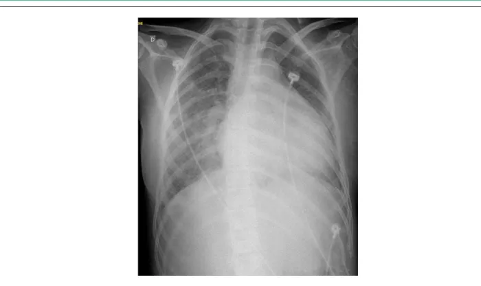
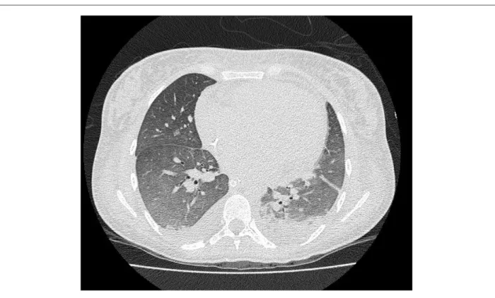
Electrocardiogram was performed and showed sinus tachycardia. The chest radiography showed cephalization of vasculature, venous congestion perihilar, presence of lung parenchyma with diffuse interstitial edema and moderate increase in heart size (Figure 1). Trans-thoracic echocardiogram showed a slight increase of the left atrium, left ventricular ejection fraction of 45% at the expense of diffuse hypokinesis, moderate mitral regurgitation and moderate pericardial effusion. At that time was considered the diagnosis of acute myopericarditis secondary to SLE in activity. Laboratory tests corroborate the hypothesis of active SLE showing leucopenia (3800 leukocytes/mm3), lymphopenia (798 lymphocytes/ mm3), homogeneous antinuclear antibodies (ANA) > 1:320, positive anti-DNA, reactive C-protein 92 mg/L, erythrocyte sedimentation rate of 67 mm and complement levels (C3 and C4) undetectable. Due to joint hypothesis of pulmonary lupus activity, we requested a chest CT scan that showed bronchocentric opacities, small areas of pulmonary consolidation, pleural effusion and extensive bilateral homogeneous ground glass (Figure 2). As a complement, it was performed bronchoalveolar lavage that showed a pattern of discrete alveolar hemorrhage, but with the absence of macrophages with hemosiderin at microscopy, slightly suggesting the presence of lung activity.
Due to the seriousness of the case associated with acute myocardial dysfunction, due to the decision to start with methylprednisolone pulse therapy 1mg/Kg/ day for three consecutive days. After two days the patient improved, being extubated and using noninvasive ventilation intermittently, remaining feverless, hemodynamically stable, with disappearance of tachycardia maintained, preserved renal function, without other complications. The chest radiography showed no changes. It was repeated the trans-thoracic echocardiogram which showed significant recovery of cardiac function (ejection fraction of left ventricle 56%) and minimal pericardial effusion. We observed increased levels of complement, and renal involvement was discarded after completion of proteinuria in 24-hour urine with no changes.
The patient was discharged asymptomatic on prednisone 60mg/day, 25mg 3x/day captopril, carvedilol 12.5 mg 2x/ day, digoxin 0.25 mg / day chloroquine 250mg/day, folic acid and sulfamethoxazole 800mg + 5mg/day trimethoprim 160 mg 3x/week. Due to cardiac involvement, it was decided to carry out monthly cyclophosphamide pulse therapy on an outpatient basis
Keywords
Pulmonary Edema; Myocarditis; Lupus Erythematosus, Systemic.
Systemic Lupus Erythematosus (SLE) is the most common systemic autoimmune disease, occurring more frequently in women, usually aged between 16 and 55 years1,2. Although classically the kidneys are the organs most affected in SLE, cardiopulmonary circulation and the heart may also be affected significantly3. In this context, the occurrence of acute pulmonary edema associated with lupus myocarditis is rare and specific immunosuppressive therapy remains unclear.
Rare Manifestation of Acute Pulmonary Edema Associated with
Acute Lupus Myocarditis
Alexandre de Matos Soeiro, Fabrício Sanchez Bergamin, Maria Carolina Feres de Almeida, Carlos Vicente Serrano
Jr., Breno Alencar de Araripe Falcão, Fernando Ganem
Instituto do Coração (InCor)- HCFMUSP, São Paulo, SP, Brazil
Mailing Address: Alexandre de Matos Soeiro •
Rua Maranhão 680, ap. 81, Santa Paula - 09530-440 - São Caetano do Sul, SP, São Paulo, Brazil
E-mail: alexandre.soeiro@bol.com.br
Manuscript received May 11, 2011; revised manuscript received August 08, 2011, accepted September 20, 2011.
Case Report
Soreiro et al Acute pulmonary edema and lupus myocarditis
Arq Bras Cardiol 2012;98(5):e78-e81 indistinguishable from other forms of pericarditis. The presence of significant pericardial effusion and cardiac tamponade cases are rare3. Pericardial effusion presents remission after appropriate immunosuppressive treatment, as in the case presented, it may evolve to constrictive pericarditis3,4.
Concerning acute myocarditis, the definitive diagnosis is anatomopathological. However, it is not necessary in most cases. The presence of chest pain and tachycardia sustained disproportionately to the presence of fever may occur as reported5. On the other hand, clinical signs and symptoms of heart failure as in our case are unusual, being present in only 5% to 10% of patients1-3,5. There are enormous difficulties in the diagnosis of myocarditis mainly due to the presence of other factors potentially responsible for myocardial damage, such as anemia, hypertension, infection and fluid retention secondary to renal disease or use of corticoids1. In addition, cardiomyopathy in patients with SLE may have different causes such as myocardial ischemia (caused by coronary arteritis, early atherosclerosis, thrombosis or embolism) and secondary to mitral or aortic failure2. The presence of renal dysfunction could delay diagnosis due to water retention. However, the absence of such manifestation attributes all the symptoms to myocardial dysfunction.
As described above, the ventricular dysfunction caused by lupus myocarditis is usually mild with few symptoms or absent4. The echocardiogram may show mainly reduced ejection fraction (present in 81% of patients) and increase in size of cardiac chambers4,5.
Discussion
The diagnosis of SLE was made based on the criteria of the American College of Rheumatology of 1997 and, thus, it was established through the presence of serositis (pleural and pericardial effusions), positive anti-dsDNA, positive antinuclear factor and lymphopenia.
The literature described the presence of some form of heart disease by 30% to 89% of patients with SLE1. Its pathogenesis is not yet fully elucidated and is currently considered as a result of immune complex deposition and complement activation. The aggression by autoimmune disease may occur as pericarditis, myocarditis, Libman-Sacks endocarditis, pulmonary arterial hypertension and coronary artery disease and it should be quickly recognized for establishing adequate immunosuppression therapy with specific cardiologic therapy. Early treatment may contribute to reducing mortality and morbidity of the disease, so that the heart disease in SLE is considered the third leading cause of death, behind nephropathy and infections1. Currently the chronic use of corticosteroids associated with chronic inflammation of the disease itself can cause premature atherosclerosis, leading to more cases of myocardial ischemia in this group of patients1.
Pericarditis is the most common form of cardiac involvement in SLE, present in up to 25% of patients. Generally, there are small pericardial effusions in asymptomatic patients by up to 54% of patients2,3. Signs such as fever, tachycardia, substernal pain and pericardial friction may be present, as well as electrocardiographic changes, which are generally
Figure 1 - Chest radiography with cephalization of vascular weft, perihilar venous congestion, presence of lung parenchyma with diffuse interstitial edema and moderate
increase in heart size.
Case Report
Soreiro et al
Acute pulmonary edema and lupus myocarditis
Arq Bras Cardiol 2012;98(5):e78-e81
Markers of inflammatory activity may be elevated in cases of lupus myocarditis. In addition, serum complement levels are usually low, reflecting the disease activity. Among all markers, the presence of anti-DNA has been associated with lupus myocarditis2. Elevation of serum markers of myocardial necrosis such as creatine kinase MB fraction (CK-MB) and/or troponin may occur in acute myocarditis, but as in other etiologies its alteration is not present in all cases and not directly related to the severity of the clinical symptoms2,3. In some studies antimyocardial antibodies were observed in the blood of patients with SLE, but their relationship with the occurrence of acute myocarditis is still uncertain1,2,6.
The acute pulmonary edema associated with acute myocarditis is rare. The good clinical response to therapy oriented with diuretics, intravenous vasodilators and noninvasive mechanical ventilation, associated to the classical clinical symptoms and to the image observed on chest radiography reinforce the diagnosis of acute pulmonary edema in our case. There seems to be an association between pulmonary venous congestion and pleural effusion on chest radiography in up to 73% of cases, even without the manifestation of acute pulmonary edema5. Moreover, even the presence of mild alveolar hemorrhage reported in bronchoalveolar lavage alone may also be secondary to pulmonary venous congestion, since there was not observed microscopically the presence of macrophages with hemosiderin. The pulmonary lupus activity is more unusual than cardiac activity, and often even
after appropriate treatment it usually leaves some degree of permanent significant impairment mainly in the form of restrictive lung disease, which has not occurred in this case2. The very improvement in chest X-ray with disappearance of reported symptoms corroborates the diagnosis of acute pulmonary edema.
The treatment described in cases of severe symptomatic ventricular dysfunction includes the use of high doses of oral steroids for 7 to 14 days, associated with specific cardiologic therapy2,4. In the case described, due to the severity of the clinical symptoms we chose to use pulse therapy with intravenous corticosteroid associated subsequently with cyclophosphamide. Some studies show reversal of cardiac involvement with this more aggressive approach even without the presence of acute pulmonary edema2-5,7. It is not established in the literature what should be done in these cases, mainly due to the rarity of the event. We obtained a good result, even with reversal of myocardial dysfunction observed at transthoracic echocardiography. Reversal of myocardial damage is reported in up to 89% of patients after 6 months of follow-up, both with the use of corticosteroids and endovenous immunoglobulin7-9. In addition, some case series have shown no recurrence of the symptoms even after four years5,10.
Conclusion
This case shows that in young patients with suggestive symptoms of acute pulmonary edema, the diagnosis of acute myocarditis should be considered as a differential.
Figure 2 - Computed tomography of the chest showed bronchocentric opacities, small areas of pulmonary consolidation, pleural effusion and extensive bilateral
homogeneous frosted glass.
Case Report
Soreiro et al Acute pulmonary edema and lupus myocarditis
Arq Bras Cardiol 2012;98(5):e78-e81
References
1. Falcão CA, Lucena N, Alves IC, Pessoa AL, Godoi ET. Cardite lúpica. Arq Bras Cardiol. 2000;74(1):55-71.
2. Jain D, Halushka MK. Cardiac pathology of systemic lupus erythematosus. J Clin Pathol. 2009;62(7):584-92.
3. Doria A, Iaccarino L, Sarzi-Puttini P, Atzeni F, Turriel M, Petri M. Cardiac involvement in systemic lupus erythematosus. Lupus. 2005;14(9):683-6.
4. Wijetunga M, Rockson S. Myocarditis in systemic lupus erythematosus. Am J Med. 2002;113(5):419-23.
5. Law WG, Thong BY, Lian TY, Kong KO, Chng HH. Acute lupus myocarditis: clinical features and outcome of an oriental case series. Lupus. 2005:14(10):827-31.
6. Caforio ALP, Daliento L, Angelini A, Bottaro S, Vinci A, Dequal G, et al. Autoimmune myocarditis and dilated cardiomyopathy: focus on cardiac autoantibodies. Lupus. 2005;14(9):652-5.
7. Chung JW, Joe DY, Park HJ, Kim HA, Park HS, Suh CH. Clinical characteristics of lupus myocarditis in Korea. Rheumatol Int. 2008;28(3):275-80.
8. Micheloud D, Calderon M, Caparros M, Cruz DP. Intravenous immunoglobulin therapy in severe lupus myocarditis: good outcome in three patients. Ann Rheum Dis. 2007;66(7):986-7.
9. Suri V, Varma S, Joshi K, Malhotra P, Kumari S, Jain S. Lupus myocarditis: marked improvement in cardiac function after intravenous immunoglobulin therapy. Rheumatol Int. 2010;30(11):1503-5.
10. Apte M, McGwin G Jr., Vilá LM, Kaslow RA, Alarcón GS, Reveille JD. Associated factors and impact of myocarditis in patients with SLE from LUMINA, a multiethnic US cohort. Rheumatology. 2008;47(3):362-7.
According to earlier description, SLE presents itself as an important condition in this context, and symptoms associated with the disease shall be investigated. The association with pericarditis and the finding of normal cardiac silhouette with left ventricular dysfunction corroborated the initial hypothesis, enabling rapid diagnosis and proper use of immunosuppressive therapy with subsequent complete reversal of myocardial dysfunction. In this patient the use of intravenous corticosteroid pulse therapy was effective and safe for the treatment of cardiac manifestation of apparent extreme lupus myocarditis.
Potential Conflict of Interest
No potential conflict of interest relevant to this article was reported.
Sources of Funding
There were no external funding sources for this study.
Study Association
This study is not associated with any post-graduation program.