AR
TIGO ORIGINAL / ORIGINAL AR
TICLE
INTRODUCTION
Helicobacter pylori (H. pylori) is one of the com-monest pathogens in humans, affecting more than 50% of the world’s population(31). Although infection rates are decreasing in pediatric populations from different regions, with prevalence ranging from less than 10% to 28%(16, 17, 18, 21, 27), H. pylori still plays an important role in the pathogenesis of peptic ulcer disease, gastric adenocarcinoma and gastric
mucosa-associated lymphoid tissue lymphoma(2) - diseases
that carry high morbidity and mortality rates. H.
pylori infection is acquired predominantly in child-hood(18, 31). Once the gastric mucosa is colonized, the bacterium is likely to remain there for decades, if not throughout the life of the host(34). However,
PREVALENCE OF INFECTION WITH
CAG
A-POSITIVE
HELICOBACTER PYLORI
STRAINS
AMONG CHILDREN AND ADOLESCENTS IN
SOUTHERN BRAZIL
Juliana Ghisleni de
OLIVEIRA
1, Cristina Helena Targa
FERREIRA
1, 2, 3,
Anna Carolina Saraiva
CAMERIN
4, Cláudia Augustin
ROTA
1,
Luíse
MEURER
5and Themis Reverbel da
SILVEIRA
1ABSTRACT – Context - Helicobacter pylori (H. pylori) has a worldwide distribution, but theprevalence of infection, virulence factors, and clinical presentation vary widely according to the studied population. In Brazil, a continental country composed of several ethnicities and cultural habits, the behavior of infection also appears to vary, as many other studies have shown. Objective - Describe the prevalence of infection with cagA-positive H. pylori strains in a group of children and adolescents who underwent esophagogas-troduodenoscopy in Porto Alegre, Rio Grande do Sul. Methods - Fifty-four gastric biopsy specimens of children and adolescents with H. pylori infection demonstrated by histology, urease test and molecular analysis were tested for the presence of cagA-positive
H. pylori strains by the polymerase chain reaction method. Results - The prevalence of cagA-positive H. pylori was 29.6% (95% conidence interval, 18 to 43.6%). There were no statistically signiicant differences in clinical or demographic characteristics or in the endoscopic and histological features of patients infected with cagA-positive strains as compared with those infected by cagA -nega-tive strains. Conclusions- The study showed a low prevalence of infection with cagA-positive H. pylori strains among children and adolescents who underwent EGD in southern Brazil, in comparison to studies conducted with children from other regions of Brazil. There was no association between the presence of cagA-positive strains and more severe clinical presentations in the studied sample.
HEADINGS - Helicobacter pylori. Helicobacter infections, genetics. Prevalence. Child.
Declared conflict of interest of all authors: none
Source of Funding: This work was supported by National Counsel of Technological and Scientific Development (CNPq) and the Incentive Fund Research (FIPE) from Hospital de Clínicas de Porto Alegre.
1 Programa de Pós Graduação em Saúde da Criança e do Adolescente, Faculdade de Medicina, Universidade Federal do Rio Grande do Sul – UFRGS; 2 Unidade de Gastroenterologia Pediátrica, Hospital de Clínicas de Porto Alegre; 3 Unidade de Endoscopia Digestiva, Hospital Moinhos de Vento, Porto Alegre; 4 Laboratório Amplicon, Porto Alegre. 5 Departamento de Patologia, Hospital de Clínicas de Porto Alegre e Medicina Digital. RS, Brasil.
Correspondence: Juliana Ghisleni de Oliveira. Rua São Luís, 920, 603, Santana - CEP 90620-170 - Porto Alegre, RS, Brasil. E-mail: [email protected]
the vast majority of infected individuals remain
asymptomatic(14). The wide spectrum of clinical
manifestations is determined by the interaction of the host’s own immune factors with environmental factors, as well as by the prevalence and expression of H. pylori virulence factors(32).
The most widely studied of these factors is
cytotoxin-associated gene A (cagA), one of the
cagA-positive strains is not the sole predictor of clinical outcomes(33). The cagA gene is diverse in its structure, especially at the 3’-terminal region, which bears a variable sequence of amino acid repetitions. This contributes to the understanding of the variety in clinical presentations observed between different individuals colonized by cag A-positive strains(20, 33).
H. pylori is ubiquitous worldwide, but the prevalence of infection and the presence of virulence factors are highly variable according to the studied population, as is the clinical presentation of H. pylori infection(15, 34). In Brazil, a continental country composed of diverse ethnicities and cultural habits, the behavior of H. pylori infection also appears to vary, as various studies have demonstrated(8, 29). So far, no published studies have used molecular analysis methods to assess H. pylori virulence factors in children from southern Brazil. The aim is to describe the prevalence of infection with cagA-positive strains of H. pylori in a group of children and adolescents who underwent esopha-gogastroduodenoscopy (EGD) in Porto Alegre, a city in Southern Brazil.
METHODS
Patients
This cross-sectional study was conducted from March 2008 to January 2011 on a sample of 400 children and adoles-cents with gastrointestinal symptoms who underwent EGD at Hospital de Clínicas de Porto Alegre and Hospital Moinhos de Vento, both located in Porto Alegre, a large city in South-ern Brazil. The inclusion criteria were age between 1 and 18 years, endoscopic nodular gastritis (which is associated with the presence of bacteria in children(3, 18)), and/or peptic ulcer and/or a positive urease test and/or presence of H. pylori on histological analysis. Patients with any contraindications to biopsy, those with a history of use of antibiotics, proton pump inhibitors, bismuth salts, or H2 blockers in the month preceding from the procedure, and those with history of non-steroidal anti-inlammatory drug and/or acetylsalicylic acid use for three days prior to the procedure were excluded from the study.
Ethical considerations
Subjects’ parents or legal guardians were instructed about the study, and written informed consent was obtained. A questionnaire designed to collect demographic and clinical data was administered to each participant. The study was approved by the Ethics Committees of both centers where it was performed.
Endoscopy
All endoscopies were performed by the same physician (CHTF). The main endoscopic indings were recorded de-scriptively(10). Five biopsy specimens were for histological evaluation (two from the body, two from the antrum, and one from the angular incisure) according to Sydney System
recommendations(9). In addition, four biopsy specimens
(two from the body and two from the antrum, one from each segment) were collected for rapid urease testing and molecular analysis respectively, in order to reduce sampling error(18).
Histological analysis
The biopsy specimens were ixed in formalin, dehydrated, and embedded in parafin wax. Sections measuring 4 µm were sliced and stained with hematoxylin-eosin (for grading of gastritis severity) or with the Giemsa stain (to detect H. pylori) per standard procedures. The classiication and grad-ing of gastritis were made in accordance with the modiied Sydney system(9). All analyses were performed by the same pathologist, who had no knowledge of the results of other tests (LM).
Rapid trease test
The rapid urease test was performed using one biopsy specimen from the body and one from the gastric antrum, which were placed into a commercially available solution containing 10% urea, 1 mL buffered potassium phosphate, and a pH indicator (Uretest®, Renylab Ltda., Minas Gerais, Brazil), at room temperature. Results were recorded up to 12 hours after inoculation. A change of color from yellow to pink was recorded as a positive reaction.
H. pylori and cagA gene detection by polymerase chain reaction (PCR)
The biopsy specimens set aside for molecular analysis (one from the body and one from the antrum) were placed in isotonic saline (0.9% NaCl solution) and deoxyribonucleic acid (DNA) was isolated directly from the specimens using the QIAamp tissue kit (Qiagen Inc., Santa Clarita, CA, USA), according to manufacturer instructions.
PCR primer sets speciic for H. pylori 16S rRNA and the ureA gene, previously tested in an adult sample of our popula-tion, with good sensitivity and speciicity(26), were used. The CagA/ConF (5’-GTGCCTGCTAGTTTGTCAGCG-3’) and CagA/ConR (5’-TTGGAAACCACCTTTTGTATTAGC-3’) forward and reverse primers were used for detection of the
cagA gene. The inal product of the 16S rRNA reaction was
examined by electrophoresis on 3% agarose gel. The ureA,
CagA/ConF e CagA/ConR amplimers were examined by electrophoresis on 2% agarose gels. Results were considered positive when products with molecular weights equivalent to those previously determined were found. Negative and positive controls were included in each assay.
Statistical analysis
RESULTS
Ninety-eight subjects was included in the study. H. pylori was identiied through molecular analysis in 54 subjects, whose specimens were therefore tested for presence of the cagA gene.
The clinical and demographic characteristics of these 54 patients are shown in Table 1.
The prevalence of infection with cagA-positive H. pylori strains in the sample was 29.6% (16 of 54 subjects, 95% conidence interval, 18%–43.6%). There were no statistically signiicant differences in demographic and clinical charac-teristics between patients infected with cagA-positive strains and those infected with cagA-negative strains. Patients with cagA-positive H. pylori infection were more likely to have a family history of peptic ulcer disease (P = 0.04). Stratiication of patients into three age groups (1 to 4 years, 5 to 10 years, and ≥10 years) failed to reveal any signiicant increase in cagA positivity with increasing age (P trend = 0.43).
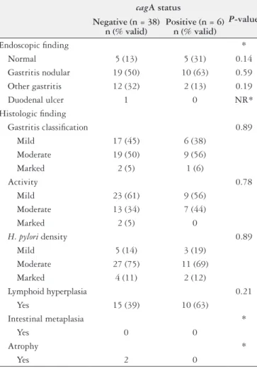
Endoscopic nodular gastritis and follicular lymphoid hy-perplasia were not different between the infected individuals by cagA-positive and cagA-negative H. pylori strains (63%
versus 50% and 63% versus 39%; P = 0.59 and P = 0.21).
Duodenal ulcer was identiied in one of the 54 subjects, who was colonized with a cagA-negative strain. By histological analysis, no between-group differences in bacterial density and inlammatory activity were found. Gastric atrophy was observed in two subjects, both of whom were infected with
cagA-negative strains. The main endoscopic and
histologi-cal indings of this sample of patients are shown in Table 2.
TABLE 1. Characteristics of 54 subjects positive for H. pylori (PCR)
Parameter Finding
Age* 9.8±4.4 (1.8–18.5)
Gender†
Male 27 (50)
Female 27 (50)
EGD indication†
Abdominal pain 18 (34.6) Vomiting/GERD 13 (24.9) Investigation of malabsorption 11 (21.2) Gastrointestinal bleeding 7 (13.4)
Others 5 (5.9)
Water supply†
Mains 47 (94)
Other 3 (6)
Sewage system†
Yes 47 (94)
No 3 (6)
Maternal education†
≤ 8 years 29 (60)
9–11 years 14 (29)
> 11 years 5 (11)
Paternal education†
≤ 8 years 33 (72)
9–11 years 7 (15)
> 11 years 6 (13)
People per room of household†‡
≤ 0.5 4 (8)
0.5–0.99 17 (35)
1–1.99 24 (49)
≥ 2 4 (8)
Family history of peptic ulcer disease†
Yes 15 (29)
No 37 (71)
Family history of stomach cancer†
Yes 10 (19)
No 42 (81)
EGD: esophagogastroduodenoscopy; GERD: gastroesophageal relux disease; PCR: polymerase chain reaction.
* Mean ± standard deviation (range); † Frequency observed (%, percentage of valid indings);
‡ Not counting bathrooms. After Staat et al.(30).
TABLE 2. Main endoscopy and histology indings of patients infected with cagA-positive and cagA-negative H. pylori strains (n = 54)
cagA status
P-value Negative (n = 38)
n (% valid)
Positive (n = 6) n (% valid)
Endoscopic inding *
Normal 5 (13) 5 (31) 0.14 Gastritis nodular 19 (50) 10 (63) 0.59 Other gastritis 12 (32) 2 (13) 0.19
Duodenal ulcer 1 0 NR*
Histologic inding
Gastritis classiication 0.89 Mild 17 (45) 6 (38)
Moderate 19 (50) 9 (56)
Marked 2 (5) 1 (6)
Activity 0.78
Mild 23 (61) 9 (56) Moderate 13 (34) 7 (44)
Marked 2 (5) 0
H. pylori density 0.89
Mild 5 (14) 3 (19)
Moderate 27 (75) 11 (69) Marked 4 (11) 2 (12)
Lymphoid hyperplasia 0.21 Yes 15 (39) 10 (63)
Intestinal metaplasia *
Yes 0 0
Atrophy *
Yes 2 0
DISCUSSION
The present study revealed a lower prevalence of cag A-positive H. pylori infection in children and adolescents from southern Brazil compared to the prevalence described in other Brazilian regions, where the frequency of these strains ranged from 67% to 78% in different studies (see Table 3)(1, 6, 12, 23). It is worth noting that some limitations hamper comparison between these studies, given the wide heterogeneity of de-mographic and clinical characteristics in their samples and the variety of diagnostic methods employed. However, the highest prevalence of cagA-positive strains infection (78%) was found in a study that included asymptomatic children, who were evaluated by enzyme-linked immunosorbent as-say for presence of the cagA gene(6). Although this result may not be comparable to our study from a methodological viewpoint, as circulating antibodies can be present for a long time even after spontaneous eradication of infection(23), this makes our results even more relevant, since our study only included patients with gastrointestinal complaints. Even with no proven association between the presence of H. pylori infection and any speciic symptom(18), one could expect to ind a higher prevalence of cagA-positive strains than we actually did (29.6%) considering that patients in our sample had more severe clinical presentations(11). Pre-vious studies with adults from southern Brazil, including from southern Brazil, including from Porto Alegre, found prevalence rates of cagA-positive infection ranging from 65% to 71%(24, 26), as well as an association between presence of this gene and duodenal ulcer(26) and gastric cancer(19). The difference between adults and children from the same population may denote a different behavior of the infection in children(18,20, 31).
Colonization by cagA-positive strains seems to increase with age(23), although we did not conirm this inding in our study. Queiroz et al.(23) argue that the susceptibility of children to colonization by cagA-positive strains may be related to differences in the expression of adhesion molecules in the gastric mucosa, which changes over time. Sgouras et al.(28) noted that children actually tend to have a higher prevalence of cagA-negative strains than adults, and believe this is a mechanism used by bacteria to allow successful colonization, as these strains induce a weaker host inlammatory response.
Another aspect discussed by Cover and Blaser(7) concern
changes in the predominant strain type in some populations. People in developing countries are predominantly colonized by cagA-positive strains, whereas those in many developed countries are colonized in almost equal proportion by cag A-positive and cagA-negative strains(7). Positive strains used to be more susceptible to antibiotic eradication, which may explain these differences(7).
The methods employed in this study can also help under-stand our indings. DNA extraction from isolates obtained from multiple colonies of H. pylori grown in culture can theoretically lead to different results compared to a technique that performs extraction directly from gastric biopsy speci-mens(22), as the one employed in our study. In two of the four Brazilian studies(1, 23) assessing cagA-positive H. pylori strains in children, molecular analysis of isolates was obtained by culture, which could explain the lower prevalence observed in our population. Direct PCR from biopsy specimens tends to underestimate the prevalence of a speciic virulence fac-tor such as cagA, especially when bacterial density is low(22). Infection with multiple H. pylori strains can also interfere with the sensitivity of the method, and even though deliberate search for other virulence factors was not performed in our
TABLE 3. Prevalence of cagA-positive strains in Brazilian children
Author, year
Genotyping method
Prevalence (%) n/
total Clinical presentation (n) and cagA gene
Place of study 95%CI
Queiroz et al.(23), 2000
PCR from isolates obtained by culture 60/80 (75) Duodenal ulcer (27) Belo Horizonte, Minas Gerais 64–81 100% cagA-positive
Ashour et al.(1), 2002
PCR from isolates obtained by culture 38/55 (69) Duodenal ulcer (24) Belo Horizonte, Minas Gerais 55–81 94.7% cagA-positive
Gatti et al.(12), 2006
Direct PCR of gastric biopsy specimen 38/57 (67) Chronic gastritis (29)
Marília, São Paulo 53–79 69% cagA-positive
Cartágenes et al.(6), 2009
Antibody anti-cagA by ELISA method 39/50 (78) Not described
Belém, Pará 64–89
Our study Direct PCR of gastric biopsy specimen 16/54 (29.6) 18–43.6 No statistically signiicant association
study, this possibility cannot be ruled out(22). Partial deletion of PAI could also yield cagA-negative results(14, 22).
In the international literature, prevalence rates similar to ours were reported in pediatric patients in Jordan and Israel, with a cagA-positive strain prevalence of 26.4 and 25.5% respectively(5, 15). Positive strains were not associated with more pronounced histological gastritis or duodenal ulcer in these studies(5, 15); this is consistent with our indings, but goes against what has been described elsewhere in the literature(1, 12, 23).
No one particular H. pylori virulence factor can be the sole determinant of clinical presentation, which is the re-sult of interactions between the predominant strain type, the host, and the environment in which they live(5). Several authors(4, 20, 28) argue that cagA polymorphisms may affect its biological function, justifying the lack of association between the presence of the gene and a more severe clinical
presentation, as was observed in this and other studies(13, 20, 28). Furthermore, the mere presence of a gene does not sufice; it must be expressed in the host for its role in pathogenesis to be fully evaluated(25).
Finally, the clinical heterogeneity of our sample, which included patients with various gastrointestinal symptoms and over a wide age range, can be considered a major limitation of this study. The low prevalence of cagA-positive strains and low overall prevalence of H. pylori-related diseases observed in our sample may account for the lack of clinical associa-tion between the presence of these strains and more severe chronic gastritis and duodenal ulcer.
In conclusion, this study found a low prevalence of in-fection with cagA-positive H. pylori strains in children and adolescents who underwent EGD in Southern Brazil. No association between colonization with cagA-positive strains and severe clinical outcomes was observed in this sample.
Oliveira JG, Ferreira CHT, Camerin ACS, Rota CA, Meurer L, Silveira TR. Prevalência da infecção por cepas de Helicobacter pyloricagA-positivo em crianças e adolescentes do Sul do Brasil. Arq Gastroenterol. 2014,51(3):180-5.
RESUMO – Contexto - Helicobacter pylori (H. pylori) tem distribuição geográica universal, embora a prevalência da infecção, os fatores de virulência, bem como a apresentação clínica, variem de acordo com a população estudada. No Brasil, um país continental composto por várias etnias e hábitos culturais diversos, o comportamento da infecção também parece variar, como muitos estudos têm demonstrado. Objetivo - Descrever a prevalência da infecção por cepas de H. pylori cagA-positivo em um grupo de crianças e adolescentes submetidos a esofagogastroduodenoscopia em Porto Alegre, Rio Grande do Sul. Métodos - Cinquenta e quatro (54) fragmentos de biópsia gástrica com presença de H. pylori demonstrada pela análise histológica, teste da urease e análise molecular foram testados para a presença de cepas de H. pyloricagA-positivo pelo método da reação em cadeia da polimerase. Resultados - A prevalência de cepas de H. pylori cagA-positivo foi de 29,6% (intervalo de coniança de 95%, 18% a 43,6%). Não houve diferenças estatisticamente signiicativas nas características clínicas e demográicas e nos achados endoscópicos e histológicos entre os pacientes infectados por cepas de H. pylori cagA-positivo em comparação com os cagA-negativo. Conclusões - O estudo demonstrou uma baixa prevalência de infecção por cepas de H. pylori cagA-positivo nas crianças e adolescentes submetidas a esofagogastroduodenoscopia no Sul do Brasil em compara-ção com os estudos realizados com crianças de outras regiões do Brasil. Não houve associacompara-ção entre a presença de cepas cagA-positivo e desfechos clínicos desfavoráveis na amostra estudada.
REFERENCES
1. Ashour AA, Gusmão VR, Magalhães PP, Mendes EN, Queiroz DMM, Rocha
GA, et al. VacA alleles, cagA, and duodenal ulcer in children in Brazil. J Bras Patol Med Lab. 2002;38:79-85.
2. Atherton JC, Blaser MJ. Coadaptation of Helicobacter pylori and humans: ancient history, modern implications. J Clin Invest. 2009;119:2475-87.
3. Bahú Mda G, da Silveira TR, Maguilnick I, Ulbrich-Kulczynski J. Endoscopic
nodular gastritis: an endoscopic indicator of high-grade bacterial colonization and severe gastritis in children with Helicobacter pylori. J Pediatr Gastroenterol Nutr. 2003;36:217-22.
4. Batista SA, Rocha GA, Rocha AM, Saraiva IE, Cabral MM, Oliveira RC, Queiroz
DM. Higher number of Helicobacter pylori CagA EPIYA C phosphorylation sites increases the risk of gastric cancer, but not duodenal ulcer. BMC Microbiol. 2011;11:61.
5. Benenson S, Halle D, Rudensky B, Faber J, Schlesinger Y, Branski D, et al.
Helicobacter pylori genotypes in Israeli children: the signiicance of geography.
J Pediatr Gastroenterol Nutr. 2002;35:680-4.
6. Cartágenes VD, Martins LC, Carneiro LM, Barile KA, Corvelo TC. Helicobacter
pyloriin children and association with CagA strains in mother-child transmission in the Brazilian Amazon region. Rev Soc Bras Med Trop. 2009;42:298-302. 7. Cover TL, Blaser MJ. Helicobacter pylori in health and disease. Gastroenterology.
2009;136:1863-73.
8. Dattoli VC, Veiga RV, da Cunha SS, Pontes-de Carvalho LC, Barreto ML,
Al-cântara-Neves NM. Seroprevalence and potential risk factors for Helicobacter pylori infection in Brazilian children. Helicobacter. 2010;15:273-8.
9. Dixon MF, Genta RM, Yardley JH, Correa P. Classiication and grading of gastri-tis. The updated Sydney System. International Workshop on the Histopathology of Gastritis, Houston 1994. Am J Surg Pathol. 1996;20:1161-81.
10. Dohil R, Hassall E, Jevon G, Dimmick J. Gastritis and gastropathy of childhood.
J Pediatr Gastroenterol Nutr. 1999;29:378-94.
11. Elitsur Y, Neace C, Werthammer MC, Triest WE. Prevalence of CagA, VacA antibodies in symptomatic and asymptomatic children with Helicobacter pylori infection. Helicobacter. 1999;4:100-5.
12. Gatti LL, de Lábio R, Silva LC, Smith MAC, Payão SLM. CagA positive Heli-cobacter pyloriin Brazilian Children Related to Chronic Gastritis. Braz J Infect Dis. 2006;10:254-8.
13. Gold BD, van Doorn LJ, Guarner J, Owens M, Pierce-Smith D, Song Q, et al. Genotypic, clinical, and demographic characteristics of children infected with Helicobacter pylori. J Clin Microbiol. 2001;39:1348-52.
14. Hsu PI, Hwang IR, Cittelly D, Lai KH, Gutierrez O, Kim JG, et al. Clinical pre-sentation in relation to diversity within the Helicobacter pylori cag pathogenicity island. Am J Gastroenterol. 2002;97:2231-8.
15. Hussein NR. Helicobacter pylori and gastric cancer in the Middle East: a new enigma? World J Gastroenterol. 2010;16:3226-34.
16. Janjetic MA, Goldman CG, Barrado DA, Cueto Rua E, Balcarce N, Mantero P, et al. Decreasing trend of Helicobacter pylori infection in children with gastro-intestinal symptoms from Buenos Aires, Argentina. Helicobacter. 2011;16:316-9. 17. Kawakami E, Machado RS, Ogata SK, Langner M. Decrease in prevalence of
Helicobacter pylori infection during a 10-year period in Brazilian children. Arq Gastroenterol. 2008;45:147-51.
18. Koletzko S, Jones NL, Goodman KJ, Gold B, Rowland M, Cadranel S, et al. Evidence-based guidelines from ESPGHAN and NASPGHAN for Helicobacter pylori infection in children. J Pediatr Gastroenterol Nutr. 2011;53:230-43. 19. Meine GC, Rota C, Dietz J, Sekine S, Prolla JC. Relationship between
cagA-pos-itive Helicobacter pylori infection and risk of gastric cancer: a case control study in Porto Alegre, RS, Brazil. Arq Gastroenterol. 2011;48:41-5.
20. Mohamed R, Hanaiah A, Rose IM, Manaf MR, Abdullah SA, Sagap I, et al. Helicobacter pylori cagA gene variants in Malaysians of different ethnicity. Eur J Clin Microbiol Infect Dis. 2009;28:865-9.
21. Oona M, Utt M, Nilsson I, Uibo O, Maaroos HI. Helicobacter pylori infection in children in Estonia: decreasing seroprevalence during the 11-year period of profound socioeconomic changes. Helicobacter. 2004;9:233-41.
22. Park CY, Kwak M, Gutierrez O, Graham DY, Yamaoka Y. Comparison of genotyping Helicobacter pylori directly from biopsy specimens and genotyping from bacterial cultures. J Clin Microbiol. 2003;41:3336-8.
23. Queiroz DM, Mendes EN, Carvalho AS, Rocha GA, Oliveira AM, Soares TF, et al. Factors associated with Helicobacter pylori infection by a cagA-positive strain in children. J Infect Dis. 2000;181:626-30.
24. Ramis IB, Fonseca TL, de Moraes EP, Fernandes MS, Mendoza-Sassi R, Ro-drigues O, et al. Molecular Basis of pathogenicity in Helicobacter pylori clinical isolates. J Clin Microbiol. 2010;48:3776-8.
25. Rick JR, Goldman M, Semino-Mora C, Liu H, Olsen C, Rueda-Pedraza E, et al. In situ expression of cagA and risk of gastroduodenal disease in Helicobacter pylori-infected children. J Pediatr Gastroenterol Nutr. 2010;50:167-72. 26. Rota CA, Pereira-Lima JC, Blaya C, Nardi NB. Consensus and variable region
PCR analysis of Helicobacter pylori 3’ region of cagA gene in isolates from individuals with or without peptic ulcer. J Clin Microbiol. 2001;39:606-12. 27. Rothenbacher D, Schultze V, Jähnig P, Scharschmidt B, Brenner H. Evidence of
a rapid decrease in prevalence of Helicobacter pylori infection in children of a high risk group living in Germany. Eur J Pediatr. 2004;163:339-40.
28. Sgouras DN, Panayotopoulou EG, Papadakos K, Martinez-Gonzalez B, Roum-bani A, Panayiotou J, et al. CagA and VacA polymorphisms do not correlate with severity of histopathological lesions in Helicobacter pylori-infected Greek children. J Clin Microbiol. 2009;47:2426-34.
29. Souto FJ, Fontes CJ, Rocha GA, de Oliveira AM, Mendes EN, Queiroz DM. Prevalence of Helicobacter pylori infection in a rural area of the state of Mato Grosso, Brazil. Mem Inst Oswaldo Cruz. 1998;93:171-4.
30. Staat MA, Kruszon-Moran D, McQuillan GM, Kaslow RA. A population-based serologic survey of Helicobacter pylori infection in children and adolescents in the United States. J Infect Dis. 1996;174:1120-3.
31. Talarico S, Gold BD, Fero J, Thompson DT, Guarner J, Czinn S, Salama NR. Pediatric Helicobacter pylori isolates display distinct gene coding capacities and virulence gene marker proiles. J Clin Microbiol. 2009;47:1680-8.
32. Tomb JF, White O, Kerlavage AR, Clayton RA, Sutton GG, Fleischmann RD, et al. The complete genome sequence of the gastric pathogen Helicobacter pylori.
Nature. 1997;388:539-47.
33. Yamaoka Y. Mechanisms of disease: Helicobacter pylori virulence factors. Nat Rev Gastroenterol Hepatol. 2010;7:629-41.
34. Yamaoka Y, Reddy R, Graham DY. Helicobacter pylori virulence factor genotypes in children in the United States: clues about genotype and outcome relationships.
J Clin Microbiol. 2010;48:2550-1.