42
INTRODUCTION
Cytomegalovirus (CMV) can cause severe disease in immunocompromised patients, either via reactivation of latent CMV infection or via acquisition of primary CMV infection. CMV infection can present in many forms. Clinical syndromes t hat may be o bserved are oesophagitis, encephalitis, pneumonitis, hepatitis, uveitis, retinitis, colitis, and graft reject ion following transplant atio n. Gastrointestinal CMV involvement may be localized or extensive and almost exclusively affects immunocompromised hosts.
Infection of the colon with CMV is increasingly being recognized as a major complication of the acquired immunodeficiency syndrome
(AIDS). The risk of CMV colitis is highest when CD4+ counts are below 50 cells/ mm3
and is rare with CD4+ counts more than 100 cells/mm3.1 Ulcers of the oesophagus, stomach,
small intestine, or colon may result in bleeding or perforation. Early diagnosis of CMV infections may thus be important in patients with AIDS. Complications of CMV infections can be prevented by early recognition and treatment.
CASE REPORT
A 46-year-old man was diagnosed to have HIV-1 infection 4 years ago. He was started on zidovudine, lamivudine and nevirapine but he was irregular with the treatment. He presented to our institute with complaints of low grade fever, abdominal pain and vomiting of 20 days
Case Report:
Cytomegalovirus colitis in a human immunodeficiency virus seropositive
individual with moderately severe immunodeficiency
M.V.S. Subbalaxmi, G. Varun Kumar, N. Chandra, Y.S.N. Raju
Department of General Medicine, Nizam’s Institute of Medical Sciences, Hyderabad
ABSTRACT
Colitis is the most common extraocular manifestation of cytomegalovirus (CMV) infection in human immunodeficiency virus (HIV) infected patients. CMV disease is usually seen in HIV-infected patients with CD4+ counts less than 50/ mm3. There are documented reports of CMV gastrointestinal disease in patients with CD4+ count greater than 50
cells/L. A 46-year-old man with HIV1 infection on irregular antiretroviral treatment presented with low grade fever, abdominal pain and vomitings. He is a known alcoholic. Physical examination revealed pallor, evidence of malnutrition and tenderness in the abdomen. Laboratory investigations revealed mild anaemia; CD4+ count was 240 cells/L. Fundus examination of the patient was normal. Contrast enhanced computed tomography (CECT) of the abdomen revealed dilated small bowel loops, thickening of wall of splenic flexure and thickening of caecal and terminal ileal wall with mild narrowing. As anti-CMV antibodies (IgM) and CMV real time-polymerase chain reaction (PCR) tested positive, patient was treated with intravenous ganciclovir for 14 days followed by oral valganciclovir and patient showed remarkable improvement. Our case highlights the fact that CMV colitis can also occur in patients with relatively preserved CD4+ counts especially if co-morbid conditions like alcoholism co-exist.
Key words: Cytomegalovirus, Colitis, Ganciclovir
Subbalaxmi MVS, Varun Kumar G, Chandra N, Raju YSN. Cytomegalovirus colitis in a human immunodeficiency virus seropositive individual with moderately severe immunodeficiency. J Clin Sci Res 2014;3:42-5.
Corresponding author:
Dr M.V.S. Subbalaxmi, Associate Professor, Department of General Medicine, Nizam’s Institute of Medical Sciences, Hyderabad, India.
e-mail: subbalaxmimvs@yahoo.com Received: 17 February, 2013.
CMV colitis in HIV-seropositive individual Subbalaxmi et al
Online access
43 duration. He gives history of pain while passing stool with passage of few drops of blood after each episode of defecation. Patient did not complain of visual disturbances. He was a known alcoholic, consumes approximately 100ml of alcohol regularly. He had no other co-morbid conditions.
On examination he was found to have mild pallor. Abdominal examination revealed tenderness in t he abdomen with no organomegaly. Digital examination of the rectum showed fissures at 6’ and 2’ oclock
position. Fundus examination was normal. Investigations revealed mild normocytic normochromic anaemia, there were no atypical cells found on peripheral smear. Liver and renal function tests were normal. Chest radiograph was normal. CD4+ count at admission was 240 cells/L.
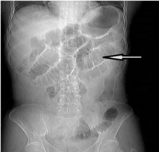
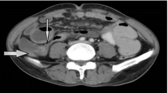
In view of vomitings and pain abdomen, we entertained possibilities of gastrooesophageal reflux disease and candidal oesophagitis. He was treated with proton pump inhibitors (PPI) and fluconazole fo r 7days. Upper gastrointestinal endoscopy (UGIE) showed mild gastric erosions and the biopsy revealed mild inflammation confirming our suspicion. Patient’s condition was worsening despite 7 days of treatment with proton pump inhibitors PPI and fluconazole. Colonoscopy was planned but could not be done as the patient refused to undergo the procedure because of severe pain due to anal fissure. Co ntrast-enhanced computed tomography (CECT) of the abdomen (Figures 1, 2A and 2B) revealed dilated small bowel loops, mild thickening of wall of splenic flexure and thickening of caecal and terminal ileal wall and mild narrowing. He was evaluated for CMV colitis and found to have Ig M CMV antibodies were positive by enzyme linked fluorescence assay (ELFA) (mini VIDAS bioMerieux, France). CMV real time polymerase chain reaction (RT-PCR) (qualitative assay) also tested positive.
Based on clinical findings, serology and imaging findings he was diagnosed to have CMV colitis and was treated with intravenous gancyclovir for 14 days and patient had remarkable relief of symptoms. Patient was continued on oral valganciclovir for another 2 weeks. He was started on teno fovir, emtricitabine and ritonavir boosted atazanavir. He was under regular follow-up; anal fissures have healed with conservative treatment and he is keeping well after 20 months of follow-up.
DISCUSSION
CMV is a common infection, with 40% to 100% of adults exhibiting evidence of past seroconversion.2 Symptoms are usually absent
or self-limited in immunocompetent hosts. Serious CMV infection occurs in patients with immune deficiency, for example, from organ transplantation, malignancy, congenital or acquired immunodeficiency syndromes, steroid therapy, or chemotherapy.
Up to 30% of patients with HIV infection develop retinitis and an additional 5%-10% develop disease in other organs.3 Chorioretinitis
commonly accounts for 80%-90% of CMV disease in patients with AIDS. Though ocular
Figure 1: CT topogram reveals dilated small bowel loops (arrow)
44 involvement is a more common presentation of CMV infection, our patient did not have involvement of eye. Colitis usually presents with diarrhoea which may be bloody or watery and is associated with vomitings and pain abdomen. But, our patient did not complain of diarrhoea rather had constipation possibly due to poor intake and painful defecation due to anal fissures. Clinically, immune competent patients with CMV colitis commonly presented with diarrhoea, fever, and abdominal pain. In a comprehensive review of the literature on CMV colitis in immunocompetent patients, diarrhoea was the most common presenting symptom being evident in 82%, with bloody and watery diarrho ea present in 53% and 29%, respectively.4
The diagnosis of CMV infection cannot be made reliably on clinical grounds alone.
Isolation of CMV or detection of its antigens or deoxyribonucleic acid (DNA) in appropriate clinical specimens is the preferred approach. In patients with suspected CMV colitis endoscopy, imaging, tissue biopsy and serology will help in diagnosis. However, in our patient we could not perform colonoscopy as the patient refused to undergo this procedure due to the presence of painful anal fissures. Though histopathology is diagnostic of CMV colitis, features may not be documented in all cases and delay in treatment can lead to complications like perforation.5 Detection of CMV antigens
(pp65) in peripheral-blood leukocytes or of CMV DNA in bloo d or tissues hast ens diagnosis.6 Such assays yield a positive result
several days earlier than culture methods. The most sensitive way to detect CMV in blood or other fluids may be by amplifying CMV DNA by PCR.7
At the time of our initial evaluation, the patient was very sick, febrile, losing weight and clinically deteriorating. Colonoscopy could not be done as the patient refused to undergo the procedure because of severe pain due to anal fissure. Waiting for the anal fissure to heal and thereafter attempt colonoscopy would result in a loss of valuable time. Furthermo re, colonoscopy while being helpful in ascertining the diagnosis is not a fool-proof method of diagnosis as false-negative results have been reported with this investigation. Since it was imperative to initiate specific therapy as early as possible at this juncture, we had no choice but to resort to other diagnostic modalities, such as, such as, serological testing and RT-PCR to confirm the diagnosis in our patient.
Given the practical clinical scenario of treating a very sick immunosuppressed HIV-sero positive patient with a serious illness, we feel perfectly justified in the practical course of action we have undertaken. While colonoscopy is a valuable diagnostic test for ascertaining CMV colitis, it can yield false-negative results
Figure 2B: CECT abdomen at the level of ileo-caecal junction; verticla arrow points to terminal ileum and horizontal arrow shows caecum: showing thickening of caecal (thick arrow) and terminal ileal wall (thin arrow) and mild narrowing. Dilated small bowel loops can be seen ventral to ileocaecal junction
Figure 2A: NCCT abdomen showing mild thickening of wall of hepatic flexure (arrow)
45 and in a clinical scenario like this it may not be always possible to carry out the same. Therefore, alternative diagnostic methods such as serology and RT-PCR can help in early diagnosis of CMV infection. It is important to develop cost effect ive algo rithms for management of opportunistic infections in HIV in a resource poor countries like India.
Treatment of CMV colitis includes a course of intravenous ganciclovir or foscarnet, for 21 to 28 days. Secondary prophylaxis is not routinely recommended for gastrointestinal disease but should be considered if relapses occur. It should be noted that an episode of CMV colitis is evidence of symptomatic HIV infection and is thus an indication for initiation of highly active antiretroviral therapy.8 Despite relatively
preserved CD4+ counts we decided to change the (antiretroviral treatment) to second-line drugs consisting of tenofovir, emtricitabine and ritonavir boosted atazanavir.
In a scenario where a HIV-infected patient presents with anal fissures as in our patient, herpes simplex virus (HSV) infection should be entertained in the differential diagnosis. However, as he was very sick it was necessary to ascertain CMV infection in a reasonably certain way so as to initiate specific treatment while managing the anal fissures. The fact that the anal fissures healed with conservative treatment alone suggests that HSV infection is an unlikely cause in our patient.
Significant alcohol use, malnutrition and candida esophagitis can predispose to CMV colitis in HIV-positive patients as in our patient.5 Although gastrointestinal
manifesta-tions of CMV in patients with advanced HIV disease are well described, our case highlights the fact that CMV colitis can also occur in
patients with relatively preserved CD4+ counts and should be included in the differential diagnosis of HIV-positive patients presenting with gastrointestinal symptoms regardless of their CD4+ counts.
REFERENCES
1. Ch akr avar ti A, Kash yap B, Matlan iM. Cytomegalovirus infection: an Indian perspective. Indian J Med Microbiol 2009;27:3-11.
2. Springer KL, Weinberg A. Cytomegalovirus infection in the era of HAART: fewer reactivations and more immunity. J Antimicrob Chemother 2004;54:582-6.
3. Mujtaba S, Varma S, Sehgal S. Cytomegalovirus co-infection in patients with HIV/AIDS in north India. Indian J Med Res 2003;117:99-103. 4. Galiatsatos P, Shrier I, Lamoureux E, Szilagyi A.
Meta-analysis of outcome of cytomegalovirus colitis in immunocompetent hosts. Dig Dis Sci 2005;50:609-16.
5. Johnstone J, Boyington CR, Miedzinski LJ, Chiu I. Cytomegalovirus colitis in an HIV-positive woman with a relatively preserved CD4count. Can J Infect Dis Med Microbiol 2009;20:e112. 6. van der Bij W, Torensma R, van son WJ, Anema J,
Sch ir m J, Tegzess AM et al. Rapid immunodiagnosis of active cytomegalovirus infection by monoclonal antibody staining of blood leukocytes. J Med Virol 1988; 25:179-88.
7.
Cassol SA, Poon M-C, Pal R, Naylor MJ, Culver-James J, Bowen TJ, et al. Primer-mediated enzymatic amplification of cytomeyalovirus (CMV) DNA. Application to the early diagnosis of CMV infection in bone marrow transplant recipients. J Clin Investig 1989;83:1109-15. 8. Centers for Disease Control and Prevention.
Treating opportunistic infections among HIV-infected adults and adolescents: Recommendations from CDC, the National Institutes of Health, and the HIV Medicine Association of the Infectious Diseases Society of America. MMWR 2009; 58 (RR-15):1-198.

