Arq Bras Oftalmol. 2009;72(2):236-8
Plasmocitoma da órbita envolvendo glândula lacrimal com transformação
secundária para mieloma múltiplo: relato de caso
Trabalho realizado no Departamento de Oftalmologia da Irmandade da Santa Casa de Misericórdia de São Paulo.
1Estagiária do Setor de Oculoplástica do Departamento de Oftalmologia da Santa Casa de Misericórdia de São Paulo - São Paulo (SP) - Brasil.
2Acadêmico do 4º ano da Faculdade de Ciências Médi-cas da Santa Casa de Misericórdia de São Paulo - São Paulo (SP) - Brasil.
3Estagiário do Setor de Oculoplástica do Departamento de Oftalmologia da Santa Casa de Misericórdia de São Paulo - São Paulo (SP) - Brasil.
4Chefe do Setor de Órbita do Departamento de Oftalmo-logia da Santa Casa de Misericórdia de São Paulo - São Paulo (SP) - Brasil.
5Professora Doutora e Chefe do Setor de Vias Lacrimais do Departamento de Oftalmologia da Santa Casa de Misericórdia de São Paulo - São Paulo (SP) - Brasil.
Endereço para correspondência: Rua Martinico Pra-do, 284 - Apto. 54 - São Paulo (SP) CEP 01224-010 E-mail: [email protected]
Recebido para publicação em 17.04.2008 Aprovado em 08.12.2008
Ivana Lopes Romero1
Felipe Augusto Garcez de Campos2
Renato Wendell Ferreira Damasceno3
José Vital Filho4
Simone Haber Duellberg Von Faber Bison5
Plasmacytoma of the orbit involving lacrimal gland
with secondary transformation into multiple
myeloma: case report
Keywords: Plasmacytoma; Orbital neoplasms/radiotherapy; Lacrimal apparatus; Immu-noglobulin kappa-chains; Neoplasm invasiveness; Multiple myeloma; Tomography, x-ray computed; Human; Female; Adult; Case reports [Publication type]
The authors presenta rare case of plasmacytoma of the orbit involving lacrimal gland with secondary transformation into multiple myeloma in a 42-year-old woman. The lesion was surgically removed and analyzed. Histopathological examination with immunostaining revealed it to be positive for immunoglobulin G and Kappa chains, demonstrating mo-noclonality. However, no abnormality was observed on serum elec-trophoresis, skeletal survey and bone marrow aspiration. Therefore, the tumor was diagnosed solitary plasmacytoma of bone. The patient was treated with external beam radiotherapy and has remained disease free for 5 years and 6 months until 2007, when she presented a patho-logical fracture due to multiple myeloma. Extensive medical work-up to rule out multiple myeloma or other malignant lymphoproliferative conditions involving orbit or ocular adnexa is needed when the diagnosis of solitary plasmacytoma of bone is suspected because treatment and prognosis are very different.
ABSTRACT
RELATOS DE CASOS
INTRODUCTION
Plasmacytoma is a solitary, localized tumor collection of clonal neo-plastic plasma cells capable of producing a monoclonal immunoglobulin(1).
These tumors are classified as either solitary plasmacytomas of the bone (SPB) or extramedullary plasmacytomas (EMP) of soft tissue(2). The
commo-nest site for EMP is the upper respiratory tract, including the oropharynx, nasal cavities, sinuses and larynx(3-5), while the SPB frequently occurs in the
axial skeleton.
Extramedullary plasmacytoma of the orbit, eyelid, ocular adnexa, as of the conjunctiva, occurs rarely(1,6), and appears to have a different natural
history than SPB, with a lower incidence of conversion to multiple myeloma. In contrast, SPB continues to convert to multiple myeloma even after 17 years. The continued conversion of SPB to myeloma suggests that these tumors are actually multiple myeloma in evolution(7).
Arq Bras Oftalmol. 2009;72(2):236-8
237 Plasmacytoma of the orbit involving lacrimal gland with secondary transformation into multiple myeloma: case report
CASE REPORT
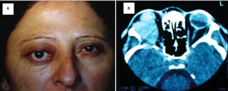
A 42-year-old woman presented proptosis in the right eye for 7 months evolving with progressive pain and vertical di-plopia (Figure 1A) in April 2002.
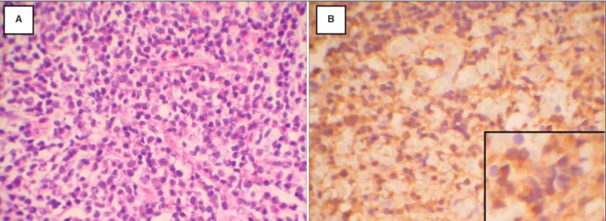
On that occasion, visual acuity was preserved and no other changes were observed either in intraocular pressure or in indirect ophthalmoscopy. B-scan ultrasonography was per-formed showing acoustic brightness of solid mass in the right orbit. Standardized A-scan ultrasonography revealed low-am-plitude internal reflectivity. Axial CT scan showed a large, globular mass occupying the lateral orbit with bone involve-ment (Figure 1B). A diagnostic biopsy was performed, display-ing proliferation of plasmacytoid cells and the histopathology was consistent with plasmacytoma (Figure 2A). Immunohis-tochemical stains were positive for IgG Kappa light chains (Figure 2B), consistent with a monoclonal proliferation.
A systemic evaluation for multiple myeloma, including bone marrow biopsy, peripheral blood test, serum and urine immunoelectrophoresis, skeletal survey and CT scans of chest and abdomen, was negative. Therefore the diagnosis of plasmacytoma of the orbit was established. The orbital mass was treated with external beam radiotherapy (total dose of 5000 cGy).
In March 2003, the tumor regressed; however, after ele-ven months local recurrence involving lacrimal gland was observed. Detailed systemic work-up was negative for mul-tiple myeloma, until October 2007, when the patient presen-ted a lumbar fracture between L2-L3, infiltrative multifocal lesions in the spine and bone marrow biopsy with 7% of plasma cells.
On the basis of these findings, the diagnosis of multiple myeloma was established.
DISCUSSION
Malignant plasma cell tumors are divided according to site of origin. They can be multicentric, such as multiple myeloma, or localized, originating at bone (SPB) or from soft tissue (EMP). The latter is a malignant tumor that is thought to be separate neoplasm from multiple myeloma and plasmacytoma of bone, with a much better prognosis(3).
Lesion size, total serum protein levels and the presence of a monoclonal spike on serum protein electrophoresis appear to correlate with risk of conversion to multiple myeloma. In addition, serum beta-2 microglobulin may be a useful prog-nostic indicator(7).
A few cases of solitary plasmacytoma of the orbit have been reported in the literature to date. Although males are affected more frequently than females in a 3 to 1 ratio and the age at presentation usually lies between the sixth and seventh decade of live(3,8), our case is related to a 42-year-old woman.
The criteria for the diagnosis of SPB include negative lymph node assessment, skeletal survey, bone marrow biopsy and computed tomography(9-10).
In addition, histopathological examination is essential to reach the diagnosis, and it is important, in particular, to de-monstrate the production of monoclonal immunoglobulin by immunostaining. Our patient was diagnosed with SPB because monoclonality of IgG and Kappa light chains had been demons-trated by immunohistochemical examination and because cli-nical, hematological, biochemical and radiological investi-gations ruled out systemic diseases until 2007, when the patient presented a pathological fracture due to multiple myeloma.
The primary method of treatment of SPB is radiation the-rapy. The treatment of choice is external beam radiation with doses between 4000 to 6000 cGy in 5 to 7 weeks(3,11).
Figure 1 - A) Clinical photograph showing 42-year-old woman with proptosis in the right eye. B) Axial CT scan showed a large, globular mass occupying the lateral orbit with bone involvement.
238Plasmacytoma of the orbit involving lacrimal gland with secondary transformation into multiple myeloma: case report
Arq Bras Oftalmol. 2009;72(2):236-8
Figure 2 - A) Plasma cells diffusely infiltrate the orbit (hematoxilin and eosin). B) Immunohistochemical stain positive for Kappa light chain. Inset shows round eccentric nuclei.
A B
In an attempt to prevent progression to multiple myelo-ma, adjuvant chemotherapy was administered in addition to local therapy for solitary plasmacytomas(7). These authors
showed chemotherapy delayed the time to conversion, al-though adjuvant chemotherapy did not affect the rate of conversion.
After treatment (radiotherapy), some patients with SPB have local recurrence, lymph node metastasis or remote me-tastasis, and some develop multiple myeloma, like our patient. Multiple myeloma is a plasma cell neoplasm characterized by plasma cell infiltration of bone marrow and by monoclonal immunoglobulin in the serum. Findings include bone pain, pathologic fractures, renal insufficiency, anemia, infection, and plasmacytomas. These patients should thus be followed carefully for a long period of time.
RESUMO
Os autores relatam um raro caso de plasmocitoma da órbita envolvendo glândula lacrimal com transformação secundária para mieloma múltiplo em uma paciente de 42 anos. A lesão foi removida cirurgicamente e o exame anatomopatológico, aliado à imuno-histoquímica, revelou positividade para imunoglo-bulina G e cadeias leve tipo Kappa. Após extensa avaliação sistêmica, nenhuma anormalidade foi observada, sendo feito o diagnóstico de plasmocitoma solitário ósseo. A paciente foi submetida à radioterapia, permanecendo livre da doença por 5 anos e 6 meses até 2007, quando apresentou uma fratura patológica devido à infiltração plasmocitária na coluna lom-bar, sendo feito diagnóstico de mieloma múltiplo. Uma vez que o plasmocitoma solitário ósseo pode ser a primeira
manifesta-ção do mieloma múltiplo, esses pacientes necessitam de ade-quado diagnóstico e seguimento a longo prazo, pois o trata-mento e prognóstico dessas duas condições é diferente.
Descritores: Plasmocitoma; Neoplasias orbitárias/radioterapia; Aparelho lacrimal; Cadeias kappa de imunoglobulina; Invasivi-dade neoplásica; Mieloma múltiplo; Tomografia computado-rizada por raios x; Humano; Feminino; Adulto; Relatos de casos [Tipo de publicação]
REFERENCES
1. Iida N, Saito K, Fukushima K. A case of extramedullary plasmacytoma arising from the lacrimal gland: a case report. Eur J Plast Surg. 2005;28(5):364-7. 2. Ahamed E, Samuel LM, Tighe JE. Extramedullary plasmacytoma of the eyelid.
Br J Ophthalmol. 2003;87(2):244-5.
3. Uceda-Montañes A, Blanco G, Saornil MA, Gonzalez C, Sarasa JL, Cuevas J. Extramedullary plasmacytoma of the orbit. Acta Ophthalmol Scand. 2000;78(5): 601-3.
4. Miller FR, Lavertu P, Wanamaker JR, Bonafede Je, Wood BG. Plasmacytomas of the head and neck. Otolaryngol Head Neck Surg. 1998;119(6):614-8. 5. Courtmans I, Pigeolet Y, Hedayat AH, Vilain J. Upper airways locations of
plasmacytoma. Acta Otorhinolaryngol Belg. 2000;54(4):487-90.
6. Seddon JM, Corwin JM, Weiter JJ, Brisbane JU, Sutula FC. Solitary extrame-dullary plasmacytoma of the palpebral conjunctiva. Br J Ophthalmol. 1982; 66(7):450-4.
7. Holland J, Trenkner DA, Wasserman TH, Finenberg B. Plasmacytoma. Treatment results and conversion to myeloma. Cancer. 1992;69(6):1513-7.
8. Moragrega-Adame E, Velasco-Barona CF, Suarez-Tata M, Rodriguez-Reyes A. Plasmocitoma orbitário com extension a glândula lagrimal: Reporte de um caso. Rev Mex Oftalmol. 2005;79(1):40-4.
9. Adkins JF, Shields JA, Shields CL, Eagle RC Jr, Flanagan JC, Campanella PC. Plasmacytoma of the eye and orbit. Int Ophthalmol. 1997;20(6):339-43. 10. De Smet MD, Rootman J. Orbital manifestations of plasmacytic
lymphoprolife-rations. Ophthalmology. 1987;94(8):995-1003.