Introduction
Adenocarcinoma is the most common histological type of cancer in various countries and has a wide spectrum of clinical, molecular, and pathological particularities, as well as of imaging patterns. Bronchioloalveolar carcinoma (BAC) is a subtype of adenocarcinoma showing an alveolar (lepidic) growth pattern along an intact interstitial framework but no vascular, stromal, or pleural invasion. These tumors can be divided into three types: nonmucinous (clear
cell subtype/type II pneumocytes); mucinous; and mixed (mucinous and nonmucinous). Nonmucinous BAC tends to present as a solitary lesion and has a better prognosis, whereas the mucinous subtype often presents with consolidation, has a propensity for dissemination, and has worse survival.(1,2)
Before the most recent World Health Organization (WHO) classification of lung neoplasms, all tumors with a lepidic growth pattern
Aspects of bronchioloalveolar carcinoma and of
adenocarcinoma with a bronchioloalveolar component:
CT findings*
Aspectos tomográficos do carcinoma bronquíolo-alveolar e dos adenocarcinomas mistos com componente bronquíolo-alveolar
Pedro Paulo Teixeira e Silva Torres, Julia Capobianco, Marcelo Eustáquio Montandon Júnior, Gustavo Souza Portes Meirelles
Abstract
Bronchioloalveolar carcinoma has various presentations and a wide spectrum of imaging patterns, as does adenocarcinoma with a bronchioloalveolar component. The objective of this essay was to describe and illustrate the CT findings that are most characteristic of these tumors. Three presentations are described: solitary pulmonary nodule, consolidation, and diffuse pattern. The last two should be included in the differential diagnosis, together with infectious diseases. Knowledge of the various presentations and the use of proper diagnostic procedures are crucial to early diagnosis and to improving survival.
Keywords: Lung neoplasms; Adenocarcinoma, bronchiolo-alveolar; Tomography, X-ray computed.
Resumo
O carcinoma bronquíolo-alveolar e os adenocarcinomas mistos com componente bronquíolo-alveolar têm apresentações variadas e múltiplos padrões por imagem. O objetivo deste ensaio foi descrever e ilustrar os achados mais característicos desses tumores em TC. São descritas três apresentações: nódulo pulmonar solitário, consolidação e apresentação difusa, sendo que as duas últimas são importantes no diagnóstico diferencial com acometimento infeccioso. O conhecimento das diversas apresentações e a utilização de propedêutica diagnóstica adequada são definitivos para o diagnóstico precoce e o aumento na sobrevida.
Descritores: Neoplasias pulmonares; Adenocarcinoma bronquíolo-alveolar; Tomografia computadorizada por raios X.
* Study carried out at Multimagem Diagnósticos, Goiânia, at Fleury Medicine and Health, São Paulo, and at the Heart Hospital, São Paulo, Brazil.
Correspondence to: Pedro Paulo Teixeira e Silva Torres Rua S-4, 452, apto. 102, Bela Vista, CEP 74823-450, Goiânia, GO, Brasil. Tel. 55 62 3212-1015. E-mail: [email protected]
Financial support: None.
diagnostic criteria.(11) Although there is a need for universally accepted criteria for subtypes of adenocarcinoma, the authors of the proposal are aware that there will be a period of adaptation to the new terms and recommend that they be initially used concomitantly with the current nomenclature.(11)
The objective of the present essay was to describe the CT findings that are most characteristic of BAC and adenocarcinoma with a bronchioloalveolar component by means of a series of illustrative cases.
Imaging patterns of BAC
and adenocarcinoma with a
bronchioloalveolar component
Pulmonary nodule and focal
ground-glass opacities
A solitary pulmonary nodule, usually in the periphery of the upper lobes, is described as the most common CT finding in BAC and adenocarcinoma with a bronchioloalveolar component.(12) Most of these nodules are semisolid, with solid and ground-glass components, as well as exhibiting irregular borders and pleural indentation.(7) Although cavitation is rare in the initial presentation, some nodules can have focal lucent areas, known as pseudocavitation (Figure 1).(7,13) There might be cavitation after chemotherapy (Figure 2).
Ground-glass opacities raise a number of differential diagnoses, which range from benign diseases, such as focal fibrosis and lesions of an infectious nature, to pre-malignant opacities, such as atypical adenomatous hyperplasia (AAH), and malignant lesions, such as BAC and adenocarcinoma with a bronchioloalveolar component.(14) The current WHO classification for lung tumors defines AAH as a pre-malignant lesion—a precursor to BAC—with particular morphological and molecular characteristics, the most common CT finding in AAH being a pulmonary nodule with ground-glass attenuation. (1) Nakata et al. studied CT characteristics that
could aid in the differential diagnosis of BAC and AAH and reported that AAH is generally < 1.0 cm and presents as pure ground-glass opacities, whereas BAC is at least 1.0 cm and can show solid components.(15) In daily practice, the follow-up evaluation of focal ground-glass were considered BAC, which accounted for 24% of
all lung tumors.(3) After the new WHO criteria for the diagnosis of BAC, BAC was found to account for less than 5% of all malignant lung neoplasms(4); most cases are now considered adenocarcinoma with a bronchioloalveolar component.(1,4)
The principal characteristics of BAC and adenocarcinoma with a bronchioloalveolar component are peripheral location, desmoplastic reaction, mucin production, high frequency of occurrence in nonsmokers, higher frequency of occurrence in women, weak trend toward lymph node/extrathoracic dissemination, and propensity for multifocality.(2,5,6) The symptoms include cough (in 35%), mucoid expectoration (in 24%), dyspnea (in 15%), weight loss (in 13%), hemoptysis (in 11%), and fever (in 8%).(7) Although bronchorrhea is known to be a characteristic of the tumor, it is uncommon and occurs in the advanced stages of the disease.(7)
The five-year survival rate for localized BAC can be as high as 100%.(8) The prognosis is more favorable for localized BAC and adenocarcinoma with a bronchioloalveolar component showing a higher proportion of ground-glass opacity (> 50%) on CT scans than for predominantly solid tumors. (3,5) There is evidence that lung neoplasms have a
shorter doubling time in smokers, which denotes increased tumor aggressiveness. This, together with the fact that many smokers present with severely impaired cardiopulmonary function, could explain the worse prognosis of lung neoplasms in these patients.(2,9)
Surgical biopsy is the gold standard for the diagnosis of BAC; when the procedure is contraindicated, the histological diagnosis can be made by percutaneous or transbronchial biopsy associated with CT findings consistent with the disease.(5)
There are three major patterns of BAC on CT scans: solitary pulmonary nodule or mass; consolidation; and multifocal disease. (2,3,10) According to the new WHO classification,
Positron emission tomography (PET) is considered a fundamental tool in the staging, treatment, and follow-up of lung neoplasms (Figure 4). However, it is of note that pure BAC might not show increased 18F-fluorodeoxyglucose (FDG) uptake,(6) given that these tumors are usually indolent, slow growing, and well differentiated. Therefore, the lack of FDG uptake does not exclude the possibility of neoplasms, such as BAC, small lesions, and indolent cancers. Martins et al.(17) studied the diagnostic accuracy of PET in the assessment of solitary pulmonary nodules and obtained, among the 32 patients studied, only one false-negative result, a case in which the final diagnosis was BAC. Therefore, CT analysis is mandatory for the diagnosis (Figure 5).
Consolidation
The consolidation pattern accounts for up to 30% of all BAC cases, being more common in the mucinous variant.(3) This pattern has also been described as the pneumonic form of BAC.(18) Given the difficulty in distinguishing this BAC pattern from that of infectious pneumonia, there is often a delay in diagnosis. Some findings, such as the angiogram sign, air bronchograms, ground-glass opacities, and air-space nodules (Figure 6), can be observed in BAC and in lobar pneumonia.(3,19) Lymph node enlargement and pleural effusion can also be found in both entities.(16) Parenchymal consolidation that remains unresolved after opacities is crucial. Although most benign lesions
tend to become smaller or disappear in three months, pre-malignant and malignant lesions can remain unchanged or grow (Figure 3).(16)
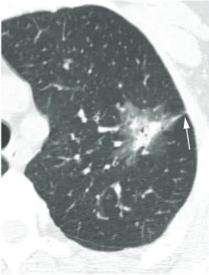
Figure 1 - Axial HRCT scan of a 63-year-old male patient with adenocarcinoma with a bronchioloalveolar component revealing a semisolid pulmonary nodule with irregular borders, pleural indentation (arrow), and internal lucent areas (pseudocavitation).
Figure 2 - CT images of a 54-year-old male patient with two foci of adenocarcinoma. In a, pre-treatment image showing focal ground-glass opacity in the left upper lobe, consistent with pure bronchioloalveolar carcinoma (open arrow). Another irregular, semisolid nodular lesion can be seen in the ipsilateral lower lobe, with pseudocavitation and pleural indentation, corresponding to adenocarcinoma with a bronchioloalveolar component (closed arrow). In b, post-treatment (three cycles of chemotherapy) image showing morphological changes in both lesions. Because of treatment-induced tumor necrosis, there was cavitation in the left lower lobe lesion (arrowhead).
Figure 3 - HRCT images of the chest of a 73-year-old male former smoker presenting with cough and mild dyspnea, as well as with focal infection. In a, image showing a semisolid nodule in the right upper lobe, with a solid central component and a ground-glass halo. In b, follow-up CT scan performed three months later showing complete resolution of the opacity and therefore confirming its infectious nature.
A B
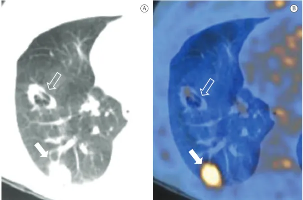
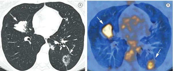
Figure 4 - CT images of a 53-year-old male patient with adenocarcinoma with a bronchioloalveolar component. In a, CT scan of the chest revealing two concomitant pulmonary nodules (foci of adenocarcinoma with a bronchioloalveolar component) in the left lower lobe (arrows). In b, positron emission tomography scan revealing minimal fluorodeoxyglucose (FDG) uptake in one of the lesions, which was cavitated (open arrow), and avid FDG uptake in the other (closed arrow), demonstrating that some of these tumors might not show radiotracer uptake.
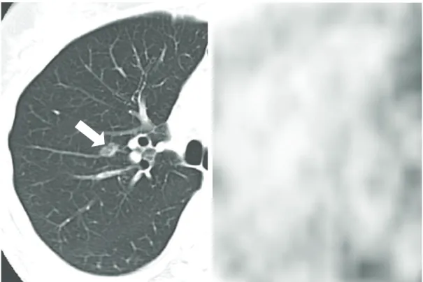
Figure 5 - CT images of a 57-year-old male patient with focal bronchioloalveolar carcinoma. In a, CT scan (lung window) showing a ground-glass nodule in the right lung (arrow). In b, positron emission tomography scan revealing no radiotracer uptake in the lesion.
Figure 6 - CT scan of the chest (lung window) of a 47-year-old male patient with persistent consolidation in the right lung, consistent with adenocarcinoma with a bronchioloalveolar component, showing consolidation in the right lower lobe, associated with small centrilobular nodules (arrows).
Figure 7 - Contrast-enhanced CT scan of the chest of a 56-year-old male patient with dyspnea and persistent cough diagnosed with bronchioloalveolar carcinoma. The image shows branching vessels (arrows) in a low-attenuating consolidation, consistent with the angiogram sign.
treatment should raise the hypothesis of lung cancer.(2,3,18)
The angiogram sign can be seen on contrast-enhanced CT scans and consists of branching pulmonary vessels in a low-attenuating consolidation (Figure 7), usually corresponding
In the assessment of parenchymal consolidations suspected of malignancy, PET can be useful (Figure 8) for the characterization of the lesion or for the staging of neoplastic consolidations. However, it should be emphasized that 18F-FDG-labeled glucose uptake might be absent in some cases of BAC or adenocarcinoma with a bronchioloalveolar component (false negatives), and that PET can show false-positive results in inflammatory or infectious processes (or both).(5)
Diffuse pattern
The diffuse form of BAC represents a multifocal origin, endobronchial dissemination, hematogenous metastases, or any combination of these patterns. Consolidation, nodules (Figure 9), ground-glass opacities, air bronchograms, and cysts (Figure 10), as well as peripheral distribution and distribution in the lower lobes, characterize this pattern. (20) Satellite lesions can be seen in BAC and in
pneumonia.(18)
Ground-glass opacities associated with septal thickening (“crazy-paving pattern”) are also described in the diffuse form of BAC (Figure 11). The differential diagnosis should include other, benign, conditions, such as alveolar proteinosis,(18) hemorrhage, and infection.
For the evaluation of patients with the diffuse pattern of BAC, PET should be used, especially for staging and post-treatment follow-up (Figure 12). It should be borne in mind that 18F-FDG-labeled glucose uptake might be absent in some cases, a careful analysis of CT scans therefore playing an important role in the differential diagnosis and staging.
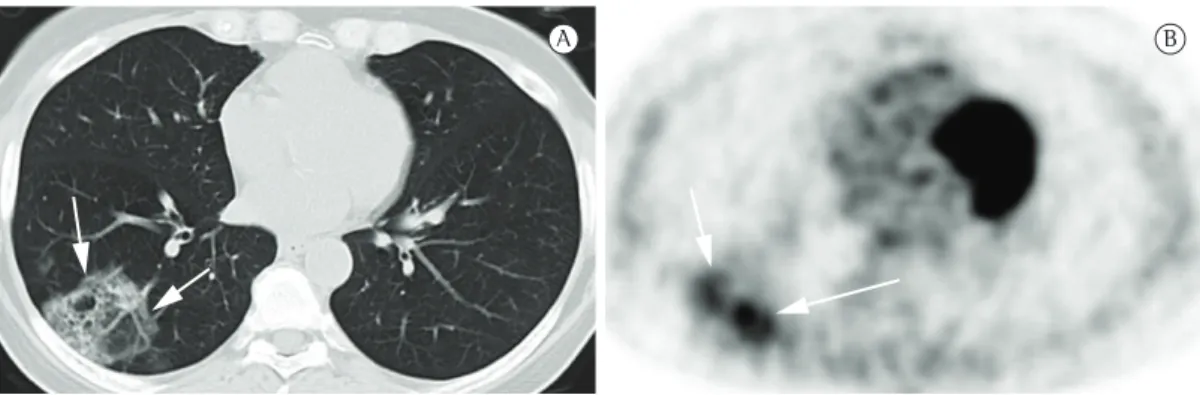
Figure 8 - Images of a 49-year-old female patient diagnosed with adenocarcinoma with a 50% bronchioloalveolar component. In a, CT scan of the chest (lung window) showing opacity in the right lower lobe, with solid and ground-glass components (arrow). In b, positron emission tomography scan showing that the lesion had moderate metabolic activity (arrows).
A B
Figure 9 - CT scan of the chest of an 81-year-old male patient with cough and dyspnea diagnosed with diffuse bronchioloalveolar carcinoma showing multiple bilateral pulmonary nodules and scattered ground-glass opacities.
However, it should be emphasized that the method can yield false-negative and false-positive results, a careful analysis of the CT findings being therefore required in all cases.
References
1. Brambilla E, Travis WD, Colby TV, Corrin B, Shimosato Y. The new World Health Organization classification of lung tumours. Eur Respir J. 2001;18(6):1059-68. PMid:11829087. http://dx.doi.org/10.1183/0903193 6.01.00275301
2. Liu YY, Chen YM, Huang MH, Perng RP. Prognosis and recurrent patterns in bronchioloalveolar carcinoma. Chest. 2000;118(4):940-7. PMid:11035660. http://dx.doi. org/10.1378/chest.118.4.940
3. Patsios D, Roberts HC, Paul NS, Chung T, Herman SJ, Pereira A, et al. Pictorial review of the many faces of bronchioloalveolar cell carcinoma. Br J Radiol. 2007;80(960):1015-23. PMid:17940131. http://dx.doi. org/10.1259/bjr/52225107
Final considerations
Both BAC and adenocarcinoma with a bronchioloalveolar component have a wide spectrum of CT patterns, such as solitary nodules or masses, ground-glass opacities, consolidation, and diffuse presentation. Knowledge of the major imaging findings in these neoplasms is of utmost importance, especially for establishing an early diagnosis and differentiating such tumors from infectious pulmonary processes. Focal opacities, parenchymal consolidations that resolve slowly or show no significant change after treatment, or a combination of the two should raise the suspicion of BAC or adenocarcinoma with a bronchioloalveolar component. In such situations, further diagnostic investigation is recommended in order to exclude lung neoplasms. For differential diagnosis or neoplasm staging, PET can be used.
Figure 11 - CT scan of the chest of a 42-year-old female patient with diffuse bronchioloalveolar carcinoma, which was initially interpreted as infection. In a, ground-glass opacity, crazy-paving pattern, air-space nodules, and foci of consolidation. In b, significant worsening of the disease after two months.
A B
Figure 12 - CT images of a 43-year-old female patient with diffuse bronchioloalveolar carcinoma. In a, CT scan of the chest revealing multiple bilateral nodules (arrows) with irregular borders and air bronchograms. In b, positron emission tomography scan revealing intense metabolic activity in the pulmonary lesions (arrows).
tomografia computadorizada de alta resolução. Radiol Bras. 2002;35(1):7-14. http://dx.doi.org/10.1590/ S0100-39842002000100004
13. Silva CI, Marchiori E, Souza Júnior AS, Müller NL; Comissão de Imagem da Sociedade Brasileira de Pneumologia e Tisiologia. Illustrated Brazilian consensus of terms and fundamental patterns in chest CT scans. J Bras Pneumol. 2010;36(1):99-123. PMid:20209314. http:// dx.doi.org/10.1590/S1806-37132010000100016 14. Oda S, Awai K, Liu D, Nakaura T, Yanaga Y, Nomori
H, et al. Ground-glass opacities on thin-section helical CT: differentiation between bronchioloalveolar carcinoma and atypical adenomatous hyperplasia. AJR Am J Roentgenol. 2008;190(5):1363-8. PMid:18430856. http://dx.doi. org/10.2214/AJR.07.3101
15. Nakata M, Saeki H, Takata I, Segawa Y, Mogami H, Mandai K, et al. Focal ground-glass opacity detected by low-dose helical CT. Chest. 2002;121(5):1464-7. PMid:12006429. http://dx.doi.org/10.1378/chest.121.5.1464
16. Li F, Sone S, Abe H, Macmahon H, Doi K. Malignant versus benign nodules at CT screening for lung cancer: comparison of thin-section CT findings. Radiology. 2004;233(3):793-8. PMid:15498895. http://dx.doi. org/10.1148/radiol.2333031018
17. Martins Rde C, Almeida SA, Siciliano AA, Landesmann MC, Silva FB, Franco CA, et al. Value of [18F]-FDG-PET/ CT as a predictor of cancer in solitary pulmonary nodule. J Bras Pneumol. 2008;34(7):473-80. PMid:18695792. 18. Jung JI, Kim H, Park SH, Kim HH, Ahn MI, Kim HS, et al.
CT differentiation of pneumonic-type bronchioloalveolar cell carcinoma and infectious pneumonia. Br J Radiol. 2001;74(882):490-4. PMid:11459727.
19. Maldonado RL. The CT angiogram sign. Radiology. 1999;210(2):323-4. PMid:10207409.
20. Akira M, Atagi S, Kawahara M, Iuchi K, Johkoh T. High-resolution CT findings of diffuse bronchioloalveolar carcinoma in 38 patients. AJR Am J Roentgenol. 1999;173(6):1623-9. PMid:10584811.
4. Travis WD, Garg K, Franklin WA, Wistuba II, Sabloff B, Noguchi M, et al. Evolving concepts in the pathology and computed tomography imaging of lung adenocarcinoma and bronchioloalveolar carcinoma. J Clin Oncol. 2005;23(14):3279-87. PMid:15886315. http://dx.doi. org/10.1200/JCO.2005.15.776
5. Arenberg D; American College of Chest Physicians. Bronchioloalveolar lung cancer: ACCP evidence-based clinical practice guidelines (2nd edition). Chest. 2007;132(3 Suppl):306S-13S. PMid:17873176. http:// dx.doi.org/10.1378/chest.07-1383
6. Brandão DS, Haddad R, Marsico GA, Boasquevisque CH. Clinicopathological aspects of and survival in patients with clinical stage I bronchioloalveolar carcinoma. J Bras Pneumol. 2010;36(2):167-74. PMid:20485936. 7. Lee KS, Kim Y, Han J, Ko EJ, Park CK, Primack SL.
Bronchioloalveolar carcinoma: clinical, histopathologic, and radiologic findings. Radiographics. 1997;17(6):1345-57. PMid:9397450.
8. Noguchi M, Morikawa A, Kawasaki M, Matsuno Y, Yamada T, Hirohashi S, et al. Small adenocarcinoma of the lung. Histologic characteristics and prognosis. Cancer. 1995;75(12):2844-52. http://dx.doi. org/10.1002/1097-0142(19950615)75:12<2844::AID-CNCR2820751209>3.0.CO;2-#
9. Hasegawa M, Sone S, Takashima S, Li F, Yang ZG, Maruyama Y, et al. Growth rate of small lung cancers detected on mass CT screening. Br J Radiol. 2000;73(876):1252-9. PMid:11205667.
10. Liebow AA. Bronchiolo-alveolar carcinoma. Adv Intern Med. 1960;10:329-58. PMid:13762021.
11. Travis WD, Brambilla E, Noguchi M, Nicholson AG, Geisinger KR, Yatabe Y, et al. International association for the study of lung cancer/american thoracic society/ european respiratory society international multidisciplinary classification of lung adenocarcinoma. J Thorac Oncol. 2011;6(2):244-85. PMid:21252716. http://dx.doi. org/10.1097/JTO.0b013e318206a221
12. Moreira LB, Marchiori E, Melo AS, Magnago M, Muniz MA, Irion K. Carcinoma bronquíolo-alveolar: aspectos na
About the authors
Pedro Paulo Teixeira e Silva Torres
Substitute Professor. Federal University of Goiás, Goiânia, Brazil.
Julia Capobianco
Radiologist. Heart Hospital and Cancer Institute of the State of São Paulo, São Paulo, Brazil.
Marcelo Eustáquio Montandon Júnior
Radiologist. Multimagem Diagnósticos and Jardim América Maternity Hospital, Goiânia, Brazil.
Gustavo Souza Portes Meirelles