Artigo Original
712
Rev Assoc Med Bras 2009; 55(6): 712-5*Correspondência: Rua Professor Miguel Couto, nº 322, complemento 1001 – Jardim Icaraí
Niterói - Rio de Janeiro CEP: 24230-240 E-mail: orsini@predialnet. com.br
Summary
Objective. To report on 9 patients presenting with sporadic motor neuron disease , who over a long period of time evolved with a symmetrical proximal brachial amyotrophic diplegia.
methOdS. Nine patients were followed-up who , displayed, since onset, a progressive limitation of arm flexion/abduction resulting in a peculiar posture with both hands hanging loosely beside the trunk. Electrophysiological test results were consistent with lower motor neuron disease. Cervical MRI was performed in all patients.
reSultS. Nine male subjects with ages ranging from 38 to 73 years at onset of symptoms, developed bilateral and symmetric paresis and atrophy of upper limb muscles. Proximal muscles were more involved than the distal groups. In most patients tendon reflexes were absent or hypoactive in the upper limbs. Needle electromyography (EMG) revealed positive sharp waves and fibrillations and high amplitude polyphasic potentials with an incomplete recruitment pattern in most upper limb muscles. EMG of lower limb muscles was normal in some cases while abnormal in others. MRC did not disclose cervical spinal cord abnormalities from C5-T1.
cOncluSiOn. Attention is called to the Man-in-the-Barrel syndrome in some motor neuron diseases, especially in patients with progressive spinal atrophy and amyotrophic lateral sclerosis
Keywords: motor neuron diseases. man-in-the-Barrel Syndrome. Brachial amyotrophic diplegia.
Progressive spinal atrophy. Amyotrophic lateral sclerosis.
man
-
in
-
the
-
barrel
SyndrOme
,
a
Symmetrical
prOximal
brachial
amyOtrOphic
diplegia
related
tO
mOtOr
neurOn
diSeaSeS
:
a
Survey
Of
nine
caSeS
marcO OrSini1*, antOniO marcOSda Silva catharinO2, fernanda martinS cOelhO catharinO3, mariana pimentel mellO4, marcOS rg de freitaS5,
marcO antôniO araújO leite6, OSvaldO j m naScimentO7
Trabalho realizado na Neuromuscular Disease Outpatient Division - Federal Fluminense University - UFF. Antônio Pedro University Hospital – HUAP, Rio de Janeiro, RJ
1. Graduando em Medicina pela Universidade - UNIGRANRIO e Doutorando em Neurociências pela Universidade Federal Fluminense – UFF, Rio de Janeiro,RJ 2. Doutorando e Mestre em Neurologia pela Universidade Federal do Estado do Rio de Janeiro – UNIRIO; Mestre em Educação pela Universidade Iguaçu – UNIG; Membro Titular
da Academia Brasileira de Neurologia e Professor adjunto de Neurologia pela Universidade Iguaçu – UNIG, Rio de Janeiro,RJ
3. Pós-graduada em Pediatria pela Pontíica Universidade Católica do Rio de Janeiro - PUC-RJ, Rio de Janeiro,RJ 4. Aluna do Programa de Iniciação Cientíica pela Universidade Federal Fluminense – UFF, Rio de Janeiro.RJ
5. Professor Titular e Chefe do Serviço de Neurologia – HUAP da Universidade Federal Fluminense – UFF,Rio de Janeiro,RJ
6. Professor de Neurologia pela Universidade - UNIGRANRIO e Neurologista Responsável pelo Ambulatório de Distúrbios do Movimento – HUAP – Universidade Federal Fluminense - UFF, Rio de Janeiro,RJ
7. Doutor em Neurologia - Professor Titular e Coordenador da Pós-Graduação em Neurologia da Universidade Federal Fluminense– UFF, Rio de Janeiro,RJ
intrOductiOn
In 1969, Mohr1 introduced the term “distal field infarction”
to describe borderzone infarcts between the large intracranial arteries caused by systemic hypoperfusion. This territory corresponds to the zone of anastomosis between the terminal arterioles of the cerebral arteries, considered sensitive to decrease of the encephalic blood stream. The borderzone between the anterior and middle cerebral arteries corresponds to the motor homunculus of the proximal segment of the
upper limbs. Therefore, bilateral ischemia of that zone explains bilateral brachial proximal paralysis 2. Secondary
causes comprise purely motor presentations of myelopathies, including cervical spinal cord infarction, cervical spondylosis and post-radiation. Routine clinical and laboratory data readily differentiate these etiologies3-5. Innumerable researchers have
published new cases of this syndrome, whose etiology differs from the classic description. In 1983, Sage2 for the first time
Man-in-the-Barrel syndroMe, asyMMetricalproxiMalBrachialaMyotrophicdiplegiarelatedto Motor neuron diseases: a surveyof 9 cases.
713
Rev Assoc Med Bras 2009; 55(6): 712-5
of the patients who seemed confined in a barrel (Figure 1). There is a very uncommon phenotype of neurogenic progressive amyotrophy involving the proximal upper limbs in adults mimicking “man-in-the-barrel”. Progress of the disease is confined to the arms for a long period of time, usually suggesting Progressive Spinal Atrophy (PSA) or Amyotrophic Lateral Sclerosis (ALS). There are few reports about this phenotype.
methOdS
Records of patients who had a neurogenic “man-in-the-barrel” phenotype documented by examination at initial evaluation were reviewed. These patients had severe bilateral upper limb neurogenic atrophy that spared, in some cases, the lower limbs, respiratory and bulbar musculature. Topics of the neurological examination were: age at onset of the disease, gender, disease duration, myotomes involved, tendon reflexes in lower limbs, electromyography of lower and upper limbs, magnetic resonance imaging (MRI) of cervical spinal cord and diagnosis. Criteria for inclusion in this study were presence of progressive motor dysfunction, a clinical examination demonstrating a pattern of bilateral weakness limited to the upper limbs for at least 12 months after onset and an electrodiagnostic study consistent with a disorder of lower motor neurons. Patients were excluded from the analysis if there was any evidence of weakness in neck, bulbar, or respiratory muscles earlier than 12 months. Patients were also excluded if there was bladder or bowel dysfunction, sensory signs or symptoms, onset in the presence of radiating pain, weakness in the distribution of individual motor nerves, or an abnormality on cervical MRI, a mass lesion, inflammation, or syrinx. All patients meeting the above criteria continue to be actively followed-up in our clinics. A term of free and informed consent was given to all patients and the objectives of the study were
described. The Research Ethic Committee of the Fluminense Federal University approved the study. Patients agreed to be photographed and approved use of the image (with a band covering the face) for publication. Evaluations were carried out at the Neuromuscular Disease Outcome Sector of the University Hospital Antonio Pedro (HUAP), Fluminense Federal University (UFF).
reSultS
Nine patients were included in the study. In the majority, a progressive severe bilateral upper limb neurogenic atrophy was found sparing the lower limbs, bulbar and respiratory muscles, resulting in a neurogenic man-in-the-barrel syndrome phenotype. The clinical and laboratory results initially suggested a diagnosis of ALS. However, later it became evident that these patients had a disorder that remained restricted to the upper limbs and failed to demonstrate pyramidal signs over time. In this report, the clinical features of this condition called, by some authors, brachial amyotrophic diplegia syndrome (BAD) 3 are described. Six of the 9 patients just had a
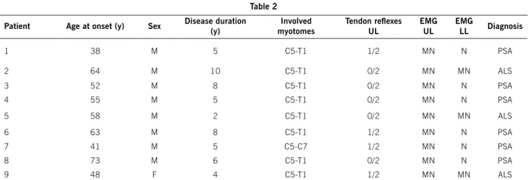
lower motor neuron disorder. During follow-up periods ranging from 2 to 10 years, only three patients developed weakness in the lower limbs, one developed respiratory or bulbar dysfunction and none lost the ability to ambulate. A higher male predominance was also observed. In our casuistry, at onset age ranged from 38 to 73 years. Some patients when admitted to our ser vice, repor ted that symptoms had started years before (2-10 years) (Table 1). The more affected myotomes, characterizing a massive compromise of the anterior horn cells in the spinal cord were the C5-C6 segments. The most affected muscles, after accurate evaluation of the muscular force were the deltoid, supraspinatus, teres minor, infraspinatus and biceps brachii (Table 2). Most of the deep reflexes were abolished or hypoactive in the upper limbs. Three patients presented, 16 months after the first evaluation, deep reflexes hyperactive and presence of the Babinski’s and Hoffmann’s signs. Thereupon they were diagnosed as having ALS according to the EL Escorial criteria 6. In all patients studied, the EMG
of the upper limbs characterized a neurogenic compromise. There was no conduction block or other evidence of demyelination. Only three patients, months later, developed a pattern of anterior horn compromise in the lower limbs. Six patients were diagnbosed as PSA and three of ALS (Table 1). Serum creatine kinase and genetic tests for mutations in the survival motor neuron gene and for X-linked spinobulbar muscular atrophy were negative.
Final Considerations
orsini M etal.
714
Rev Assoc Med Bras 2009; 55(6): 712-5Myotomes Cases (Left/Right)
C1 C2 C3 C4 C5 C6 C7 C8 C9
C5 – Deltoid 2/1 2/2 2/2 2/2 1/1 1/1 2/1 1/1 0/0
C5 – Teres minor/Infraspinatus 2/2 2/1 1/2 2/2 2/2 2/2 4/3 2/2 0/0
C5 – Supraspinatus 1/2 1/2 1/2 2/1 3/3 2/2 2/2 2/2 2/3
C5 – Biceps brachii 2/2 2/3 2/1 2/2 2/2 2/2 3/3 2/2 0/0
C6 – Extensor carpi radialis 3/2 3/3 3/3 2/0 3/3 3/2 4/4 3/3 3/3
C7 – Triceps brachii 4/4 4/3 4/3 3/3 3/3 2/3 4/4 3/3 4/4
C8 – Fingers lexors 5/4 5/5 4/3 3/2 3/4 3/3 5/5 4/4 3/3
T1 – Dorsal and Palmar Interosseous 5/4 4/4 4/3 1/0 3/3 4/3 5/5 4/4 3/3
L2 – Iliopsoas 5/5 5/4 5/4 4/4 4/4 4/4 5/5 4/4 4/4
L3 – Quadriceps femoris 4/4 4/4 4/5 5/5 4/4 4/4 5/5 4/4 4/4
L4 – Tibialis Anterior 4/4 4/4 4/4 4/5 5/5 3/4 5/5 5/5 4/4
L5 – Extensor Hallucis Longus 5/4 5/5 5/5 5/5 5/5 4/4 5/5 5/5 4/4
S1 – Ankle Plantar Flexors 5/5 5/5 5/5 5/5 5/5 5/5 5/5 5/5 5/5
Patient Age at onset (y) Sex Disease duration (y)
Involved myotomes
Tendon relexes
UL
EMG UL
EMG
LL Diagnosis
1 38 M 5 C5-T1 1/2 MN N PSA
2 64 M 10 C5-T1 0/2 MN MN ALS
3 52 M 8 C5-T1 0/2 MN N PSA
4 55 M 5 C5-T1 0/2 MN N PSA
5 58 M 2 C5-T1 0/2 MN MN ALS
6 63 M 8 C5-T1 1/2 MN N PSA
7 41 M 5 C5-C7 1/2 MN N PSA
8 73 M 6 C5-T1 0/2 MN N PSA
9 48 F 4 C5-T1 1/2 MN MN ALS
Table 1 - Characteristics of symmetrical proximal brachial amyotrophic diplegia
Table 1 - Compromise of muscular force in the lower limbs according to Medical Research Council7 (0. No movement; 1. Palpable contraction, no visible movement; 2. Movement
but only with gravity eliminated; 3. Movement against gravity; 4. Movement against resistance but weaker than normal; 5. Normal power).
financial SuppOrt:
CNPQ
_______________________ Conlict of interest: none
reSumO
Síndrome do homem no Barril, uma diplegia amiotrófica braquial proximal simétrica relacionada a doença do neurônio motor: a propósito de nove casos.
ObjetivO.Relatar nove pacientes com a doença do neurônio
de motor esporádica que apresentam progressiva e simétrica diplegia braquial amiotrófica.
MétOdOs. Acompanhamos nove pacientes que exibiram,
desde o começo, uma limitação progressiva de flexão/abdução do membro superior resultando em uma postura peculiar com ambas as mãos pendentes ao longo do tronco. Os resultados dos testes eletrofisiológicos foram consistentes com achados de doença do neurônio motor inferior. Ressonância magnética (RM) cervical foi realizada em todos pacientes.
2. (M - male; y - years; UL - upper limbs; LL - lower limbs; EMG- electromyography; MN - motor neuron involvement; N - normal; PSA - Progressive Spinal Amyotrophy; ALS - Amyotrophic Lateral Sclerosis; Tendon relexes: 0 - absent, 1 - hypoactive, 2 - normal)
Man-in-the-Barrel syndroMe, asyMMetricalproxiMalBrachialaMyotrophicdiplegiarelatedto Motor neuron diseases: a surveyof 9 cases.
715
Rev Assoc Med Bras 2009; 55(6): 712-5
ResultadOs. Nove pacientes do sexo masculino com idades
entre 38-73 anos no início dos sintomas desenvolveram paresia e a atrofia bilateral simétrica dos músculos dos membros superiores. Os músculos proximais foram mais comprometidos do que os distais. Os reflexos tendíneos dos membros superiores encontravam-se abolidos ou hipoativos na maioria dos pacientes. A eletromiografia (EMG) revelou ondas positivas, fibrilações e potenciais polifásicos de alta amplitude com padrão incompleto de recrutamento na maioria dos músculos dos membros superiores. A EMG dos membros inferiores foi normal em alguns casos e anormal em outros. A RM não evidenciou anormalidades da medula espinhal cervical de C5-T1.
COnClusãO. Atentamos para a síndrome do homem do
Barril em algumas doenças do neurônio de motor, especial-mente na atrofia espinhal progressiva e esclerose lateral amiotrófica. [Rev Assoc Med Bras 2009, 55(6): 712 - 5]
uniterMos: Doença do neurônio motor. Síndrome do homem do Barril. Diplegia braquial amiotróica.Atroia espinhal progressiva. Esclerose lateral amiotróica.
referenceS
1. Mohr JP. Distal field infarction. Neurology. 1969;19:279.
2. Sage JL. Man-in-the-barrel syndrome after cerebral hypoperfusion: clinical description, incidence, and prognosis. Ann Neurol. 1983;14:131. 3. Katz S; Wolfe GI; Andersson PB, Saperstein DS, Elliott JL, Nations SP,
et al. Brachial amyotrophic diplegia: a slowly progressive motor neuron disorder. Neurology. 1999;53:1071.
4. Crisostomo EA, Suslavich FJ. Man-in-the-barrel syndrome associated with closed head injury. J Neuroimaging. 1994;4:116-7.
5. Soubrier M, Demarquilly F, Urosevic Z, Zbadi K, Dubost JJ, Risotri JM, et al. Cervical epidural infection. Four case-reports. Rev Rhum Engl Ed. 1995;62:29-34.
6. Brooks BR, Miller RG, Swash M, Munsat TL. World Federation of Neuro-logy Research Group on Motor Neuron Diseases. El Escorial revised: revised criteria for the diagnosis of amyotrophic lateral sclerosis. Amyo-troph Lateral Scler Other Motor Neuron Disord. 2000;1:293-9. 7. Medical Research Council. Aids to the investigation of peripheral nerve
injuries. War Memorandum. 2nd ed. London: HMSO; 1943.