*Correspondence Rua Dr. Cesário Mota Jr, nº 112
São Paulo - SP - Brazil CEP: 01221-020
AbstrACt
Objective. To evaluate aromatase enzyme expression in invasive ductal carcinoma (IDC) and ductal carcinoma in situ (DCIS) present in the same breast in adjacent epithelium and stroma.
Methods. Forty-five surgical samples were collected from mastectomies and quadrantectomies from
patients with simultaneous stage I and II IDC and DCIS. Aromatase enzyme expression analysis used anti-aromatase polyclonal antibodies. The samples were classified by number and intensity of stained cells.
Results. Aromatase expression was positive in 32 (71%) IDC cases and negative in 13 (29%). The same results were obtained in the DCIS, showing a perfect positive correlation. In normal epithelium, aromatase expression was positive in 19 (42.2%) and negative in 26 (57.8%) cases, a statistically significant positive correlation when compared to IDC and DCIS (p < 0.01). Analysis of normal stroma revealed only 7 (15.5%) of the 45 cases of positive expression, showing no correlation with any variables analyzed for aromatase expression. As for tumor stroma, aromatase expression was positive in 36 (80%) and negative in 9 (20%) of cases, a statistically significant correlation with IDC (p < 0.01) and DCIS (p < 0.01) expression. No statistically significant diffe-rences were found by comparing aromatase expression results in IDC, DCIS, normal epithelium and tumor stroma with nuclear grade, histological grade, tumor size and age.
cOnclusiOn. Results showed high levels of correlation between aromatase expression in IDC, DCIS, normal epithelium and tumor stroma, suggesting the enzyme has a possible autocrine and paracrine mechanism in breast cancer.
Keywords: Aromatase. Breast neoplasms. Ductal carcinoma. Carcinoma in situ.
A
ROmAtAse
expRessiOn
in
invAsive
And
in
situ
ductAlinOmAs
pResent
in
the
sAme
bReAst
vilmAR mARquesde OliveiRA1*, lecticiAde siqueiRA RibeiRO2, liA mARA ROssi3, mARiA AntOnietA lOngO gAlvãO silvA4, jOsé mendes AldRighi5, FábiO bAgnOli6, jOsé FRAnciscO RinAldi7, tsutOmu AOki8
Study conducted at Irmandade da Santa Casa de Misericórdia de São Paulo, School of Medical Sciences of Santa Casa de São Paulo
1. Doutor em Medicina - Professor assistente da Faculdade de Ciências Médicas da Santa Casa de São Paulo, São Paulo, SP 2. Especialista e pós-graduanda em Tocoginecologia pela Faculdade de Ciências Médicas da Santa Casa de São Paulo, São Paulo, SP 3. Mestre em Ciências - Pós-graduanda em Ciências da Saúde pela Faculdade de Ciências Médicas da Santa Casa de São Paulo, São Paulo, SP
4. Doutora em Medicina - Professor doutor pela Faculdade de Ciências Médicas da Santa Casa de São Paulo e do departamento de Anatomia Patológica da Santa Casa de Misericórdia de São Paulo, São Paulo, SP
5. Livre-docente - Professor titular da Faculdade de Ciências Médicas da Santa Casa de São Paulo do departamento de Obstetrícia e Ginecologia da Santa Casa de Misericórdia de São Paulo, São Paulo, SP
6. Especialista e pós-graduando em Tocoginecologia pela Faculdade de Ciências Médicas da Santa Casa de São Paulo, São Paulo, SP
7. Doutor em Medicina - Professor auxiliar da Faculdade de Ciências Médicas da Santa Casa de São Paulo do departamento de Obstetrícia e Ginecologia e chefe da Clínica de Mastologia da Santa Casa de Misericórdia de São Paulo, São Paulo, SP
8. Doutor em Medicina - Professor adjunto da Faculdade de Ciências Médicas da Santa Casa de São Paulo; chefe do departamento de Obstetrícia e Ginecologia da Santa Casa de Misericórdia de São Paulo, São Paulo, SP
i
ntROductiOnThe aromatase enzyme, a member of the cytochrome
P450 family and a product of gene CYP19, acts as a catalyst in the biosynthesis of estrogen. The protein is responsible for bonding the androgenic steroid C19 to its substrate, as well as for catalyzing a series of reactions that wind up producing the phenolic A ring typical of estrogens.1
Menopausal women with hormonally responsive breast cancer may synthesize estrogen through the aromatase enzyme in
peripheral tissues, such as muscles, the liver and adipose tissue, from whence the steroid enters the circulation and may, through endocrine mechanisms, affect mammary tumors. However, local estrogen production in the tumor tissue, or tissue adjacent to it, as well as cell growth stimulated by autocrine or paracrine mechanisms, may be more important in determining tumor growth than the action of peripheral hormones.2
could allow for an effective approach to the enzyme, thus enabling a strategy for preventing and treating breast cancer.3
The objective of this study was thus to examine its expression in IDC, DCIS, tumor stroma, and normal epithelium and stroma, classifying aromatase expression according to tumor size and to age group.
m
ethOdsThis was a retrospective study conducted at Hospital Central da Irmandade da Santa Casa de Misericórdia de São Paulo (ISCMSP) over 22 months, comprehending patients submitted to mastectomies and quadrantectomies for to stages I and II breast cancer. The study was approved by the Research Ethics Committee of ISCMSP’s School of Medical Sciences. The following cases were excluded from the study: patients undergoing chemotherapy, radiotherapy or hormonotherapy treatment during the eight weeks preceding the surgery; preg-nant and lactating women; the morbidly obese; and patients with metabolic disorders.
Of the 45 surgical specimens selected for the study, 23 (51%) came from conservative surgeries (quadrantectomies or setorectomies), while 22 (49%) of specimens came from mastectomies. The surgical specimens were submitted to histopathological studies, followed by immunohistoche-mistry at the Pathological Anatomy service of ISCMSP’s department of Pathological Sciences. The same slides contained samples of IDC, DCIS, normal epithelium, normal stroma, and tumor stroma.
All cases were evaluated by two examiners and their reports were issued by the ISCMSP Pathological Anatomy service, follo-wing World Health Organization standards. The reports were reviewed and histopathological diagnoses conirmed, conirming the presence of DCIS and IDC.
Aromatase enzyme expression was analyzed by using anti-aromatase polyclonal antibodies, obtained from rabbit serum (3599-100, Biovision research Products), diluted 1:50. The samples were processed simultaneously, using negative controls. Aromatase enzyme immunohistochemical expressions were scored following the same criteria as Ristimäki et al.4 The criteria
assessed to determine the score were: Score 0 - no stained cells; Score 1 - diffuse, weak staining in cytoplasm and cell membrane (less than 10 percent of cells strongly stained); Score 2 - mode-rate to strong granular cytoplasmic and cell membrane staining (10 to 90 percent of cells strongly stained); Score 3 - over 90 percent of cells strongly stained.
Immunohistochemistry was assessed quantitatively by counting 100 cells under 200x direct magnification, directly under the microscope, in which tumors were considered either positive or negative for the antibodies analyzed. Results were assessed using Statistical Package for Social Sciences (SPSS) software application version 14.0 for Micro-soft Windows. The only parametric variable assessed was age; the study calculated its median, average variation and standard deviation.
Nonparametric variables were assessed using Spearman’s rank correlation, while the Kruskal-Wallis test was applied to assess histological and nuclear grades. The Mann-Whitney test was used to assess the presence or absence of comedonecrosis;
the objective of this test was to verify possible differences between the positive percentages of the categories. The study also used the chi-square test to verify possible differences between age groups and tumor diameters.
R
esultsPatients in this study ranged from 31 to 85 years of age, with mean age of 54.33 years, standard deviation of 12.78 years and median age of 50 years. Of the 45 analyses for aromatase expression in IDC, 32 (71%) cases were positive. The same rela-tion was found for DCIS, providing perfect positive associarela-tion. Immunohistochemical aromatase expression in the various histological compartments tested was of 71 percent (n = 32) both for IDC and DCIS; 42.5 percent (n = 19) for normal epithe-lium, 79 percent (n = 36) for tumor stroma, and 15.5 percent (n = 7) for normal stroma. Expression in normal epithelium had a statistically signiicant positive association (p < 0.01) when compared to IDC and DCIS; the same was true for tumor stroma (p < 0.05). Analysis of normal stroma revealed that aromatase expression in seven cases had no relation to any variables analyzed for aromatase expression. Presence of aromatase in tumor stroma had a statistically signiicant association with expression in IDC (p < 0.001), DCIS (p < 0.01), and normal epithelium (p < 0.05).
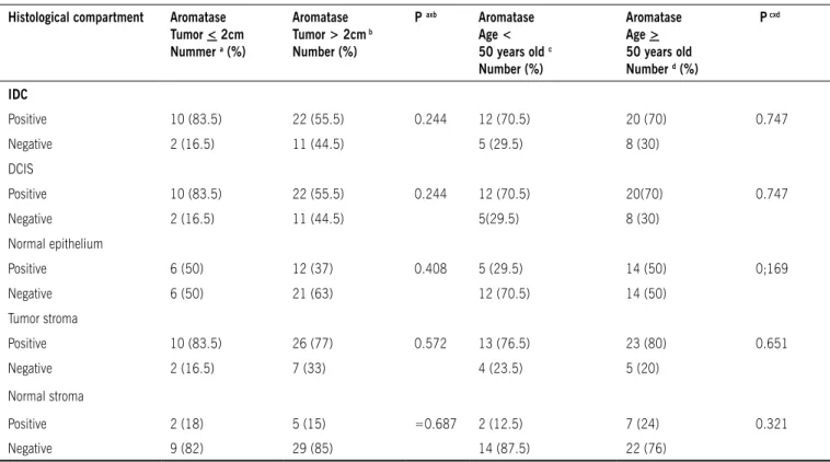
The study also analyzed aromatase expression according to tumor size and according to age group; the results can be found in Table 1. By comparing aromatase expression in IDC and DCIS with histopathological parameters (nuclear grade and presence or absence of comedonecrosis), as well as tumor size (larger or smaller than two centimeters) and age greater or lower than 50 years old, we found no statistically significant differences (Tables 2 and 3). When comparing aromatase enzyme expression in the ductal carcinomas in situ
with nuclear grade, we found expressions rates of 60 percent for nuclear grade I (p = 0.272), 60 percent for nuclear grade II (p = 0.010), and 76 percent for nuclear grade III (p = 0.001). In the presence of comedonecrosis, there was enzyme expression in 74 percent of cases (p = 0.01), while in its absence the expression rates reached 61 percent (p = 0.001). For invasive ductal carcinomas, there were expression rates of 60 percent for nuclear grade I (p = 0.272), 50 percent for nuclear grade II (p = 0.016), and 86 percent for nuclear grade III (p = 0.006). As for histological grade, aromatase expression reached 56.5 percent for grade I (p = 0.178), 73.5 percent for grade II (p = 0.022), and 60 percent for histological grade III (p = 0.272).
d
iscussiOnAromatase enzyme expression has been related to breast cancer and is gaining increasing therapeutic importance for this form of neoplasm. Despite its seemingly clear relationship to the carcinogenesis and progression of breast cancer, the way it happens is still not fully understood.
table 1 - Analysis of aromatase expression according to tumor size and aromatase expression according to age group
Histological compartment Aromatase tumor < 2cm Nummer a (%)
Aromatase tumor > 2cm b
Number (%)
P axb Aromatase
Age < 50 years old c
Number (%)
Aromatase Age > 50 years old Number d (%)
P cxd
IDC
Positive 10 (83.5) 22 (55.5) 0.244 12 (70.5) 20 (70) 0.747 Negative 2 (16.5) 11 (44.5) 5 (29.5) 8 (30)
DCIS
Positive 10 (83.5) 22 (55.5) 0.244 12 (70.5) 20(70) 0.747 Negative 2 (16.5) 11 (44.5) 5(29.5) 8 (30)
Normal epithelium
Positive 6 (50) 12 (37) 0.408 5 (29.5) 14 (50) 0;169
Negative 6 (50) 21 (63) 12 (70.5) 14 (50)
Tumor stroma
Positive 10 (83.5) 26 (77) 0.572 13 (76.5) 23 (80) 0.651
Negative 2 (16.5) 7 (33) 4 (23.5) 5 (20)
Normal stroma
Positive 2 (18) 5 (15) =0.687 2 (12.5) 7 (24) 0.321
Negative 9 (82) 29 (85) 14 (87.5) 22 (76)
IDC - invasive ductal carcinoma; DCIS - ductal carcinoma in situ (Chi-square test with Fisher’s exact test)
table 2 - Immunohistochemical aromatase expression in 45 DCIs cases by nuclear grade and presence or absence of
comedocarcinoma.
DCIs Aromatase Number (%)
Value of p
NGI
Positive 3 (60) =0.272 Negative 2 (40)
NGII
Positive 9(60) =0.010* Negative 6 (40)
NGIII
Positive 19 (76) <0.001* Negative 6 (24)
Comedo
Positive 20 (74) =0.001* Negative 7 (26)
n-comedo
Positive 11 (61) =0.001* Negative 7 (39)
(*): Statistically signiicant correlation (Spearman’s Correlation). The differences for NG (Kruskal-Wallis test) and comedonecrosis (Mann-Whitney test) percentages in the columns were not statistically signiicant.
table 3 - Immunohistochemical aromatase expression in 45 IDC cases by nuclear and histological grade.
IDC Aromatase
Number (%) Value of p NGI
Positive 3 (60) =0.272 Negative 2 (40)
NGII
Positive 9 (50) =0.016* Negative 9 (50)
NGIII
Positive 19 (86) =0.006* Negative 3 (14)
HGI
Positive 4 (56.5) =0.178 Negative 2 (43.5)
HGII
Positive 25 (73.5) =0.022* Negative 9 (26.5)
HGIII
Positive 3 (60) =0.272 Negative 2 (40)
this study, while the presence of aromatase messenger RNA was noted by hybridization in situ.5
Some studies have found aromatase in breast cancers, with aromatase expression in 72 percent of cases assessed.6 Similar
numbers were found in other studies, which found aromatase in 63 percent (91 of 145)7 and 69 percent (78 of 113) of cases.8
Our studies found aromatase expression in 71 percent of tumors, both for invasive and in situ components. Other researchers found similar data, with signiicant aromatase activity ranging from 52 to 72 percent in invasive carcinoma samples.3-6, 8-10
As for studies that tried to assess aromatase enzyme expression in DCIS, there seems to be discrepancies in the literature. Aromatase expression analyses in 61 cases of pure DCIS found higher rates than for 101 cases of IDC. This discrepancy may be explained by paracrine mechanisms, since the presence of both components would lead to higher expression rates for the invasive component.7 However, the
present study found no such difference.
Assessments of aromatase expression in ductal carcinoma in situ (DCIS) and invasive ductal carcinoma (IDC) in 162 cases using semi-quantitative immunohistochemistry found aromatase expression both in tumor cells and in adjacent stroma, with signiicantly higher positive values for DCIS than for IDC.11
In turn, using monoclonal antibodies, positive results for aromatase expression were found in 58 of 102 cases of stages III and IV breast cancer of another study. Follow-up analysis found no relation between presence of aromatase expression and responsiveness to hormonal treatment.12
When studying 83 cases of IDC, other researchers found aromatase expression in 47 percent of cases, especially in the stromal component of tumor tissue. No relation was found between being positive for aromatase and clinical-pathological parameters such as age, menopause, tumor size, lymph node status, histological type, and estrogen receptors.13 In another
study,14 aromatase expression was found in 70 percent of cases
of invasive ductal carcinoma and ductal carcinoma in situ, with expression in adjacent epithelium for 42.5 percent of cases and in tumor stroma for 79 percent.
Aromatase expression in tumor stroma was found in 80 percent of cases, a higher rate than those found in IDC and DCIS. The data coincide with those found in the literature and seem to be related to the very origin of the enzyme, found much more often in the stroma than in mammary epithelium. The results show that estrogen synthesis should be more expressive in tissues adjacent to the tumor, modulating tumor growth through paracrine, autocrine and intracrine mechanisms.15, 16
Though we found a trend towards greater aromatase expres-sion in cases of nuclear grade III DCIS with comedonecrosis, the data were not statistically significant and similar to other findings described in the literature.7 Greater aromatase
expres-sion has been been found in cases of nuclear grade III IDCs (p = 0.03),4 similar to what we found in analyzing IDCs (p
= 0.05). However, the same results were not found in histo-logical grade analysis.
The assessment of aromatase expression according to patient age (over or under 50 years of age) found no statis-tically significant difference in the analysis of IDC, DCIS and tumor stroma, unlike other studies, which report higher
expression rates for patients over the age of 50 (p = 0.012).7
As for tumor size, there were positive aromatase expression percentages for tumors smaller than or as large as 2 cm (IDC and DCIS) compared to larger tumors, but the difference was not statistically significant (p = 0.224). Assessments of IDC alone found no statistically significant difference in aromatase expression by tumor size, but did find a tendency towards greater expression in smaller tumors.9
c
OnclusiOnOur results showed high levels of correlation between aromatase expression in IDC, DCIS, normal epithelium and tumor stroma, suggesting the enzyme has a possible autocrine and paracrine mechanism in breast cancer. The regulation of aromatase activity is extremely complex. Tumors seem to grow in areas with high aromatase expression. Growth is also enabled by stimulating aromatase activity in adjacent tissues. This seems to be related to factors intrinsic to mammary tissue, but the process is not yet fully understood.17 Also, aromatase superexpression seems to be
related to worseprognosis for breast cancer, another reason to understand exactly how this happens. The data from this study might contribute to advancing the knowledge about aromatase inhibitors in breast cancer therapies.
No conflicts of interest declared concerning the publication of this article.
R
eFeRences1. Subramanian A, Salhab M, Mokbel K. Oestrogen producing enzymes and mammary carcinogenesis: a review. Breast Cancer Res Treat. 2008;111:191-202.
2. Santner SJ, Pauley Rj, Tait L, Kaseta J, Santen RJ. Aromatase activity and expression in breast cancer and benign breast tissue stromal cells. J Clin Endocrinol Metab. 1997;82:200-8.
3. Díaz-Cruz ES, Shapiro CL, Brueggemeier RW. J Clin Endocrinol Metab. 2005;90:2563-70.
4. Ristimäki A, Sivula A, Lundin J. Prognostic signiicance of elevated cyclooxy -genase-2 expression in breast cancer. Cancer Res. 2002;62:632-35. 5. Lu Q, Nakmura J, Savino A, Yue W, Weisz J, Dabbs DJ, et al. Expression of
aromatase protein and messenger ribonucleic acid in tumor epithelial cells and evidence of functional signiicance of locally produced estrogen in human breast cancers. Endocrinology. 1996;137:3061-8.
6. Miller WR. Aromatase activity in breast cancer. J Steroid Biochem Mol Biol. 1991;39(5B):783-90.
7. Silva MC, Rowlands MG, Dowsett M, Gusterson B, McKinna JA, Fryatt I. Intratumoral aromatase as a prognostic factor in human breast carcinoma. Cancer Res. 1989;49:2588-91.
8. Lipton A, Santen RJ, Santner SJ, Harvey HA, Sanders SI, Matthews YL. Prog-nostic value of breast cancer aromatase. Cancer. 1992;70:1951-55. 9. Brodie AM, Lu Q, Long BJ, Fulton A, Chen T, Macpherson N, et al. Aromatase
and COX-2 expression in human breast cancers. J Steroid Biochem Mol Biol. 2001;79:41-7.
10. Shibuya R, Suzuki T, Miki Y, Yoshida K, Moriya T, Ono K, et al. Intratumoral concentration of sex steroids and expression of sex steroid-producing enzymes in ductal carcinoma in situ of human breast. Endocr Relat Cancer. 2008;15:113-24.
11. Zhang Z, Yamshita H, Toyama T, Hara Y, Omoto Y, Sugiura H, et al. Semi-quantitative immunohistochemical analysis of aromatase expression in cuctal carcinoma of the breast. Breast Cancer Res Treat. 2002;74:47-53 12. De Jong PC, Blankenstein MA, Nortier JW, Slee PH, Van De Ven J, Van Gorp
Biochem Mol Biol. 2003;87:149-55.
13. Yamamoto Y, Yamashita J, Toi M, Muta M, Nagai S, Hanai N, et al. immuno-histochemical analysis of estrone sulfatase and aromatase in human breast cancer tissue. Oncology Rep. 2003;10:791-96.
14. Oliveira VM, Piato S, Silva MA. Correlation of cyclooxygenase-2 and aromatase immunohistochemical expression in invasive ductal carcinoma, ductal carcinoma in situ, and adjacent normal epithelium. Breast Cancer Res Treat. 2006;95:235-41. 15. Talbott KE, Gammon MD, Kibriva MG, Chen Y, Teitelbaum SL, Long CM, et al.
A CYP19 (aromatase) polymorphism is associated with increased premeno-pausal breast cancer risk. Breast Cancer Res Treat. 2007;111:481-7.
Artigo recebido: 13/03/08 Aceito para publicação: 21/07/09
16. Hudelist G, Wüling P, Kersting C, Burger H, Mattsson B, Czerwenka K, et al. Expression of aromatase and estrogen sulfotransferase in preinvasive and invasive breast cancer. J Cancer Res Clin Oncol. 2008;134:67-73. 17. Cheung KL. Endocrine therapy for breast cancer: an overview. Breast.