ABSTRACT
BACKGROUND AND OBJECTIVES: he objective of this
study is to describe the restrospective analysis of medical records of patients with temporomandibular disorder in a healthcare service of a Brazilian public university. he prevalence of signs and symp-toms of temporomandibular disorder, associated factors, diagnosis and observations related to the treatment were recorded.
METHODS: Two hundred and thirteen medical records were assessed by one single surveyor from March 2013 to December 2014. Information about sociodemographic factors, prevalen-ce of symptoms of temporomandibular disorder and treatment need were collected (Fonseca Anamnestic Index), clinical exami-nation, diagnosis, treatments and referral to other professionals. RESULTS: he majority of patients were female (81.7%), sin-gle (53.0%), students (23.3%) between 20 and 29 years of age (26.8%). Pain was reported by 50.4% of patients. According to FAI, 41.8% of patients were classiied with severe synptoms of temporomandibular disorder and 73.2% identiied with the need of treatment. Presence of temporomandibular disorder symptoms (p = 0.001) and need of treatment (p <0.001) were signiicantly associated to the female gender. he most prevalent diagnosis was muscle temporomandibular disorder (41.5%) and the most afected muscle was the masseter (21.3%). he most common treatments were occlusal splint (27.6%) and counse-ling (22.6%).
CONCLUSION: he greater demand for temporomandibular disorder treatment came from young patients, single, female, complaining from pain. he prevalence of temporomandibular disorder symptoms was high, muscular disorders was the most
Retrospective review of patients referred to a temporomandibular
dysfunction care setting of a Brazilian public university
Revisão retrospectiva de pacientes encaminhados a um serviço de disfunção
temporomandibular de uma universidade pública brasileira
Wellington Pereira de Moura1, Pâmela Lopes Pedro da Silva2, George Azevedo Lemos3, Paulo Rogério Ferreti Bonan4, Robinsom
Viégas Montenegro2, André Ulisses Dantas Batista2
1. Universidade Federal da Paraíba, João Pessoa, PB, Brasil.
2. Universidade Federal da Paraíba, Departamento de Restauração Dental, João Pessoa, PB, Brasil. 3. Universidade Estadual de Campinas, Campinas, SP, Brasil.
4. Universidade Federal da Paraíba, Departamento Clínico e Odontologia Social, João Pes-soa, PB, Brasil.
Submitted in October 24, 2016. Accepted for publication in May 15, 2017. Conlict of interests: none – Sponsoring sources: none.
Correspondence to:
Cidade Universitária, s/n - Castelo Branco 58051-900 João Pessoa, PB, Brasil. E-mail: [email protected]
© Sociedade Brasileira para o Estudo da Dor
prevalent indings and most of the treatments were reversible and conservative. he frequency of referral to other specialties related to temporomandibular disorder was low.
Keywords: Temporomandibular disorder, Orofacial pain, Epi-demiology.
RESUMO
JUSTIFICATIVA E OBJETIVOS: O objetivo deste estudo foi descrever a análise retrospectiva de prontuários referentes a um serviço de atendimento a pacientes com disfunção temporoman-dibular em uma clínica de ensino de uma universidade pública brasileira. A prevalência de sinais e sintomas de disfunção tem-poromandibular, fatores associados, diagnósticos e observações relacionadas ao tratamento foram registrados.
MÉTODOS: Duzentos e treze prontuários foram avaliados por um único examinador no período de março de 2013 a dezembro de 2014. Coletou-se informações sobre fatores sócio-demográi-cos, prevalência de sintomas de disfunção temporomandibular e necessidade de tratamento (índice anamnésico de Fonseca), exa-me clínico, diagnósticos, trataexa-mentos e encaminhaexa-mentos para outros proissionais.
RESULTADOS: A maioria dos pacientes era do sexo feminino (81,7%), solteira (53,0%), estudantes (23,3%) e entre 20 e 29 anos (26,8%). A dor foi relatada por 50,4% dos pacientes. De acordo com o índice FAI, 41,8% dos pacientes foram classii-cados com sintomas graves de disfunção temporomandibular e 73,2% identiicados com necessidade de tratamento. Presença de sintomas de disfunção temporomandibular (p = 0,001) e necessidade de tratamento (p <0,001) foram signiicativamente associadas ao sexo feminino. O diagnóstico mais prevalente foi disfunção temporomandibular muscular (41,5%) e o músculo mais afetado foi o masseter (21,3%). Os tratamentos mais co-muns foram placa oclusal (27,6%) e aconselhamento (22,6%). CONCLUSÃO: A maior demanda por tratamento para disfun-ção temporomandibular foi de pacientes jovens, solteiros, do sexo feminino, com queixa de dor. A prevalência de sintomas de disfunção temporomandibular foi alta, os distúrbios musculares foram os achados mais prevalentes e a maioria dos tratamentos foi reversível e conservadora. A frequência de encaminhamentos para outras especialidades relacionadas à disfunção temporoman-dibular foi baixa.
Descritores: Desordem temporomandibular, Dor orofacial, Epi-demiologia.
INTRODUCTION
According to the American Academy of Orofacial Pain (AAOP), temporomandibular disorder (TMD) is described as a group of clinical problems that afect the masticatory muscles, the temporo-mandibular joint (TMJ), and related structures1. It is characterized
as pain and fatigue of the masticatory muscles, TMJ pain, headache, otalgia, clicking, and limitation of mandibular movements2,3.
he etiology of TMD is multifactorial, with numerous con-tributing factors, such as parafunctional habits (e.g. gum chewing, “jaw play”, leaning of the head on the palm of the hand or arm and biting objects)4-6, direct and indirect traumas,
psychosocial and psychological factors, and genetic factors1,6-10.
Other factors, such as sleep bruxism (SB)11,12,awake bruxism
(AB)13, sleeping in the lateral decubitus position14 and some
occlusal factors have also been associated with the presence of
TMD signs and symptoms15.
he study of this disorders in public health has gained prominence due to its increasing and early incidence in the population, besides its association with the psychological aspects and its capacity to afect the quality of life of the patients9,16. Regarding its prevalence,
cross-sectional epidemiological studies have shown that approxi-mately 40 to 75% of the adult population has at least one clinical sign of TMD1.Moreover, studies have shown that due to the wide
variety of signs and symptoms2,3,17,this disorder may cause
func-tional and psychosocial harm, such as a decrease in quality of life in afected individuals18,19,thus making it necessary to promote
and expand access to adequate treatment for these patients5,20.
herefore, the evaluation of the services aimed at the treatment of patients with TMD is critical to enabling a better understan-ding of the epidemiological characteristics of the afected popu-lation, to improve planning strategies regarding the provision of services and the academic training on both theory and practice, and to foster strategies aimed at expanding care toward post-gra-duate services21,22. Moreover, these data may contribute to the
generation of scientiic knowledge through research to improve the understanding of the characteristics of this disorder2,3,23.
hus, the objective of the present study was to perform a retros-pective analysis of patient records referred to a temporomandi-bular disorder service in a healthcare setting of a Brazilian pu-blic university that ofers diagnostic and treatment services to patients with TMD and other orofacial pain disorders, as well as to describe the prevalence of signs and symptoms of TMD associated factors, diagnosis and treatment related observations.
METHODS
his study was performed at the TMD school clinic of the Divi-sion of OccluDivi-sion, Temporomandibular Disorder and Orofacial Pain, Department of Restorative Dentistry, Federal University of Paraíba (UFPB), João Pessoa, Paraíba, Brazil. his was a re-trospective study that followed an inductive approach with a research method based on indirect documentation through the analysis of patient records3,24.
A total of 213 patient records of patients attended at the service from March 2013 to December 2014 were evaluated. Patient
re-cords were numbered and audited by a single examiner and all iel-ds were analyzed, including those with incomplete or absent data. hose that contained only the patient’s identiication were exclu-ded. At the study site, patients are screened based on the Fonseca’s Anamnestic Index (FAI). he FAI index enables the evaluation of the severity of TMD symptoms as well as the need for treatment based on the symptoms reported8,11,25,26.In contrast, the
classiica-tion of TMDs was established based on medical history, clinical exam, and diagnostic imaging, as suggested by the AAOP1,20.
Cli-nical exam consisted of measuring the maximum mouth opening (mm) with a caliper, and values were added to the overlap of the anterior incisive teeth, and mouth opening was classiied as either normal (40-60 mm), restricted mouth opening (<40mm) and hypermobility (>60mm); the presence of joint sounds (clicking, popping or “thud” and crepitus); tenderness on TMJ palpation (lateral and posterior pole palpation under a pressure of approxi-mately 0.5 kg/cm2); tenderness on palpation of the masseter,
tem-poralis, medial pterygoid, sternocleidomastoid, trapezius, and pos-terior cervical muscles (pressure of approximately 1 kg/cm2);and
performance of the provocation test or functional manipulation of lateral pterygoid muscles (resistive protrusion)4,6,8,10,15,27–29.
SB was diagnosed based on the criteria of the American Academy of Sleep Medicine (AASM), as presented by Carra, Huynh and Lavigne30 and Ommerborn et al.31 according to patient history
(recent patient, parent, or sibling report of tooth-grinding soun-ds occurring during sleep for 6 months) and clinical evaluation (one or more of the following: abnormal tooth wear; hypertro-phy of the masseter muscles on voluntary forceful clenching; discomfort, fatigue, or pain in the jaw muscles and transient, morning jaw-muscle pain or headache). AB was evaluated using the question: ‘During the day, do you grind your teeth or clench
your jaw?’ (Brazilian-Portuguese RDC/TMD questionnaire).32
he clinical criteria proposed by Lobbezoo et al.33, using a
diag-nostic grading system of ‘possible’, ‘probable’ and ‘deinitive’ was employed, and, the diagnosis of SB or AB was categorized as “possible” (self-report) and “probable” (use of self-report plus the inspection by a clinical examination and the absence of polysom-nographic and electromyographic records).
Other data were also collected from the analysis of patient recor-ds: sociodemographic factors (gender, age, marital status, profes-sion, and city of residence); major reported complaints; prevalen-ce of TMD symptoms and need for treatment by an anamnestic index (FAI index); self-report of parafunctional habits; sleeping position; occlusal characteristics (tooth wear, lateral and anterior guidance); TMD clinical evaluation; TMD diagnoses, according to the AAOP/IHS criteria; previous treatments and referral to professionals of other areas.
To analyze the subjective data obtained from medical records, such as the major complaint and diagnosis, a theme-categorical methodology with discursive analysis was used, which consists of transforming and grouping the narrative data into units, consi-dering their relevance, frequency, and meaning34.
Statistical analysis
Data were assessed using the Statistical Package for the Social Sciences (SPSS) software, version 22.0, and analyzed descripti-vely, with the frequency and percentages of the study variables computed. he chi-square (x2) or Fisher’s Exact test was used to
test the associations between the study variables. For both tests, we set p<0.05 as the statistical signiicance level.
RESULTS
Table 1 shows the sociodemographic characteristics of the evalu-ated sample. he majority of the patients were female (81.7%), between 20 and 29 years of age (26.8%), single (53%), students (23.3%), and residents of the city of Joao Pessoa (79.3%) or a metropolitan region. Pain (50.4%) and TMJ clicking (14.9%) were among the most prevalent complaints reported by the
sub-Table 1. Sociodemographic characteristics of the study sample (n=213)
Variables n %
Gender
Female 174 81.7
Male 39 18.3
Age range
13-19 18 8.5
20-29 57 26.8
30-39 35 16.4
40-49 38 17.8
50-59 42 19.7
60-69 18 8.5
70-80 5 2.3
Marital status
Single 113 53.0
Married 83 39.0
Divorced 15 7.0
Widower 1 0.5
Unknown* 1 0.5
Profession
Student 50 23.3
Housewife 26 12.2
Civil servant 23 10.8
Retired 17 8.0
Teacher 13 6.1
Other professions** 78 36.8
Unknown* 6 2.8
City
Joao Pessoa 169 79.3
Santa Rita 10 4.7
Bayeux 8 3.8
Cabedelo 4 1.9
Other cities*** 22 10.3
* Absent data in the medical record; ** Professions reported by less than 10 patients; *** Cities reported by less than three patients.
Table 2. Frequency of the major reported complaints
Reported complaints n* %
Pain 155 50.4
TMJ clicking 46 14.9
Tooth wear 24 7.8
Chewing dificulty 18 5.9
Fatigue 16 5.2
Tooth grinding 15 4.9
Jaw locking 10 3.2
Dificulty in opening the mouth 5 1.6
Tingling sensation 3 1.0
Aperture deviation 2 0.6
Poorly adapted prosthesis 2 0.6
Tinnitus 2 0.6
Displaced mandible 1 0.3
Tooth crowding 1 0.3
Unknown ** 8 2.7
* The sum is greater than 213 because a patient could report more than one complaint; ** Absent data in the medical record.
jects (Table 2). Notably, the present study grouped pain com-plaints into a single category that combined muscle pain, joint pain or headache.
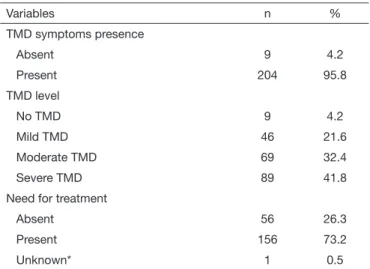
Regarding TMD symptoms prevalence, 95.8% of the patients had TMD symptoms, in an initial trial performed by FAI Ana-mnestic Index. According to the FAI index, the majority presen-ted “severe TMD” (41.8%). In the study sample, 73.2% of the patients were classiied as “in need for treatment” (Table 3). he presence of TMD symptoms (p=0.001) and need for treatment were signiicantly associated with the female gender (p<0.001). Regarding self-reported parafunctional habits, 58.2% (n=124) of the patients reported having such habits, with the most prevalent being ingernail biting (28.6%, n=39), leaning the head on the palm of the hand or arm (22.0%, n=30), object biting (20.6%, n=28), lip/cheek biting (16.2%, n=22), and gum chewing (8.8%, n=12). In addition, patients also reported tongue biting
Table 3. Prevalence of temporomandibular disorders symptoms and the need for treatment according to the Fonseca index
Variables n %
TMD symptoms presence
Absent 9 4.2
Present 204 95.8
TMD level
No TMD 9 4.2
Mild TMD 46 21.6
Moderate TMD 69 32.4
Severe TMD 89 41.8
Need for treatment
Absent 56 26.3
Present 156 73.2
Unknown* 1 0.5
(1.5%, n=2), tongue thrust (1.5%), and prosthesis dislocation (0.7%). he sum of the habits above is greater than 124 because some patients could have reported more than one habit. Regarding the occlusion assessment, 33.7% of the patients had evidence suggestive of tooth wear compatible with sleep bruxism (SB) (n=66) and 18.4% had evidence of tooth wear suggestive of AB (n=36). Functional facets and nail biting were present in 6.1% (n=13) and 0.9% (n=2) of patients, respectively. A
sub-Table 4. Frequency of data related to temporomandibular disorder clinical evaluation
Clinical exam n %
Maximum mouth opening
Normal 156 73.2
Restricted mouth opening 45 21.2
Hypermobility 5 2.3
Unknown* 7 3.3
Mouth opening pattern
Straight (normal) 68 31.9
Deviation 92 43.1
Delection with aperture restriction 31 14.6
Delection without aperture restriction 11 5.2
Unknown* 11 5.2
TMJ sounds
Absent 84 39.4
Clicking 67 31.5
Popping 37 17.4
Clicking and popping 10 4.7
Crepitus 3 1.4
Clicking and crepitus 2 0.9
Unknown* 10 4.7
Tenderness on TMJ palpation
Absent 95 44.6
Left and right TMJ 60 28.2
Only left TMJ 27 12.7
Only right TMJ 26 12.2
Unknown* 5 2.3
Tenderness on muscle palpation
Absent 66 31.0
Present** 140 65.7
Masseter 101 21.3
Lateral pterygoid 85 17.9
Sternocleidomastoid 76 16.0
Temporal 74 15.6
Trapezius 66 13.9
Posterior cervical 42 8.8
Medial pterygoid 24 5.0
Occipitofrontalis 7 1.5
Unknown* 7 3.3
* Absent data in the medical record; ** The sum of patients with tenderness on muscle palpation is higher than 140 because a patient could report tenderness of more than one muscle.
Table 5. Prevalence of joint and muscle disorders (medical history, clinical evaluation, and diagnostic imaging)
Joint and muscle disorders n %
No clinical TMD diagnosis 19 8.9
With diagnosis* 137 64.3
Masticatory Muscle disorders** 83 41.5
Disc displacement with reduction 39 19.5
Capsulitis/synovitis 26 13.0
Retrodiscitis 16 8.0
Disc displacement without reduction 10 5.0
Subluxation (Hypermobility) 9 4.5
Adherence 8 4.0
Osteoarthritis 7 3.5
Osteoarthrosis 2 1.0
Absent information*** 57 26.8
* The sum of the speciic diagnoses is higher than 137 because a patient could have more than one diagnosis; ** Includes the following TMD muscle disorders: protective co-contraction, local muscle soreness, myospasm, myofascial pain and chronic centrally mediated myalgia; ***Absent data in the medical record; ** Data were absent or the patient did not know.
set of 28.6% of the patients did not exhibit tooth wear facets (n=61), and in 8.5% of the charts, this information was absent or the patient was unsure of this information (n=18). he preva-lence of “possible” AB diagnoses (self-reported) was reported by 19.7% (n=42) of the patients and “possible” SB by 8.0% (n=17), although the diagnosis of “probable” (self-report plus clinical examination) resulted in smaller values of prevalence: 4.3% (n=9) for SB and 8.05% (n=17) for AB.
Canine guidance was the most prevalent disocclusion pattern for both the right and left sides (n=70, 32.9% for both sides), follo-wed by incomplete group function on the right (n=65, 30.5%) and left (n=66, 31.0%) sides. he anterior guidance pattern was considered to be normal (including only the incisor teeth) for 38.5% of the sample (n=82). Regarding the sleep position pattern, 126 patients reported sleeping in the lateral decubitus position (59.2%), 36 slept in the prone position (16.9%), and 27 slept in the supine position (12.7%). his information was absent or the patient was unsure of this information in 24 of the charts (11.2%). Table 4 presents the data related to the TMD clinical exam. he majority of the patients had a normal maximum mouth ope-ning (73.2%) and aperture pattern with deviation (43.1%). Ar-ticular sounds were present in 55.9% of the patients, with cli-cking (31.5%) and popping (17.4%) being the most prevalent. Tenderness at TMJ palpation was present in 53.1% of the pa-tients, with most of these patients reporting pain in both TMJs (28.2%). Regarding muscle tenderness, 65.7% of the patients reported pain. he muscles that were most commonly afected were the masseter (21.3%), lateral pterygoid (17.9%), and ster-nocleidomastoid (16.0%).
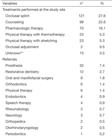
he treatments and referrals are reported in Table 6. Occlusal splint (27.6%) and counseling (22.6%) were the most common treatments, while dental prosthesis (7.4%) and restorative dentis-try (2.7%) were the most common referrals. Of important note is that the referrals to dental specialties were not necessarily for the treatment of TMD but rather due to the patient’s needs in each speciic area. Referrals to other specialties related to TMD, including physical therapy and speech therapy, were usually few.
Table 6. Frequency of treatments and referrals to other specialties
Variables n* %
Treatments performed at the study site
Occlusal splint 121 27.6
Counseling 99 22.6
Pharmacologic therapy 70 16.1
Physical therapy with thermotherapy 23 5.3
Physical therapy with stretching 23 5.3
Occlusal adjustment 2 0.5
Unknown** 15 3.5
Referrals
Prosthesis 32 7.4
Restorative dentistry 12 2.7
Oral and maxillofacial surgery 8 1.8
Orthodontics 6 1.4
Physical therapy 6 1.4
Endodontics 4 0.9
Speech therapy 4 0.9
Rheumatology 3 0.7
Neurology 3 0.7
Orthopedics 2 0.5
Otorhinolaryngology 2 0.5
Periodontics 1 0.2
* The sum is higher than 213 because a patient could receive more than one treatment and/or referral; ** Data were absent or the patient did not know.
DISCUSSION
In agreement with the current literature, the present study reve-aled that the majority of the patients were women2-4,24 in the age
range of 20 to 25 years (young adults)8,35.Moreover, women were
signiicantly associated with the presence of TMD symptoms and with the need for treatment according to the FAI index. he reasons for the higher female TMD population are still contro-versial, although a few factors are suggested in the literature, such as the greater perception of pain among females, the higher inci-dence of psychological factors among females, physiological and hormonal diferences, muscle structure diferences, and women’s greater concern about their own health compared with men1,8,35.
he majority of patients in the sample were students, single, and residents of João Pessoa or its metropolitan area. A similar
socio-demographic proile was demonstrated by Pimentel et al.23.
Con-versely, Dantas et al.3 observed a higher prevalence of TMD among
individuals in the age range of 41 to 60 years and with formal
em-ployment. hese authors conducted their study in a teaching hos-pital where most of the patients were referred by dentists or physi-cians from primary care clinics and private practices. In contrast, our study was conducted at the teaching clinic of the Division of Occlusion, which is embedded in an academic environment. his location explains the greater search of the service by the population of interest. Several studies have demonstrated a high prevalence of TMD among university students9,15,35,36 suggesting that this
popula-tion is exposed to risk factors that promote the development of these disorders, such as emotional stress and anxiety8,35.
Regarding the complaints reported, results are in agreement with previous studies, which showed pain as the most prevalent com-plaint2,3,37.his inding is relevant, as the current literature shows
that the presence of pain is associated with a higher degree of im-pairment of individual and psychosomatic characteristics among patients with TMD, which negatively inluences their quality of life related to oral health3,18,19.
he FAI index data revealed that most patients presented severe TMD with need for treatment. hese indings are explained by the fact that the present study was performed in a patient lation. In contrast, epidemiological studies in non-patient popu-lations have shown a high prevalence of mild TMD and lower values of patients in need of treatment8,35,36,38.
Regarding the presence of habits, a large percentage of the pa-tients reported at least one parafunctional habit (58.2%), with ingernail biting, leaning the head on the palm of the hand or arm and object biting being the most prevalent. Corroborating these indings, other studies also observed a high prevalence of parafunctional habits in patients with TMD37,39.Branco et al.39
observed that 76.9% of patients with TMD had some
parafunc-tional habit, while Carvalho et al.37 showed a lower frequency
(47%). Moreover, epidemiological studies in non-patient po-pulations have also found a high prevalence of parafunctional habits in individuals with signs and symptoms of TMD4-6,16,36.
Data from the present study also show that diferent prevalence values were found for “possible” and “probable” sleep and awake--bruxism. he diagnose of “possible” AB was reported by 19.7% of the patients, and “possible” SB by 8.8%, and the diagnoses of “probable” AB and SB was reported only by 8.05% and 4,3% of the patients, respectively. he prevalence of “possible” AB and SB were similar12 or lower than others reported in the literature.11,40
he prevalence of sleep bruxism varies widely in the literature, and is stated to be more prevalent in children (40%), with an average of 8% prevalence during adulthood30,41, and this is
pro-bably the result from diferent strategies for bruxism diagnosis and classiication (e.g. questionnaires, oral history, clinical exa-mination); the characteristics of the study population (e.g. chil-dren, adults, general or patient population) and because many studies failed to distinguish between awake-time and sleep-rela-ted bruxism30,33,41. he prevalence of awake-bruxism also varies
in the literature, but it tends to increase with age, ranging from an estimated prevalence of 12% in children to more than 20% in adults30. Considering this, it is possible to verify that both
pre-sence of signs and symptoms of TMD and the diagnosis of sleep
bruxism and/or awake bruxism (tooth clenching)7,42, although
this inding is not a consensus 43. Notably, in the present study
setting, the diagnoses of SB and AB were determined based on the clinical exam (presence of tooth wear) and medical history, similar to previous studies4,6,31. Currently, the gold standard for
the evaluation of sleep bruxism and awake clenching is poly-somnography and electromyography,30,33 however, this is still a
high-cost technique with limited availability in most Brazilian orofacial pain and TMD public health care services6.
Nowadays, there is a consensus in the literature that dental occlu-sion should not be considered a major factor in the TMD etiology. Recent studies have shown a lack of scientiic evidence supporting the relationship between occlusal factors and TMD15,44,45.Lemos
et al.15 suggested that occlusal factors may play a role as co-factors
in predisposing individuals to or perpetuating this disorder but that they should not be considered as primary etiologic agents. Supporting the current literature, the indings of the present study revealed that the majority of patients had a normal lateral and an-terior guidance pattern. he prevalence of tooth wear in the study sample was high. However, the diagnoses of sleep bruxism and tooth clenching were only observed in a small number of patients. his enforces the opinion that the prevalence of SB and AB should not rely only on the presence of tooth wear, since they may be the result of a previous activity and may overestimate the actual prevalence12. Tooth wear may could also be related to many other
factors that can induce attrition and erosion on dental surfaces, like ageing, loss of posterior teeth, occlusal conditions, diet, medi-cations or alimentary disorders30.
Regarding the sleep position pattern, most patients reported sleeping preferably in the lateral decubitus and prone position. In the literature, few studies investigated the sleep position in patients with TMD14,46, but the results of this studies suggest
that sleeping in the lateral decubitus position may be a contri-buting factor to TMJ anterior disc displacement, suggesting that due to gravity, the mandibular position may change, leading to the deviation of the ipsilateral condyle posterolaterally and the
contralateral one anteromedially14. he prone position was also
associated with the development of TMJ dysfunction, in pa-tients with unilateral obstructive nasal septal deviation46, which
suggests that these habitual postures during sleep may act as a
predisposing factor of TMD1,46. Further studies are needed to
elucidate this relationship.
he clinical evaluation of TMD revealed a high frequency of patients with articular sound, with clicking being the most pre-valent. he incidence of tenderness on TMJ palpation was also high, and most patients reported pain in both TMJs. hese re-sults support previous studies that demonstrated a similar preva-lence pattern of joint signs in individuals with TMD2,4,8,35,37.In
contrast, the high number of subjects with tenderness in both TMJs agrees with the high number of patients with severe TMD and a need for treatment observed in our study, indicating that joint pain may be associated with a greater severity of TMD and an increased demand for treatment18,19.
Corroborating previous studies, the incidence of tenderness on muscle palpation was high2,37,with the masseter, lateral pterygoid,
sternocleidomastoid, and temporal muscles being the most afec-ted muscles. he literature has shown greater involvement of the mandible elevator muscles in TMDs, especially the masseter and
temporal muscles1,7,47.he involvement of these muscles may
be associated with muscle hyperactivity, ischemia, sympathetic relexes, and fusimotor relexes, which alter the blood supply, muscle tone, and emotional and psychological status in patients
with TMD1.
he high involvement of the lateral pterygoid muscle in the pre-sent study may be associated with its function, as it is the only muscle that is directly attached to the TMJ. he upper and lower heads of the lateral pterygoid insert into the articular disc and condyle, respectively, and are responsible for the movements of protrusion, laterality, and mouth opening1,48.hus, it has been
suggested that the uncoordinated activity of this muscle or hype-ractivity of its upper heads may lead to intra-articular disorders, such as disc displacements48,49.
he sternocleidomastoid muscle is one of the main muscles invol-ved in the support of the skull and cervical region, and it could be afected in the presence of abnormal stomatognathic function in patients with TMD50.Studies that investigate the activity of the
sternocleidomastoid in patients with TMD are necessary to im-prove the understanding of its participation in this disorder. he data related to the diagnosis of TMD are in agreement with previous studies conducted in populations of patients, which demonstrated a higher prevalence of masticatory muscle disor-ders, followed by disc displacements with and without reduction and TMJ degenerative disease (osteoarthritis/osteoarthrosis)17,51.
However, studies in non-patient populations have demonstra-ted a higher prevalence of joint disorders compared to muscular alterations4,6,17,52. Regarding treatments, we observed a greater
prevalence of reversible therapies, including patient education, self-management, use of medications, interocclusal splints, pos-tural training, physical therapies, and behavioral intervention. hese indings are consistent with the current literature, which recommends the use of conservative practices for the treatment of TMDs1,21,22.
Concerning referrals to TMD-related areas, we found only a small frequency of referrals, with physical therapy and speech therapy being the most common. hese data are not in agreement with the literature, which suggests a multidisciplinary and integrative approach in the treatment of TMDs1,2,15,21,22.hese results can be
explained by the fact that the study setting is part of an academic division and is not located in the outpatient clinic of the university hospital, which centralizes most of the specialized medical care. he data also showed a high frequency of referrals to other dentistry specialties, especially prosthodontics and restorative dentistry. As our study setting provides on-demand service with no screening, it re-ceives patients with problems that are not directly related to TMD, thus explaining the large number of referrals to those specialties.
CONCLUSION
duals in the age range of 20 to 29 years, students, single indivi-duals, and individuals with pain complaints. he prevalence of severe symptoms of TMD was high according to the anamnestic index, and the clinical and diagnostic imaging evaluations reve-aled muscular disorders as the most prevalent indings. he vast majority of therapies were conservative and reversible, and the frequency of referrals to other TMD-related specialties was low.
REFERENCES
1. De Leeuw R. Dor Orofacial - Guia de Avaliação, Diagnóstico e Tratamento. 4ª ed. São Paulo: Quintessence; 2010. 315p.
2. Donnarumma MD, Muzilli CA, Ferreira C, Nemr K. Disfunções temporomandibula-res: sinais, sintomas e abordagem multidisciplinar. Rev CEFAC. 2010;12(5):788-94. 3. Dantas AM, Santos EJ dos, Vilela RM, Lucena LB de. Peril epidemiológico de pa-cientes atendidos em um Serviço de Controle da Dor Orofacial. Rev Odontol UNESP. 2015;44(6):313-9.
4. Gavish A, Halachmi M, Winocur E, Gazit E. Oral habits and their association with signs and symptoms of temporomandibular disorders in adolescent girls. J Oral Reha-bil. 2000 Jan;27(1):22-32.
5. Oliveira CB, Lima JA, Silva PL, Forte FD, Bonan PR, Batista AU. Temporomandi-bular disorders and oral habits in high-school adolescents: a public health issue? RGO - Rev Gaúcha Odontol. 2016;64(1):8-16.
6. Paulino MR, Moreira VG, Lemos GA, Silva PL, Bonan PR, Batista AU. Prevalência de sinais e sintomas de disfunção temporomandibular em estudantes pré-vestibulandos: associação de fatores emocionais, hábitos parafuncionais e impacto na qualidade de vida. Ciência e Saúde Coletiva. 2015; Article in press. Available from: http://www. cienciaesaudecoletiva.com.br/artigos/artigo_int.php?id_artigo=15413
7. Lauriti L, Motta LJ, de Godoy CH, Biasotto-Gonzalez DA, Politti F, Mesquita-Ferrari RA, et al. Inluence of temporomandibular disorder on temporal and masseter muscles and occlusal contacts in adolescents: an electromyographic study. BMC Musculoskelet Disord. 2014;15(1):123.
8. Lemos GA, Silva PL, Paulino MR, Moreira VG, Beltrão RT, Batista AU. Prevalência de disfunção temporomandibular e associação com fatores psicológicos em estudantes de Odontologia. Rev Cubana Estomatol. 2015;52(4):22-31.
9. Bezerra BP, Ribeiro AI, Farias AB, Farias AB, Fontes LB, Nascimento SR, et al. Preva-lência da disfunção temporomandibular e de diferentes níveis de ansiedade em estu-dantes universitários. Rev Dor. 2012;13(3):235-42.
10. Okeson JP. Tratamento das desordens Temporomandibulares e Oclusão. 7ª ed. Rio de Janeiro: Elsevier; 2013. 512p.
11. Yalçın Yeler D, Yılmaz N, Koraltan M, Aydın E. A survey on the potential rela-tionships between TMD, possible sleep bruxism, unilateral chewing, and occlusal factors in Turkish university students. Cranio. 2016;6:1-7. [Epub ahead of print]. 12. Cortese SG, Fridman DE, Farah CL, Bielsa F, Grinberg J, Biondi AM. Frequency of
oral habits, dysfunctions, and personality traits in bruxing and nonbruxing children: a comparative study. Cranio. 2013;31(4):283-90.
13. Kobs G, Bernhardt O, Kocher T, Meyer G. Oral parafunctions and positive clinical examination indings. Stomatol Balt Dent Maxillofac J. 2005;7(3):81-3.
14. Hibi H, Ueda M. Body posture during sleep and disc displacement in the temporo-mandibular joint: a pilot study. J Oral Rehabil. 2005;32(2):85-9.
15. Lemos GA, Moreira VG, Forte FD, Beltrão RT, Batista AU. Correlação entre sinais e sintomas da Disfunção Temporomandibular (DTM) e severidade da má oclusão. Rev Odontol da UNESP. 2015;44(3):175-80.
16. Al-Khotani A, Naimi-Akbar A, Albadawi E, Ernberg M, Hedenberg-Magnusson B, Christidis N. Prevalence of diagnosed temporomandibular disorders among Saudi Arabian children and adolescents. J Headache Pain. 2016;17(1):41.
17. Manfredini D, Guarda-Nardini L, Winocur E, Piccotti F, Ahlberg J, Lobbezoo F. Research diagnostic criteria for temporomandibular disorders: a systematic review of axis I epidemiologic indings. Oral Surg Oral Med Oral Pathol Oral Radiol Endod. 2011;112(4):453-62.
18. Dahlström L, Carlsson GE. Temporomandibular disorders and oral health-related quality of life. A systematic review. Acta Odontol Scand. 2010;68(2):80-5. 19. Lemos GA, Paulino MR, Forte FD, Beltrão RT, Batista AU, Lemos GA, et al.
Inluen-ce of temporomandibular disorder presenInluen-ce and severity on oral health-related quality of life. Rev Dor. 2015;16(1):10-4.
20. Carrara SV, Conti PC, Barbosa JS. Termo do 1o Consenso em Disfunção
Temporo-mandibular e Dor Orofacial. Dental Press J Orthod. 2010;15(3):114-20. 21. Ommerborn MA, Kollmann C, Handschel J, Depprich RA, Lang H, Raab WH-M. A
survey on German dentists regarding the management of craniomandibular disorders. Clin Oral Investig. 2010;14(2):137-44.
22. Reissmann DR, Behn A, Schierz O, List T, Heydecke G. Impact of dentists’ years sin-ce graduation on management of temporomandibular disorders. Clin Oral Investig. 2015;19(9):2327-36.
23. Pimentel PH, Coelho Júnior LG, Caldas Júnior AF, Kosminsky M, Aroucha JM. Peril de-mográico dos pacientes atendidos no Centro de Controle da Dor Orofacial da Faculdade de Odontologia de Pernambuco. Rev Cir Traumatol Buco-Maxilo-Facial. 2008;8(2):71-8.
24. Wedel A, Carlsson GE. Retrospective review of 350 patients referred to a TMJ clinic. Community Dent Oral Epidemiol. 1983;11(1):69-73.
25. Fonseca DM, Bonfante G, Valle AL, Freitas SF. Diagnóstico pela anamnese da disfun-ção craniomandibular. RGO (Porto Alegre). 1994;42(1):23-4, 27-8.
26. Chaves TC, Oliveira AS De, Grossi DB. Principais instrumentos para avaliação da disfunção temporomandibular, parte I: índices e questionários; uma contribuição para a prática clínica e de pesquisa. Fisioter Pesqui. 2008;15(1):92-100.
27. DuPont JS, Brown CE. Provocation testing to assist craniomandibular pain diagnosis. Cranio. 2010;28(2):92-6.
28. Fricton JR, Schifman EL. he craniomandibular index: validity. J Prosthet Dent. 1987;58(2):222-8.
29. Pehling J, Schifman E, Look J, Shaefer J, Lenton P, Fricton J. Interexaminer reliability and clinical validity of the temporomandibular index: a new outcome measure for temporomandibular disorders. J Orofac Pain. 2002;16(4):296-304.
30. Carra MC, Huynh N, Lavigne G. Sleep bruxism: a comprehensive overview for the den-tal clinician interested in sleep medicine. Dent Clin North Am. 2012;56(2):387-413. 31. Ommerborn MA, Giraki M, Schneider C, Fuck LM, Handschel J, Franz M, et al.
Efects of sleep bruxism on functional and occlusal parameters: a prospective control-led investigation. Int J Oral Sci. 2012;4(3):141-5.
32. Dworkin SF, LeResche L. Research diagnostic criteria for temporomandibular disor-ders: review, criteria, examinations and speciications, critique. J Craniomandib Di-sord. 1992;6(4):301-55.
33. Lobbezoo F, Ahlberg J, Glaros AG, Kato T, Koyano K, Lavigne GJ, et al. Bruxism deined and graded: an international consensus. J Oral Rehabil. 2013;40(1):2-4. 34. Oliveira DC. Análise de conteúdo temático-categorial: uma proposta de
sistematiza-ção. Rev Enferm UERJ. 2008;16(4):569-76.
35. Pedroni CR, De Oliveira AS, Guaratini MI. Prevalence study of signs and symptoms of temporomandibular disorders in university students. J Oral Rehabil. 2003;30(3):283-9. 36. Medeiros SP, Batista AUD, Forte FDS. Prevalência de sintomas de disfunção
tem-poromandibular e hábitos parafuncionais em estudantes universitários. Rev Gaucha Odontol. 2011;59(2):201-8.
37. Carvalho LP, Piva MR, Santos TS, Ribeiro CF, Araújo CR, Souza LB. Estadiamento clínico da disfunção temporomandibular: estudo de 30 casos. Odontol Clínico Cien-tíica. 2008;7(1):47-52.
38. Campos J, Carrascosa A, Bonafé F, Maroco J. Epidemiology of Severity of Tem-poromandibular Disorders in Brazilian Women. J Oral Facial Pain Headache. 2014;28(2):147-52.
39. Branco RS, Branco CS, Tesch RD, Rapoport A. Freqüência de relatos de parafunções nos subgrupos diagnósticos de DTM de acordo com os critérios diagnósticos para pesquisa em disfunções temporomandibulares (RDC/TMD). Rev Dent Press Ortod e Ortop Facial. 2008;13(2):61-9.
40. Fernandes G, van Selms MK, Gonçalves DA, Lobbezoo F, Camparis CM. Factors associated with temporomandibular disorders pain in adolescents. J Oral Rehabil. 2015;42(2):113-9.
41. Lobbezoo F, Ahlberg J, Manfredini D, Winocur E. Are bruxism and the bite causally related? J Oral Rehabil. 2012;39(7):489-501.
42. Sato F, Kino K, Sugisaki M, Haketa T, Amemori Y, Ishikawa T, et al. Teeth contacting habit as a contributing factor to chronic pain in patients with temporomandibular disorders. J Med Dent Sci. 2006;53(2):103-9.
43. Camparis CM, Formigoni G, Teixeira MJ, Bittencourt LR, Tuik S, Siqueira JT. Sleep bruxism and temporomandibular disorder: Clinical and polysomnographic evalua-tion. Arch Oral Biol. 2006;51(9):721-8.
44. de Sousa ST, de Mello VV, Magalhães BG, de Assis Morais MP, Vasconcelos MM, de França CJ, et al. he role of occlusal factors on the occurrence of temporomandibular disorders. Cranio. 2014;33(3):211-6.
45. Türp JC, Schindler H. he dental occlusion as a suspected cause for TMDs: epidemio-logical and etioepidemio-logical considerations. J Oral Rehabil. 2012;39(7):502-12. 46. Yalçınkaya E, Cingi C, Bayar Muluk N, Ulusoy S, Hanci D. Are temporomandibular
disorders associated with habitual sleeping body posture or nasal septal deviation? Eur Arch Oto-Rhino-Laryngology. 2016;273(1):177-81.
47. Woźniak K, Lipski M, Lichota D, Szyszka-Sommerfeld L. Muscle fatigue in the tem-poral and masseter muscles in patients with temporomandibular dysfunction. Biomed Res Int. 2015;2015: Article ID 23734, 1-6.
48. Murray G, Phanachet I, Uchida S, Whittle T. he human lateral pterygoid muscle: A review of some experimental aspects and possible clinical relevance. Aust Dent J. 2004;49(1):2-8.
49. Oliveira AT, Camilo AA, Bahia PR, Carvalho AC, DosSantos MF, da Silva JV, et al. A novel method for intraoral access to the superior head of the human lateral pterygoid muscle. Biomed Res Int. 2014;2014:432635.
50. Milanesi JD, Corrêa EC, Borin GS, Souza JA, Pasinato F. Atividade elétrica dos mús-culos cervicais e amplitude de movimento da coluna cervical em indivíduos com e sem DTM. Fisioter Pesqui. 2011;18(4):317-22.
51. Blanco-Hungria A, Blanco-Aguilera A, Blanco-Aguilera E, Serrano-del-Rosal R, Bie-dma-Velazquez L, Rodriguez-Torronteras A, et al. Prevalence of the diferent Axis I clinical subtypes in a sample of patients with orofacial pain and temporomandibular disorders in the Andalusian Healthcare Service. Med Oral Patol Oral y Cir Bucal. 2016;21(2):e169-77.