Copyright © 2014 Revista Latino-Americana de Enfermagem
This is an Open Access article distributed under the terms of the Creative Commons Attribution Non-Commercial License (CC BY-NC).
This license lets others distribute, remix, tweak, and build upon your work non-commercially, and although their new works must also acknowledge you and be non-commercial, they don’t have to license their derivative works on the same terms.
DOI: 10.1590/0104-1169.3143.2385 www.eerp.usp.br/rlae
Corresponding Author:
Patrícia de Albuquerque Sarmento
Universidade Federal de Alagoas. Escola de Enfermagem e Farmácia Rod. 104 Norte, Km 96
Bairro: Tabuleiro do Martins CEP: 57072-970, Maceió, AL, Brasil E-mail: [email protected]
Patrícia de Albuquerque Sarmento
2Terezinha da Rocha Ataíde
3Ana Paula Fernandes Barbosa
4João Xavier de Araújo-Júnior
5Ingrid Martins Leite Lúcio
6Maria Lysete de Assis Bastos
61 Paper extracted from master’s thesis “Evaluation of antimicrobial activity and cytotoxicity in vitro and in vivo of crude ethanol extract of the
stem Zeyheria tuberculosis (vell) Bur. (Bignoniaceae): perspective of a supplement for wound healing”, presented to Faculdade de Nutrição, Universidade Federal de Alagoas, Maceió, AL, Brazil. Supported by Conselho Nacional de Desenvolvimento Científico e Tecnológico (CNPq), process # 474023/2010-9.
2 Doctoral student, Rede do Nordeste de Biotecnologia, Carpina, PE, Brazil. Assitant Professor, Escola de Enfermagem e Farmácia, Universidade
Federal de Alagoas, Maceió, AL, Brazil.
3 PhD, Associate Professor, Faculdade de Nutrição, Universidade Federal de Alagoas, Maceió, AL, Brazil. 4 PhD, Adjunct Professor, Faculdade de Medicina, Universidade Federal de Alagoas, Maceió, AL, Brazil.
5 PhD, Associate Professor, Escola de Enfermagem e Farmácia, Universidade Federal de Alagoas, Maceió, AL, Brazil. 6 PhD, Adjunct Professor, Escola de Enfermagem e Farmácia, Universidade Federal de Alagoas, Maceió, AL, Brazil.
Objectives: to evaluate the antimicrobial, cytotoxic and healing activities of the ethanolic extract
of the stems of Z. tuberculosa via topical use and/or oral ingestion. Method: antimicrobial assays
in vitro using the disk diffusion method, the Artemia salina toxicity test, and in vivo assays with
Wistar rats. From these was collected clinical, histological and biochemical data for evaluating the
healing process. Results: in vitro antimicrobial testing showed activity in relation to Streptococcus
pyogenes, Staphylococcus aureus and Staphylococcus epidermidis, with zones of inhibition of 18, 14 and 10 mm, respectively. The best minimum inhibitory concentration was 62.5 μg/ml for S. aureus, this bacteria being chosen for the in vitro assays. Animals treated with the ointments
with the extract of Z. tuberculosa showed the best results in the reduction of the wound diameter,
data confirmed by the presence of re-epithelialization in the histological samples. Conclusion:
the extract was shown to be promising for the continuation of studies which may identify the
active ingredients responsible for the pharmacological activity and its mechanism of action in the
process of wound healing, so as to develop a product which may be used as an alternate means
in the repair of infected cutaneous wounds.
Descriptors: Wound Healing; Biological Assay; Vegetal Extracts; Microbial Sensitivity Tests.
Rev. Latino-Am. Enfermagem 2014 Jan.-Feb.;22(1):165-72.
Introduction
The process of the scarring of wounds involves a cascade of complex events, involving the inlammatory, proliferative and remodeling phases. These phases
overlap continuously and temporally. The treatment’s
success is directly related to the choice of the correct
substance to act in each of these stages. However, when
there is infection in the wound bed it is necessary to
eliminate the infectious process in order to continue with
the therapy(1).
In spite of the allopathic hegemony and the
predominance of synthetic substances for local and
systemic use in wound treatment, one may observe
a growing interest in naturalist alternatives, which
promote the healing of the wounds. If the therapeutic
use of medicinal plants in the care was previously
was marginalized by the health institutions, today it
is overcoming these barriers to be legitimized in this
environment(2).
Brazil is one of the countries which have so-called
megadiversity, and possesses approximately 120,000
species of plant. Only 10% of these species, however,
have been studied from the phytochemical and biological
viewpoint(3). The plants of the Bignoniaceae family, in
which the species Zeyheria tuberculosa (Z. tuberculosa)
is included, are considered important because among
their constituents they present bio-active ingredients
and various pharmacological activities, and are included
in traditional medicine for treatment of conditions such
as cancer, snakebite, skin diseases, gastro-intestinal
disorders, respiratory tract disorders, gynecological
conditions, hepatic disorders, epilepsy, cholera, pain,
urinary problems, malaria, cardiac problems, and
sexually transmitted diseases(3-4).
The perspective of use of the extract of the
stems of Z. tuberculosa arose from a study(5) in
which was proven its antimicrobial activity regarding
Staphylococcus aureus and Candida albicans, in in vitro bioassays. That research also isolated and identiied four lavonoids, three of which were described for the irst time for this genus. The lavonoids are compounds found in some foods, barks, stems, roots, stalks, and lowers, possessing innumerable pharmacological activities, such as oxidants, antimicrobials, anti-inlammatories, analgesics, vasodilators, wound-healing and regenerating cartilage and bone(5-6). With the
objective of extending the possibilities for therapeutic
resources for the treatment of cutaneous wounds,
based on experimental research, this study proposes to
evaluate the in vitro antimicrobial and in vitro
wound-healing activity of the raw ethanolic extract of the stems
of Z. tuberculosa by topical and/or oral means.
Method
The plant and the preparation of the extracts
The plant species Zeyheria tuberculosa (Vell.) Bur.
(Bignoniaceae) was collected by the research group and identiied by the Institute for the Environment of the State of Alagoas (IMA), where the exsiccata was
deposited under number 23,819.
For the preparation of the extract, the stem was
dried at ambient temperature, ground, and placed in
ethanol 90%. After 15 days, the solvent was evaporated
in a rotary evaporator, under vacuum, at a maximum
temperature of 45 °C, until it reached a constant volume.
This process of rotary evaporation took place through
three cycles, until a clear residue was obtained, which
remained exposed in a fume hood until all the solvent
contained had volatilized and the raw concentrated
extract was obtained.
Evaluation of the in vitro antimicrobial activity
and the Determination of the minimum inhibitory concentration (MIC)
The antimicrobial activity of the extract of the stem
was tested in triplicate, by the disk diffusion method(7),
using bacterial and fungal strains standardized and
distributed by the American Type Cell Collection
(ATCC). The bacterial strains tested were Streptococcus
pyogenes (ATCC 19615), Staphylococcus aureus (ATCC
25923), Staphylococcus epidermidis (ATCC 12228),
Pseudomonas aeruginosa (ATCC 27853), Proteus
mirabilis (ATCC 49565), Klebsiella pneumoniae (ATCC
31488) and Escherichia coli (ATCC 14942). As a fungal
strain, Candida albicans (ATCC 10231) was tested. The
minimum inhibitory concentration (MIC) was determined
from the sample which presented activity and was
chosen for continuation of the study, the MIC being deined based on serial dilutions of the active extract. The experiments were undertaken in triplicate.
Toxicity for Artemia salina Leach (TAS)
The bioassay with Artemia salina was undertaken
in accordance with the literature(5). Artemia salina L. is
a microcrustacean which can be used in the laboratory
in preliminary bioassays to determine the toxicity of
action potential. The test is carried out with second
instar larvae (nauplii), extracts being considered toxic
if they induce mortality greater than or equal to 30%.
Experimental animals or groups
The in vitro assays were undertaken with 24
adult male rats (Rattus norvegicus albinus – Wistar
strain), respecting the Ethical Principles in Animal
Experimentation. The study was approved by the
Research Ethics Committee of the Federal University of
Alagoas (Nº 009880/2009-77).
The rats were weighed and separated by the
probabilistic method of random sampling into six groups (n= 4), identiied based on the therapy: Positive Control (PC); Negative Control (NC); Topical Extract (TE); Topical
and Oral Extract (TOE); Oral Extract (OE) and Vehicle
Control (VC). The animals were kept in polyethylene
cages, one animal in each cage, each lined with sawdust,
in a photoperiod of 12 hours of light and darkness,
with minimal noise, and at an ambient temperature of
21 ± 1°C, maintained by air conditioning. The animals
were fed with commercial feed (Labina®, Purina, Brasil),
with monitoring of the ingestion, and water “ad libitum”.
Ointments for topical use
The ointments used in this experiment were
prepared by a pharmacist and all were produced from a
non-ionic base without preservatives. For the PC, 0.1%
gentamycin was added to the above-mentioned base, in
the standard concentration for use with humans. In the
TE and TOE groups, 5% of the extract was added to the
ionic base, and for the NC and OE groups, the
non-ionic base alone was used.
Extract for oral use
A tincture was prepared with the raw ethanolic
extract of the stem of Z. tuberculosa, in the concentration
of 1mg/ml, a value determined based on the results of
the MIC and the TAS to be added to the food of the
animals in groups TOE and OE, which received the
extract orally.
1mL of tincture was added to each pellet of feed in
these groups, and after 24 hours of evaporation of the
ethanol, in a drying oven at a temperature of 25ºC, was
offered to the animals daily, with their ingestion being
monitored.
Biological assays for evaluation of wound healing and antimicrobial activity in vivo
The body weight of each animal was ascertained
for the monitoring and calculation of the anesthesia.
Sodium thiopental was used at 40 mg/Kg of weight,
via intraperitoneal injection. Following this, their temperatures were veriied rectally, epilation was undertaken of their backs, skin antisepsis with
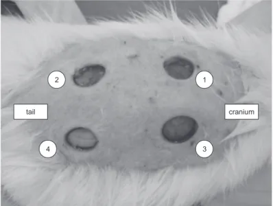
performed with chlorhexidine 2%, and, with a metal
punch, four 12 mm para-vertebral excision wounds
were made, based on the medial dorsal line, down
to the level of the aponeurotic tissue, as shown in
Figure 1.
Figure 1 - Lesions on the back of animal n. D0
1
3 2
4
Rev. Latino-Am. Enfermagem 2014 Jan.-Feb.;22(1):165-72.
After the excisions were made, each lesion received
0.25 µL of a suspension containing 1.5 x 10⁶ CFU/mL of
S. aureus, to infect the aponeurotic tissue supericially. Following that, the lesions were covered with gauze and
sterile dressings, and the researchers waited 24 hours to
carry out a culture swab of the four lesions and to start
the therapy.
The animals were assessed every 24 hours for 14
days, for clinical observation of the lesions, in relation to
the presence of: adverse effects, perilesional irritation,
scar retraction, and dressing the wounds, in accordance
with the group’s treatment. All the data was recorded in
pre-established protocols.
On the 3rd, 7th, 11th and 14th post-operative days
(POD) the wound areas were measured with digital
calipers, and on each occasion, removing a fragment
of one of the lesions for histopathological evaluation.
On the last day of the experiment, all the animals were
anesthetized and euthanized with thoracotomy, and
cardiac puncture was undertaken, removing 4 ml of blood
for analysis of glucose, cholesterol and triglycerides,
in addition to the liver, for microscopic analysis of the
hepatotoxicity of the extract.
Histological analysis
All the material removed for microscopic examination was numbered, without the identiication of the group to which it belonged, and was ixed using a 10% formaldehyde solution. The samples were
dehydrated and diaphanized in alcohol and xylol, included and embedded in warmed parafin. After hardening, the blocks were taken for microtomy, for
the obtaining of sections of 5 µm, these being collected
on glass slides and stained with Hemotoxylin and
Eosin stain.
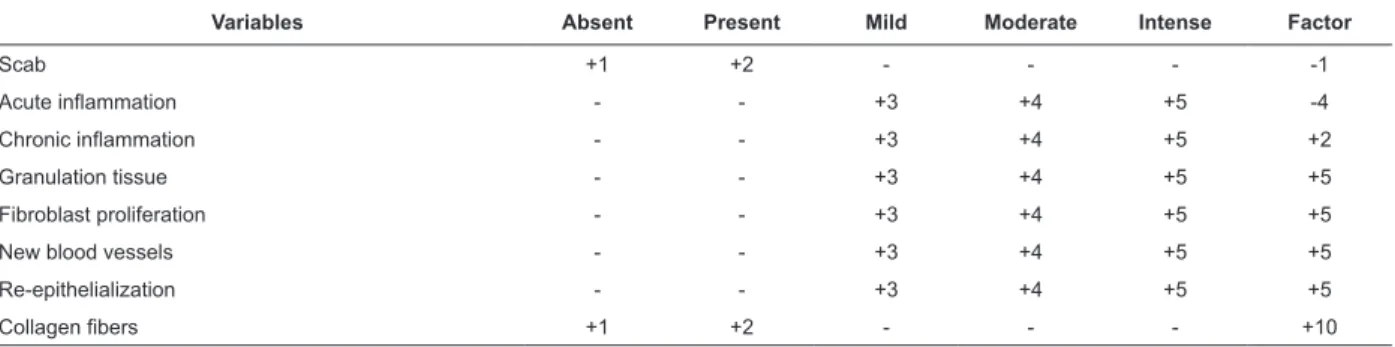
The analysis of the material used a reference points the inlammatory, proliferative and remodeling phases, which make up the process of healing. For analysis of
the results, Table 1 was prepared, with scores based
on the literature(5,8). The intensity of the variables
(1+ to 5+) was multiplied by positive or negative
factors based on their importance for the healing.
The sum of these products corresponded to the
total score for each animal, which was later
multiplied by 4 (n).
Table 1 - Scores used for evaluation of the histopathological test
Variables Absent Present Mild Moderate Intense Factor
Scab +1 +2 - - - -1
Acute inlammation - - +3 +4 +5 -4
Chronic inlammation - - +3 +4 +5 +2
Granulation tissue - - +3 +4 +5 +5
Fibroblast proliferation - - +3 +4 +5 +5
New blood vessels - - +3 +4 +5 +5
Re-epithelialization - - +3 +4 +5 +5
Collagen ibers +1 +2 - - - +10
Statistical tests
The data was analyzed using the program
GraphPad InStat®. The numerical variables were
evaluated using the ANOVA test with two interaction
factors, with Tukey’s post-hoc test for analysis of the
effect between the groups. For the non-parametric
data, the Mann-Whitney test was used. The level of signiicance established was 5% (p < 0.05).
Results
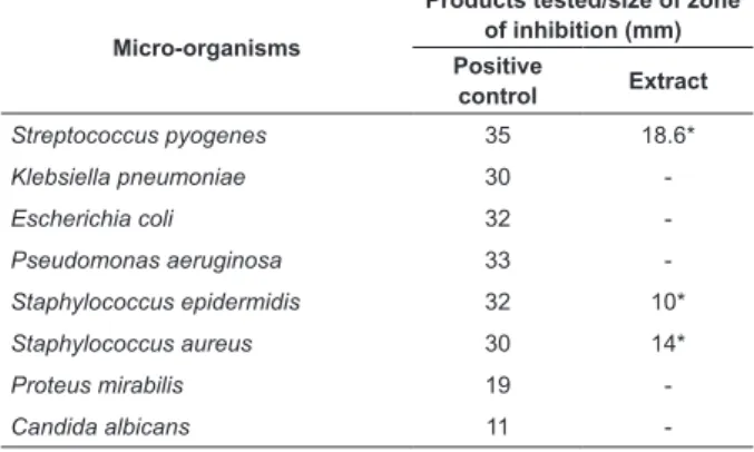
Evaluation of the antimicrobial activity and determination of the MIC
The raw extract of the stem of Z. tuberculosa did
not demonstrate activity for the fungus tested, but it
was active against the S. pyogenes, S. aureus and S.
epidermidis bacteria, as shown in Table 2. S. aureus
was the microorganism chosen for continuation of
the tests, because it is important in the occurrence
of nosocomial infections, mainly in integumentary
infections, as well as presenting a zone of inhibition
with approximately 50% of the size of the positive
control’s zone of inhibition(9-10). The extract’s MIC for
this microorganism was 62.5 μg/mL.
Toxicity in relation to Artemia salina (TAS)
Following the determination of the MIC, the
researchers proceeded to the TAS. According to the
criteria determined in this research, the extract was
Table 2 - Result of disk diffusion with the raw extract of
the stem of Z. tuberculosa for bacterial and fungal strains
(-) Did not present a zone of inhibition of growth *Value of the mean of the triplicate
Micro-organisms
Products tested/size of zone of inhibition (mm) Positive
control Extract
Streptococcus pyogenes 35 18.6*
Klebsiella pneumoniae 30
-Escherichia coli 32
-Pseudomonas aeruginosa 33
-Staphylococcus epidermidis 32 10*
Staphylococcus aureus 30 14*
Proteus mirabilis 19
-Candida albicans 11
-μg/ml) and in this study’s TAS are in consonance for safe use with this study’s animals, this being legitimized
by the absence of mortality among the animals during
the experiment.
Evaluation of the healing process in vivo
During the experiment, the animals were
monitored in relation to their temperature and weight,
and the lesions were observed macroscopically,
including the measuring of the lesions’ diameters.
Regarding temperature, on the day of the surgery, (D0)
all of the animals had temperatures within the normal
range of 36.2 to 37 ºC. From the 3rd to the 7th POD, only
the animals in the groups TE and NC presented rises in
temperature. The animals’ mean temperature varied between 36.2 and 38ºC, not showing a signiicant difference between the groups (p=0.1116).
The animals in all the groups presented weight
oscillation over the 14 days of the experiment. The
mean maximum weight was 265.1 grams and the
minimum, 208.4 grams. This difference was shown to be signiicant between the groups NC and TOE, and the groups NC and OE. However, the groups TOE, OE and
PC presented a similar pattern.
It was observed that in the pre-operative period,
the animals maintained a mean daily ingestion of feed
of 24g/day. After the undertaking of the lesions on
the backs, however, there was oscillation, the mean
ingestion being 18.5 g/day per animal.
In the evaluation of the lesions’ diameters,
contraction of the wounds was observed from the 3rd
POD, although the groups NC and TE did not have
this characteristic. From the 7th POD, reduction in the
diameter was observed, which was accentuated on the
11th and 14th days. The TOE and OE groups had better
responses in terms of the contraction of the wound.
The highest mean over the experiment was observed
in the TE group, with 7.46 mm, and the lowest, in the
TOE group, with 5.21 mm. The Tukey test showed a signiicant difference between groups NC and TOE, and TE and TOE, as well as between TE and OE.
The macroscopic study allowed the researchers
to observe differences between the groups regarding
the lesion’s color. The lesion’s aspect, in relation to the color, clinically relects the development of the healing process. Yellow coloration indicates infection.
This data indicates that the groups treated with
Z. tuberculosa had a smaller quantity of animals
with this characteristic, showing the extract’s
antimicrobial activity, whether topically and/or orally.
This is corroborated by the scores obtained in the
histological study.
From the 11th day onward, yellow or pink coloration
was observed in 100% of the animals in the groups
PC, TE, TOE and OE, and in 75% of those in group
NC, indicating that the phases of granulation and
re-epithelialization had started. No animal presented
brown coloration, which is an indication of cell death
and necrosis.
The phlogistic signs of inlammation, such as perilesional rubor or hyperemia, edema and the
presence of exsudate were not observed macroscopically
in any of the groups, evidencing that the extract used
in the research does not provoke irritation in the
perilesional skin, or in the cutaneous lesion. This data,
moreover, is corroborated by the scores obtained in the
histological study.
Furthermore, macroscopically, in relation to the
occurrence of granulation tissue, it was observed that
on the 3rd POD, the animals in group TE had 100%
of granulation, in those in groups PC and TOE it was
75%, and in those in group OE, it was 50%. These indings conirm that group TE had the best response, which was also legitimized by the scores of the
histological study.
Histological evaluation of the lesions
The histological data was collected in accordance
with the phases of healing, and was organized in accordance with the scores for quantiication. In Table 3 it is possible to observe that the highest means in the
scores were obtained by the groups which received the
Rev. Latino-Am. Enfermagem 2014 Jan.-Feb.;22(1):165-72.
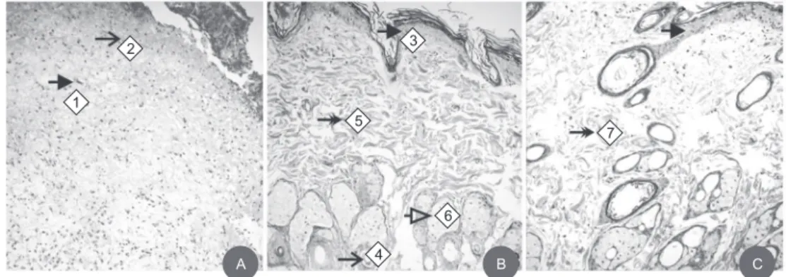
The analysis of the lesions of the groups TE, TOE
and OE treated with the extract of Z. tuberculosa evidenced important indings in the healing process, visualized in Figure 2. On the 3rd day, one can observe
acute inlammation with presence of macrophages (2A); on the 7th day, one can identify the granulation
tissue, with formation of neovascularization, collagen ibers and the start of epithelialization (2B); on the 11th day, one can see emphasized the
collagen ibers (2C).
Post-operative days
Scores
Positive control
Negative control
Topical extract
Topical and oral extract
Oral extract
3rd 87 87 634 372 229
7th 493 293 300 272 842
11th 670 420 1231 1150 812
14th 704 634 1400 1004 1324
Mean 488.5 358.5* 891.25* 699.5 801.8
Table 3 - The scores of the histopathological study of the
wounds infected by S. aureus
Figure 2 - (A) Photomicrography of a cutaneous wound, topical extract, 3rd day post-surgery (1 macrophages,
2 acute inlammation); (B) Photomicrography of a cutaneous wound, topical and oral extract, 7th day post-surgery
(3 epithelialization, 4 neovascularization, 5 collagen ibers, 6 granulation tissue) and (C) Photomicrography of a cutaneous
wound, oral extract, 11th day post-surgery (7 collagen ibers) (Magniication 10x, HE)
*p<0.05
A B C
1
2 3
4 5
6
7
The histological analysis of the liver showed an
absence of hepatotoxicity in all the groups studied.
In the biochemical evaluation, the data was not given
as it presented similar parameters for cholesterol,
triglycerides and glucose in all the animals in the
experiment, it being impossible to establish correlations between these indings and the use of the extract of Z. tuberculosa.
Discussion
Caring for wounds should be a constant concern
for the nurse, as caring for cutaneous lesions with the
resources currently existing remains a challenge. In spite
of the technological advances, few have access to these,
due to their high cost and their limitation to the major
urban centers(11). Studies show that the use of plants
is becoming a necessity for broadening the therapeutic arsenal in this area and in the reduction in the inancial resources spent for the treatment(12). In the present
study, the raw ethanolic extract of Z. tuberculosa showed
antimicrobial activity in vitro for S. aureus, was shown to
be atoxic by the test with Artemia salina, and in vivo had
a healing effect for infected excision wounds in rats, this
data being promising for the continuation of studies which
may elucidate the extract’s mechanism of action, as this
pathogen is responsible for the majority of infections of
the tegumentary system, and is resistant to the majority
of the antimicrobials used in clinical practice(10).
In the in vivo experiment, the animals’ temperature
was little-changed, not passing 38ºC. Even with local
infection, proven by the culture of the lesions, the
animals did not have systemic consequences. The rise
observed was due to the mechanism of healing itself,
which leads to an increase in the vascularization through the local inlammatory response, thus increasing the body temperature(13).
Checking the weight is an important nutritional
parameter in various experimental studies, being
related to wound healing alterations(14). The difference
in weight can be directly related to reduction in the daily
ingestion of feed, as also by the daily handling, with
wound dressing and biopsies undertaken on the 3rd, 7th,
The association of weight loss with the inlammatory process itself was described in a study asserting that inlammation produces inlammatory cytokines and Tumor Necrosis Factor (TNF), which act as mediators of the inlammation and of the immunity(6). With the
serum levels of TNF raised, there is stimulation in the
production of a protein called leptin, a rise in which is
related to the feeling of fullness. Increased levels of this
protein induce the organism to spend energy and reduce
the consumption of food, causing lack of appetite and,
consequently, weight loss.
The coloring, as a parameter for evaluation of the
condition of the wounds, showed that these presented
clinical progression within that expected for each stage
of wound healing. In the groups which were treated,
there was a predominance of pink coloration, which is characteristic of epithelialization, this being the inal phase of the tissue repair. This data legitimized the indings which used the barbatimão (stryphnodendron barbatimam) and Pink Ipê (tabebuia impetiginosa) cream
in wounds in rats(15) and differed from the study with gel
and the glycolic extract of the leaf of the Common Guava
(Psidium guajava L.) which did not observe signiicant
difference between the groups treated and that which
used saline solution(16).
Macroscopically, the total closure of the lesions did
not occur, in all the groups, over the 14 days of the experiment. There was, however, a signiicant reduction in the diameter in the treated animals, data conirmed by the presence of re-epithelialization in the histological
samples. In animals whose skin adheres less to the
deeper layers (such as rats), the contraction can reach
80 to 90% closure of cutaneous lesions(14).
The histological analysis allows one to infer that the
groups treated with the extract of Z. tuberculosa obtained
the best results, when compared with the positive
and negative control groups. This is due to the lower inlammatory reaction of the groups treated with the extract. A certain degree of inlammation is necessary, but a heightened inlammatory reaction is harmful, as there can be compromising of the microcirculation and it can also inhibit the formation of ibroblasts(17). The
literature also presents similar results with the evaluation
of the effects of the topical treatment with cream with a
pequi-oil base (Caryocar coriaceum Wittm)(18) and with
essential oils of some species of Juniper (Juniperus) a
traditional plant in folk medicine in Turkey(19).
The biochemical values of the animals in experiments
can be changed, due to the environmental conditions
and the stress induced by the procedure itself(20). The
results of triglycerides, cholesterol and glucose in the
animals studied had similar results to those presented
in the literature.
Conclusion
The raw ethanolic extract of Z. tuberculosa has
antimicrobial action in vitro for Streptococcus pyogenes,
Staphylococcus epidermidis and Staphylococcus aureus.
The groups treated with the extract had a result which
was similar to that of the positive control in the process
of wound healing. Histologically, the animals treated
topically had a greater quantity of cells for closing
the lesions. This research validates previous studies
with this extract and advances by demonstrating the
potential healing activity of Z. tuberculosa. This provides
encouragement for the continuation of studies, with a view to the identiication of the active ingredients which are responsible for the pharmacological activity, and its
mechanism of action in the process of wound healing.
This will contribute to the development of a product
which may be used as an alternative in the repair of
infected cutaneous wounds.
References
1. Mendonça RJ, Coutinho-Netto J. Cellular aspects of wound healing. An Bras Dermatol. 2009; 84(3):257-62.
2. Alvim NAT, Ferreira MA, Cabral IE, Almeida AJ Filho. The use of medicinal plants as a therapeutical resource: from the influences of the professional formation to the ethical and legal implications of its applicability as an extension of nursing care practice. Rev. Latino-Am. Enfermagem. 2006;14(3):316-23.
3. Pagano MC, Maria SM. Effect of Phosphorus Fertilization on Arbuscular Mycorrhizal Colonization of Zeyheria tuberculosa a Native Species in Brazil’s Forest. Middle-East J Sci Res. 2010;6(6):604-11.
4. Rahmatullah M, Rahman MA, Haque MZ, Mollik AH, Miajee EU, Begum R, et al. A survey of medicinal plants used by folk medicinal practitioners of station purbo para village of Jamalpur Sadar Upazila in Jamalpur district, Bangladesh. Am-Eurasian J Sustain Agric. 2010;4:122-35.
5. Bastos MLA, Lima MRF, Conserva LM, Andrade VS, Rocha EM, Lemos RP. Studies on the antimicrobial activity and brine shrimp toxicity of Zeyheria tuberculosa (Vell.) Bur. (Bignoniaceae) extracts and their main constituents. Ann Clin Microbiol Antimicrob. 2009;8:1-16.
Rev. Latino-Am. Enfermagem 2014 Jan.-Feb.;22(1):165-72.
Received: Feb. 14th 2013
Accepted: Aug. 22nd 2013
healing in surgically-induced clean wounds in Wistar rats. Semina Cienc Biol Saude. 2008;29(1):65-74. 7. Ayres MCC, Brandão MS, Vieira-Junior GM, Menor JCAS, Silva HB, Soares MJS, et al. Antibacterial activity of useful plants and chemical constituents of the roots of Copernicia prunifera. Braz J Pharmacogn. 2008; 18(1):90-7.
8. Medeiros AC, Ramos AMO, Dantas Filho AM, Azevedo RCF, Araujo FLFB. Topic treatment of rat burns with hyaluronic acid. Acta Cir Bras. [Internet]. 1999 [acesso 8 jun 2012];14(4). Disponível em: http://www.scielo.br/ scielo.php?script=sci_arttext&pid=S0102-86501999000 400010&lng=en&nrm=iso
09. Vitorino Filho RNL, Batista MCS, Verçosa BLA, Silva SMMS, Bonfim JM, Brandão AAC, et al. Avaliação do uso de pomada à base de sementes de jaqueira (Artocarpus heterophyllus Lam) na terapêutica tópica de feridas. J Basic Appl Pharm Sci. 2007;28(3):279-86.
10. Moura JP, Pimenta FC, Hayashida M, Cruz EDA, Canini SRMS, Gir E. Colonization of Nursing Professionals by Staphylococcus aureus. Rev. Latino-Am. Enfermagem. 2011;19(2):325-31.
11. Lima ACB, Guerra DM. Avaliação do custo do tratamento de úlceras por pressão em pacientes hospitalizados usando curativos industrializados. Ciênc Saúde Coletiva. 2011;16(1):267-77.
12. Majewska I, Gendaszewska-Darmach E. Proangiogenic activity of plant extracts in accelerating wound healing – a new face of old phytomedicines. Acta Biochim Pol. 2011;58(4):449-60.
13. Carvalho LH Júnior, Santos RL, Mendonça CJA, Campos CT, Andrade MAP. Evalution of skin temperature, reactive c protein, and hemosedimentation speed variation in uncomplicated primary knee total arthroplasty. Acta Ortop Bras. 2006;14(3):161-4. 14. Ono MCC. Influência de dieta imunomoduladora na
cicatrização cutânea em ratos [dissertação de mestrado]. Santa Catarina (PA): Universidade Federal do Paraná; 2009. 15. Coelho JM, Antoniolli AB, Silva DN, Carvalho TMMB, Pontes ERJC, Odashiro AN. Effects of silver sulfadiazine, ipê roxo (Tabebuia avellanedae) extract and barbatimão (stryphnodendron adstringens) extract on cutaneous wound healing in rats. Rev Col Bras Cir. 2010;37(1):45-51.
16. Okamoto MKH. Estudo das atividades cicatrizante e antimicrobiana do extrato glicólico e gel de Psidium guajava L. e estudo da estabilidade do gel [tese de doutorado]. São Paulo (SP): Faculdade de Ciências Farmacêuticas da Universidade de São Paulo; 2010. 17. Lucena PLH, Ribas Filho JM, Mazza M, Czeczko NG, Dietz UA, Correa Neto MA, et al. Evaluation of the aroreira (Schinus terebinthifolius Raddi) in the healing process of surgical incision in the bladder of rats. Acta Cir Bras. 2006;21(2):46-51.
18. Batista JS, Silva AE, Rodrigues CMF, Costa KMFM, Oliveira AF, Paiva ES, et al. Avaliação da atividade cicatrizante do óleo de pequi (caryocar coriaceum wittm) em feridas cutâneas produzidas experimentalmente em ratos. Arq Inst Biol. 2010;77(3):441-7.
19. Tumem I, Suntar I, Keles H, Küpeli AE. A therapeutic approach for wound healing by using essential oils of cupressus and juniperus species growing in Turkey. Evid Based Complement Alternat Med. [Internet]. 2012 [acesso 9 dez 2012]; 2012. Disponível em: http:// www.hindawi.com/journals/ecam/2012/728281/abs/ doi:10.1155/2012/728281
20. Dantas JA, Ambiel CR, Cuman RKN, Baroni S, Amador CAB. Valores de referência de alguns parâmetros fisiológicos de ratos do Biotério Central da Universidade Estadual de Maringá, Estado do Paraná. Acta Sci Health Sci. 2006;28(2):165-70.
Erratum
Issue v22n1, page 165
For
Patrícia de Albuquerque Sarmento2
Terezinha da Rocha Ataíde3
Ana Paula de Souza e Pinto4
João Xavier de Araújo-Júnior5
Ingrid Martins Leite Lúcio6
Maria Lysete de Assis Bastos6
Read
Patrícia de Albuquerque Sarmento2
Terezinha da Rocha Ataíde3
Ana Paula Fernandes Barbosa4
João Xavier de Araújo-Júnior5