(1) Departamento de Fonoaudiologia da Universidade Federal de Santa Maria – UFSM – Santa Maria (RS), Brasil. (2) Universidade Federal de Santa Maria –
UFSM - Santa Maria (RS), Brasil. (3) Departamento de Fonoaudiologia da
Universidade Federal de Santa Catarina – UFSC – Florianópolis (SC), Brasil. Study conducted at the Speech-Language Pathology Course, Federal University of Santa Maria (RS), Brazil.
Conlict of interest: non-existent
Avaliação clínica e eletromiográfica da mastigação
nos diferentes padrões de crescimento facial
Clinical and electromyographic evaluation of mastication
within different facial growth patterns
Luciele da Silva Prates(1) Marjana Gois(1) Luana Cristina Berwig(2) Ana Paula Blanco-Dutra(3) Angela Ruviaro Busanello-Stella(3) Ana Maria Toniolo da Silva(1)
Received on: May 12, 2015 Accepted on: July 05, 2015
Mailing address:
Luciele da Silva Prates
Avenida Roraima, 1000, Prédio 26, 1433. Camobi, Km 9
Santa Maria - RS - Brasil CEP: 97105-900
E-mail: [email protected]
doi: 10.1590/1982-021620161817015
ABSTRACT
Purpose: to analyze masticatory function within different facial types through clinical and electromyogra-phic evaluation of masseter and anterior temporalis muscles.
Methods: sixty-ive children aged six to 12 years old, males and females, who met the study criteria, were
selected. The clinical evaluation of mastication was performed based on the MBGR protocol, as well as the electromiography of masseter and anterior temporalis muscles, on the right and left side in directed
continued mastication. The data were analyzed considering the signiicance level of 5%.
Results: in the clinical evaluation of mastication, no signiicant differences between the three facial types
studied were observed, except for a tendency to signiicance in unexpected muscle contractions, with the highest occurrence observed in dolichofacials (66.67%), followed by mesofacials (46.67%) e bra
-chyfacials (26.83%). Through electromyographic evaluation, no signiicant differences between the three
groups studied were observed. Comparing the muscles within each facial type, a higher electrical activity of the right masseter muscle over the right temporal, and a higher electrical activity of the left temporal
muscle over the right temporal muscle (p=0.049) was veriied.
Conclusion: the results of clinical and electromyographic evaluations of mastication did not differ within
the facial types, suggesting that the pattern of facial growth itself is not a determinant in the modiications of the masticatory function and these modiications might be related to other variations not taken into
account in this study.
Keywords: Face;Cephalometry; Stomatognathic System; Mastication; Electromyography
RESUMO
Objetivo: analisar a função mastigatória nos diferentes padrões faciais de crescimento por meio da
ava-liação clínica e eletromiográica dos músculos masseteres e temporais anteriores.
Métodos: foram selecionadas 65 crianças entre seis e 12 anos de idade, de ambos os sexos, que se
adequaram aos critérios do estudo. Foi realizada avaliação clínica da mastigação tendo por base o pro
-tocolo MBGR e a avaliação eletromiográica dos músculos masseteres e temporais anteriores, mediante mastigação contínua direcionada. Os dados foram analisados considerando nível de signiicância de 5%.
Resultados: na avaliação clínica da mastigação, não foi observada diferença signiicante entre os três
padrões faciais estudados, apenas tendência à signiicância para as contrações musculares não espera
-das, sendo observada maior ocorrência nos dolicofaciais (66,67%), seguido dos mesofaciais (46,67%) e braquifaciais (26,83%). Na avaliação eletromiográica da mastigação, não foi observada diferença sig
-niicante entre os três grupos estudados. Ao comparar os músculos para cada padrão facial, veriicou-se nas crianças braquifaciais maior atividade elétrica do músculo masseter direito em relação ao temporal direito e maior atividade elétrica do músculo temporal esquerdo em relação ao músculo temporal direito (p=0,049).
Conclusão: os resultados das avaliações clínica e eletromiográica da mastigação não se diferiram nos
padrões faciais, sugerindo que o padrão por si só não é determinante nas modiicações da função masti
-gatória e que estas podem estar relacionadas a outras variáveis não consideradas neste estudo.
Descritores: Face; Cefalometria; Sistema Estomatognático; Mastigação; Eletromiograia Original articles
INTRODUCTION
Facial patterns are closely linked to stomatognathic functions of each individual, such as speech, chewing,
breathing and swallowing. For the eficient performance
of these functions, it is necessary craniofacial structures to be in harmony1.
Characteristics that compose the craniofacial skeleton also constitute the type of face through the relationship between vertical and horizontal growth and variation of the facial type. A frequently described
classiication in literature sorts the face in three
different facial patterns: brachyfacial, in which there is a tendency towards horizontal growth; mesofacial, prone to balanced growth of the facial thirds; and dolicho-facial, with a tendency to vertical growth2,3.
Some studies2,4,5 indicate that the main clinical
characteristics of brachyfacial are the higher horizontal growth; reduction of the lower third; thicker and powerful mandibular elevator muscles; ease of labial closure; tongue position supported all over the palate; greater potential for overbeat and bruxism; and wider nasal-pharyngeal functioning spaces, favoring nasal breathing. The mesofacial, on the other hand, presents balanced growth of the facial thirds; appropriate distri-bution of functioning space and accommodation of structures of soft tissue; and absence of adaptations of orofacial functions. The dolichofacial present mainly
vertical growth; increase in the lower third; dificulty of tongue support on hard palate; dificulty with labial
closure; stretched and less powerful mandibular elevator muscles; narrow nasal-pharyngeal functioning spaces, favoring oral breathing.
Mastication is a complex physiological function that depends on the development of the craniofacial complex, on the dental occlusion and on the central nervous system. Moreover, it is considered a functional unit, where teeth, jaw, temporomandibular joints, jaw muscles, lips and tongue and vascular and nervous systems of these tissues are involved6.
For an effective chewing, the activities of the masti-catory, buccinator and suprahyoid muscles must be
synchronized7. Besides, it must be composed by
bilateral alternating movements, with the occluded lips and mandibular rotation. These characteristics enable the distribution of the masticatory strength of these structures, alternating periods of work and rest for muscles and joints, leading to muscle and functional balance8.
Due to the importance of this function, the evaluation thereof has been showing an improvement in clinical
practice. This appears as a result of new features that allow complementation of qualitative with quantitative data, as the electromyographic evaluation and orofacial myofunctional evaluation based on MBGR protocol.
In this context, surface electromyography (SEMG) is an important tool for understanding the muscle behavior of the main functions of the stomatognathic system. It is also considered an objective and quanti-tative method, enabling the easier obtainment of the parameters for the diagnosis and contributing to the therapeutic process9.
The orofacial myofunctional evaluation (MBGR) furthers our understanding of the anatomical and functional conditions of the stomatognathic system, contributing to the diagnostic process and thera-peutic reasoning. It is a subjective method containing some objective parameters (quantitative) that require subjective analysis by the evaluator10.
As aforementioned, this study aims to analyze the chewing function in different facial growth patterns through clinical and electromyographic evaluation of the masseter and anterior temporal muscles, so that information of masticatory function in different types of face may be provided.
METHODS
This study presented quantitative and transversal character and was linked to a major project conducted in the Orofacial Motricity Laboratory of the Speech-Language Pathology Course at Federal University of Santa Maria (UFSM). It was approved by the Research Ethics Committee (REC) of that institution (no. CAAE 08105512.0.0000.5346).
Subjects
The study population consisted of children from public schools located in a country town of the State of Rio Grande do Sul (RS) and from the Clinical School of the Speech-Language Pathology Course of UFSM. All subjects composing the sample agreed to participate and had parental consent obtained by the signature of the written informed consent form (WICF).
In order to be selected, children of both genders should be aged between 6-12 years, present erupted
irst permanent molars and be classiied as eutrophic according to the clinical classiication of body mass
index (BMI).
Exclusion criteria included history of speech therapy
as long as in even tooth positions; signs indicating pathological bruxism diagnosed by dental evalu-ation; syndromes or craniofacial malformations; as well as incidence of neurological damage or any sign suggesting it. To check the suitability of the subjects concerning the study criteria, anamnesis and dental evaluation were performed, as well as the calculus of the BMI.
The interview with parents was held taking into account the medical history, assessed by the Myofunctional Orofacial Assessment Protocol
(MBGR)10. This protocol addresses issues related
to pregnancy and birth complications, motor
devel-opment, motor dificulties, health problems and treat -ments performed, among others.
The dental evaluation aimed to detect signs suggesting pathological bruxism (non-physiological), as dental wear for example, and verify the presence of
the upper irst permanent molars and dental laws. This
assessment was conducted by a single dental surgeon. For the evaluation of BMI status, the subjects had their height and weight measured with a tape attached to the wall and an anthropometric weight scale (ToledoTM) respectively. In both procedures, the children were standing barefoot and the approximate weight of their clothes was deducted. The BMI consisted on the ratio between weight (in kilograms) and the square of
height (in meters)11. Since the sample was composed
by children, the parameters adopted were the ones suggested by the World Health Organization12. Hence, only eutrophic children, i.e. with BMI status between 5-85%, were included.
The sample comprised 65 children, being 34 girls and 31 boys. They were assigned into groups according to their facial growth pattern, which was determined by the cephalometric evaluation.
The cephalometric evaluation was performed in a radiology and orthodontic imaging center. The procedure consisted on the lateral teleradiography,
with a 18x24 cm ilm (KodakTM) mounted on chassis coated with Kodak Lanex regular screen and was held
in the X-Mind unityTM. This device was equipped with a cephalostat in order to standardize the position of the subject’s head in relation to the emitted radiation at a distance of 1.5 m. In the lateral radiography obtained, a computerized cephalometric analysis was held, as well as the Ricketts Analysis.
The VERT index, obtained from the cephalometric
analysis, is based on ive cephalometric variables
(facial axis angle, facial depth, mandibular plane angle,
lower height and mandibular arch) and enables the
classiication of facial types13. According to this index, facial types were classiied in brachyfacial (VERT index
>0.5); mesofacial (VERT index between -0.5 and +0.5); and dolichofacial (VERT index <0.5).
Clinical evaluation of mastication
The speech-language evaluation was based on the
MBGR protocol10 and was applied always by the same
speech-language pathologist in order to achieve a better standardization and a greater control of the test. Regarding the information presented by this protocol, the following aspects concerning mastication were considered: incision of food; grinding of food;
masti-catory eficiency; mastimasti-catory pattern; labial closure;
mastication noise; unexpected muscle twitching and average masticatory time.
The evaluation of chewing performance was carried out with the child sitting with the hip at 90º. Each child was given three portions of French bread and should eat them according to his/her usual habits. The activity was video recorded and independently analyzed by three speech-language pathologists with experience in orofacial motricity. The results should be common for at least two speech-language pathologists in order to be considered. In the situations where there was no agreement between the judges, a new collective analysis was conducted.
Electromyographic evaluation of mastication
The electromyographic evaluation was performed always by the same speech-language pathologist, avoiding, hence, deviations and differences in the collection procedure. The masseter and anterior temporal muscles were evaluated, in both right and left sides. The subjects were familiarized to the exam procedures, as well as to the local of collection and equipment used. Also, they were trained concerning the collection procedure. They remained seated comfortably with the hip, knees and ankles at 90º, oriented by the Frankfurt plane. The muscles were then assessed at rest and at directed continuous chewing.
The rest stage lasted for 10 seconds and aimed to standardize the electrical activity during the baseline condition.
since this material well resembles the food, but does not deteriorate or produce any residues, which could affect the evaluation. Initially, subjects were asked to chew the gum for 40 seconds in order to diminish and standardize its hardness. After a rest of 1 minute, children were instructed to chew rhythmically synchro-nized according to a digital metronome (Cherub – WSM 001A), set at 80 bmp14, until they felt tired.
Three collections were performed in both test situa-tions, respecting an interval of 2 minutes of rest for
muscle recovery15. The analysis selected was always
the one presenting better signal quality (analysis by FFT). After choosing the signal, chewing analysis was carried out by excluding the time before the beginning
of muscle activity, as well as the irst 0.5 seconds of
the activity, in order to homogenize the evaluated
sections16. The following 15 seconds of mastication
were selected and analyzed. The electromyographic signal was measured in terms of amplitude (RMS – microvolts) and normalized by the maximum peak value.
Regarding the sensors, the international standards where followed17,18. Miotec sensors with differential input
were placed on the muscle bellies15 and connected
to DOUBLE type Ag/AgCl electrodes (Hal Indústria e Comércio Ltda.) The devices had discotic format (10 mm diameter). A 20 mm distance therebetween was kept. Other characteristics were: conductive gel present
in a ixed amount; 20X gain; 10 GΩ input impedance
and common mode rejection rate >100 dB. Also, in order to prevent electromyographic interference, a reference electrode was placed in the patient’s glabellar region (connected to the ground).
In order to detect, condition and amplify the electro-myographic signal, the Miotool equipment (Miotec) was used with 8 input channels, 14 bit A/D converter in the electromyographic signal acquisition channels, 5000 V electrical isolation, data acquisition capability of 2000 samples/second/channel, 20 Hz high-pass and 500 Hz
low-pass ilter. The collection and signal processing
were performed by Miograph 2.0 software and saved in a HP Pavilion dv5-204br laptop with 500 GB HD and 4 GB of RAM, with no connection to the power grid.
The skin was previously cleaned with a 70% ethanol solution and gauze in order to diminish its impedance. The physical location where the collections were performed, including both the equipment and the chair
evaluation area, was carefully treated: the loor was
covered in rubberized Pavilex. Moreover, all equipment
that would electromagnetically interfere with the exam was turned off and kept at a safe distance15.
Statistical Analysis
After tabulating the data, statistical analysis was performed using the Statistical Analysis System (SAS), version 9.2. Chi-square and Fisher’s exact tests were used to compare categorical variables of clinical evalu-ation, being the latter applied when expected values were lower than 5. To compare numerical variables, non-parametric tests were used due to the absence of a normal distribution, indicated by the Shapiro-Wilk test.
The Kruskal-Wallis test was used to compare the
numerical variables of clinical and electromyographic evaluations among the three groups. The Friedman test was used for comparison between muscles in the electromyographic evaluation within each facial
type. In all tests, the level of signiicance was set at 5%
(p<0.05).
RESULTS
Table 1 shows the frequency distribution concerning the gender, age and facial type of the studied group. A similar distribution between genders and ages was observed. Regarding the facial pattern, on the other hand, more than half of the children evaluated were considered brachyfacial (63.08%).
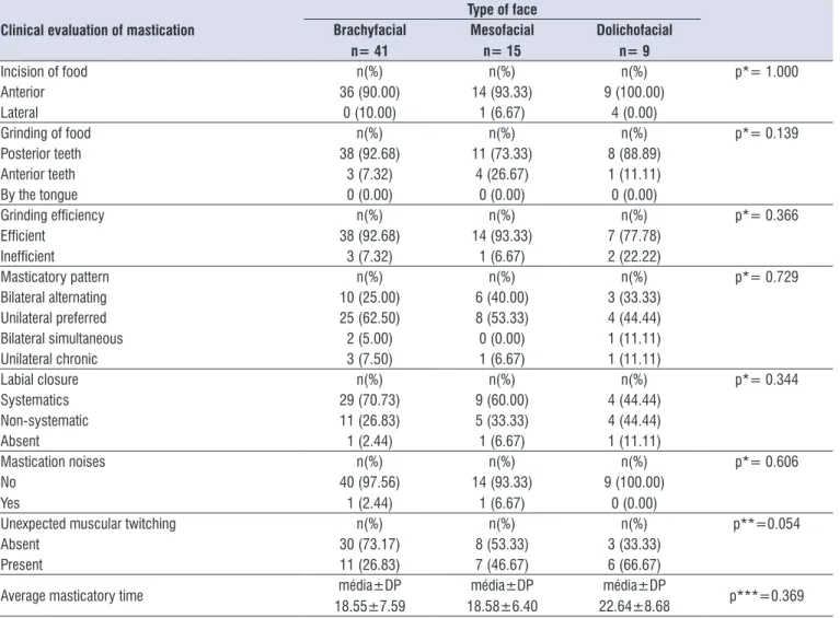
The relationship between facial types and clinical evaluation of chewing performance is shown in Table
2. There was no signiicant difference among the three groups assessed. However, a tendency to signiicance
in respect to muscle contractions not usually expected during mastication was observed. This trend was most marked in dolichofacial (66.67%), followed by mesofacial (46.67%).
The results of the electromyographic evaluation of mastication associated to facial patterns are presented in Table 3. While the analysis shows there are no
signiicant differences between the three groups, the
Table 1. Characterization of the subjects (n=65 children)
Frequency (n)
Percentage (%)
Gender Female
Male
34
31
52.31 47.69
Age group
6 – 7 years 8 – 9 years 10 – 12 years
19
25
21
29.23
38.46
32.31
Type of face
Brachyfacial Mesofacial Dolichofacial
41 15
9
63.08 23.08 13.85 Key: n - number of children; % - percentage.
Table 2. Clinical evaluation of mastication and type of face of the subjects
Clinical evaluation of mastication
Type of face Brachyfacial
n= 41
Mesofacial n= 15
Dolichofacial n= 9
Incision of food n(%) n(%) n(%) p*= 1.000
Anterior 36 (90.00) 14 (93.33) 9 (100.00)
Lateral 0 (10.00) 1 (6.67) 4 (0.00)
Grinding of food n(%) n(%) n(%) p*= 0.139
Posterior teeth 38 (92.68) 11 (73.33) 8 (88.89)
Anterior teeth 3 (7.32) 4 (26.67) 1 (11.11)
By the tongue 0 (0.00) 0 (0.00) 0 (0.00)
Grinding eficiency n(%) n(%) n(%) p*= 0.366
Eficient 38 (92.68) 14 (93.33) 7 (77.78)
Ineficient 3 (7.32) 1 (6.67) 2 (22.22)
Masticatory pattern n(%) n(%) n(%) p*= 0.729
Bilateral alternating 10 (25.00) 6 (40.00) 3 (33.33)
Unilateral preferred 25 (62.50) 8 (53.33) 4 (44.44)
Bilateral simultaneous 2 (5.00) 0 (0.00) 1 (11.11)
Unilateral chronic 3 (7.50) 1 (6.67) 1 (11.11)
Labial closure n(%) n(%) n(%) p*= 0.344
Systematics 29 (70.73) 9 (60.00) 4 (44.44)
Non-systematic 11 (26.83) 5 (33.33) 4 (44.44)
Absent 1 (2.44) 1 (6.67) 1 (11.11)
Mastication noises n(%) n(%) n(%) p*= 0.606
No
Yes
40 (97.56)
1 (2.44) 14 (93.33)1 (6.67) 9 (100.00)0 (0.00)
Unexpected muscular twitching n(%) n(%) n(%) p**=0.054
Absent 30 (73.17) 8 (53.33) 3 (33.33)
Present 11 (26.83) 7 (46.67) 6 (66.67)
Average masticatory time média±DP
18.55±7.59 18.58±6.40média±DP 22.64±8.68média±DP p***=0.369
Table 3. Electromyographic evaluation of mastication and type of face of the subjects
Electromyographic evaluation of mastication
Type of face
p** Brachyfacial
n= 41
Mesofacial n= 15
Dolichofacial n= 9
average±SD average±SD average±SD
Right masseter (RM) 14.14±3.41 15.37±3.19 13.61±3.23 0.378
Right temporal (RT) 13.20±3.03 13.78±3.10 12.78±2.03 0.615
Left masseter (LM) 13.88±3.19 15.01±4.27 12.97±2.76 0.359
Left temporal (LT) 14.41±3.07 14.87±2.88 13.40±2.51 0.464
p*** comparison between muscles evaluated in each type of face.
0.049* TD ≠ MD
TD ≠ TE 0.575 0.589
Key: n – number of children; SD- Standard deviation; *Friedman test p<0.05; ** Kruskal-Wallis’ test; ***Friedman test.
DISCUSSION
The aim of this study was to analyze the relationship between chewing and facial patterns, regardless of gender and age. Another study showed no statistically
signiicant differences between chewing performance
on boys and girls in the intertransitional period during mixed dentition period19.
The age group considered is the same as from other studies in literature20,21 which consisted of children aged between 6-12 years who presented erupted irst
permanent molars.
The choice of the age group is justiied by the fact
that at an age of approximately 6, transformations occur
in the oral cavity as a result of the irst permanent molars
eruption. At 12, the second permanent molars erupt20, which is an important condition for occlusal stability, as well as for electromyographic signal stability.
Regarding the facial growth pattern, it was veriied a
higher incidence percentage of brachyfacial, followed by mesofacial and dolichofacial respectively (Table 1). These results were not expected, since most studies report a greater incidence of mesofacial type22,23.
The higher frequency of brachyfacial compared to
other facial types may be justiied by the characteristics
of the region where the study was conducted. The
signiicant ethnic mix present in this region reinforces
the importance of genetic factors in the establishment of the facial patterns5.
There was no statistically signiicant difference in
most of the variables studied on the clinical evaluation of chewing performance when comparing different facial patterns. However, there was a trend towards
signiicance on the aspect “unexpected muscle
twitching”, which was more marked in dolichofacial, followed by mesofacial subjects (Table 2). According to
the literature, dolichofacial subjects may present such
contractions, as well as ineficient grinding of food,
which might be related to the hypofunctional masti-catory muscles24. This ineficiency was also reported by other authors25 who associated it to a smaller occlusal contact area. There was also no statistically signiicant
difference amongst the three types of face regarding other variables assessed by the evaluation, such as
incision of food, masticatory eficiency, mastication
noise and lip closure (Table 2).
These results anticipate that other variables not considered in this study, such as mixed dentition
period, severe open-bite, presence of dental laws,
poor dental state, malocclusion, temporomandibular disorders and bruxism may be related to changes in clinical characteristics of mastication and not to the facial type26-29.
The results found in the electromyographic
evalu-ation of this study reinforce our indings of the clinical
evaluation, i.e. the greater need of muscle contraction during chewing on dolichofacial individuals. Although
there was no signiicant difference between groups,
dolichofacial children presented lower average electrical activity in all muscles assessed (Table 3).
Despite having methodologies based on mandibular rest and isometric conditions, other studies corroborate our results, showing lower electrical activity values in masseter muscles in this group25,30-32.
patterns. This suggests that the facial growth pattern by
itself is not determinant in modiications of the masti -catory function, which therefore may be related to other variables not considered in this study.
There was a tendency to unexpected muscular twitching in mastication, mainly in dolichofacial children followed by mesofacial.
Brachyfacial children presented higher electrical activity of the right masseter when compared to the left one and higher electrical activity of the left temporal when compared to the right one.
REFERENCES
1. Rizzo MC, Akaishi NMM, Naspitz N. Multidisciplinary aspects of oral breathing in children Allergie Suppl. 2001;1(2):59-60.
2. Berwig LC, Silva AMT, Côrrea ECR, Moraes AB, Montenegro MM, Ritzel RA. Análise quantitativa do palato duro em diferentes tipologias faciais de respiradores nasais e orais. Rev CEFAC. 2012;14(4):616-25.
3. Bolzan GP, Berwig LC, Prade LS, Weinmann ARM, Moraes AB, Silva AM. Concordância entre método
antropométrico e cefalométrico na classiicação do
tipo facial. Rev CEFAC. 2014;16(1):222-7.
4. Bianchini EMG. Avaliação fonoaudiológica da motricidade oral – distúrbios miofuncionais orofaciais ou situações adaptativas. Rev Dental Press OrtodonOrtop Facial. 2001;6(3):73-82.
5. Busanello-Stella AR. Fadiga Muscular: Análise dos Músculos Orbiculares da Boca e Mastigatórios de Crianças de 6 A 12 anos de idade [tese]. Santa Maria (RS): Universidade Federal de Santa Maria; 2014.
6. Rahal A, Gofi-Gome MVS. Estudo eletromiográico do músculo masseter durante o apertamento dentário e mastigação habitual em adultos com oclusão dentária normal. Rev Soc Bras Fonoaudiol. 2009;14(2):160-4.
7. Rahal A, Gofi-Gomez MVS. Avaliação
eletromiográica do músculo masseter em pessoas
com paralisia facial periférica de longa duração. Rev CEFAC. 2007;9(2):207-12.
8. Pizzol KE. Inluência da Mastigação Unilateral no desenvolvimento da Assimetria Facial. Rev UNIARA. 2004;15:215-9
9. Mangilli LD, Sassi FC, Sernik RA, Tanaka C.
Caracterização eletromiográica e ultrassonográica
da função mastigatória em indivíduos com oclusão normal. J Soc Bras Fonoaudiol. 2012;24(3):211-7. morphology affected the electrical activity of the
masti-catory muscles studied25.
When comparing the electrical activity of the muscles evaluated in each facial pattern group, a
statis-tically signiicant difference (p = 0.049 *) was veriied
only for the brachyfacial group. This highlights the higher activity of the right masseter muscle in relation to the left one and the opposite situation for the temporal
muscle, i.e. higher activity on the left side (p = 0.049).
As reported in literature, brachyfacial individuals show a greater thickness of the masseter muscle, a wider nasal airway and a lower third of the lower face. Meso and dolichofacial individuals, on the other hand, present thinner masseter and shorter endurance time24,33. Therefore, the higher activity of the masseter in comparison to the temporal muscle in this group may have been affected by this morphophysiological feature.
These results are in disagreement with another study34, which evaluated the activity of the masticatory muscles of children throughout a day of activity, excluding feeding and sleeping time. The authors observed that the temporal muscle showed a greater activity than the masseter, regardless of the facial growth pattern.
Also, the electrical activities of the left and right temporal muscles for brachyfacial group are different, as indicated by our results.
Studies state that brachyfacial individuals may present oral pathologies, such as bruxism and temporomandibular disorders26. These characteristics, which were not investigated in this study, together with the unilateral chewing side preference may have
inluenced the asymmetry observed in the electromyo -graphic evaluation.
Once this subject enables the awareness of the particularities of the masticatory function in different types of face and given its importance for clinical practice, further investigations with more balanced samples regarding groups constitution are suggested. Also, some not yet considered variables that might interfere with the chewing performance of individuals with different facial growth pattern should be included, as suggested by other authors32.
CONCLUSION
According to the results obtained in this study, it is possible to infer that:
esquelético facial em indivíduos com oclusão normal natural. Ortodontia. 2010;43(3):237-42. 24. Ramires RR, Ferreira IP, Marchesan IQ, Cattoni
DM, Silva MAA. Tipologia facial aplicada à Fonoaudiologia: revisão de literatura. Rev Soc Bras Fonoaudiol. 2010;15(1):140-5.
25. Farias Gomes SG, Custodio W, Jufer JSM, Cury AADB, Cunha R, Garcia MR. Mastication, EMG Activity and Occlusal Contact Areain Subjects with Different Facial Types. The journal of craniomandibular practice. 2010;4(28):274-9.
26. Busanello-Stella AR, Berwig LC, Almeida FL, Silva AMT, Mello FM. Aspectos do sistema estomatognático de indivíduos bruxistas. Salusvita. 2011;1(30):7-20.
27. Andrada e Silva Ma, Natalini V, Ramires RR, Ferreira LP. Análise comparativa da mastigação de crianças respiradoras nasais e orais com dentição decídua. Rev CEFAC. 2007;1(9):190-8.
28. Weber P, Corrêa ECR, Bolzan GP, Ferreira FDS, Soares JC, Silva AMT. Mastigação e deglutição em mulheres jovens com desordem temporomandibular. Codas. 2013;1(25):375-80. 29. Pastana SG, Costa SM, Chiappetta ALML. Análise
da mastigação em indivíduos que apresentam mordida cruzada unilateral na faixa-etária de 07 a 12 anos. Rev CEFAC. 2007;3(9):339-50.
30. Rodrigues KA, Rahal A. A inluência da tipologia
facial na atividade eletromiográica do músculo
masseter durante o apertamento dental em máxima intercuspidação. Rev. CEFAC. 2003;2(5):127-30. 31. Farella M, Michelotti A, Carbone G, Gallo LM,
Palla S, Martina R. Habitual daily masseter activity of subjects with different vertical craniofacial morphology. Eur J Oral Sci. 2005;113(5):380–5. 32. Vianna-Lara M.S, Caria PHF, Tosello DO, Lara
F, Amorim MM. Electromyographic activity of masseter and temporal muscles with different facial types. Angle Orthod. 2009;3(79):515-20.
33. Lione R, Franchi L, Noviello A, Bollero P, Fanucci E, Cozza P. Three-Dimensional Evaluation of Masseter Muscle in Different Vertical Facial Patterns: A Cross-Sectional Study in Growing Children. Ultrasonic Imaging. 2013;35(4):307–17.
34. Ueda HM, Miyamoto K, Saifuddin MD, Ishizuka Y,
Tanne K. Masticatory muscle activity in children and
adults with different facial type. American Journal of Orthodontics and Dentofacial Orthopedics. 2000;1(118):63-8.
10. Genaro KF, Berretin-Felix G, Rehder MIBC, Marchesan IQ. Avaliação miofuncional orofacial – protocolo MBGR. Rev CEFAC. 2009;11(2):237-55. 11. Rosner B, Prineas R, Loggie J, Daniels SR.
Percentiles for body mass index in U.S. children 5 to 17 years of age. J Pediatrics. 1998;132(2):211-22. 12. Organização Mundial da Saúde. Padrões de
Crescimento Infantil. 2006. Disponível em: </http: www.saude.org.br/>. Acesso em: 30 abril. 2014. 13. Ricketts RM. Orthodontic diagnosis and planning
their roles in preventive and rehabilitative dentristy. Denver: Rocky Mountain Orthodontics. 1982;1:269. 14. Mendonça RG, Oliveira AS, Pedroni CR, Bérzin
F, Gastaldi AC. Electromyography assessment of chewing induced fatigue in temporomandibular disorders patients. Braz J Oral Sci. 2005;4(15):894-8. 15. De Luca C.J. The use of surface electromyography in biomechamics. Journal of applied biomechanics. 1997;13(2):135-63.
16. Silva SRD, Gonçalves M. Comparação de
protocolos para veriicação da Fadiga Muscular pela Eletromiograia de Superfície. Motriz.
2003;9(1):51-8.
17. Merletti R. Standards for Reporting EMG data.
Journal of Electromyography and Kinesiology.
1999;1:3-4.
18. Hermens HJ, Freriks B, Disselhorst-Klug C, Rau G. Development of recommendations for SEMG sensors and sensor placement procedures. Journal of electromyography and kinesiology. 2000;10(5):361-74.
19. Motta AR. Descrição da mastigação no período intertransicional da dentição mista. [mestrado]. São Paulo (SP): Pontifícia Universidade Católica de São Paulo. 2002:32-7.
20. Cattoni DM, Fernandes FDM, Marchesan IQ, Latorre MRDO. Medidas antropométricas faciais em crianças segundo períodos da dentição mista. Rev CEFAC. 2003;(5):21-9.
21. Bolzan GP, Silva AMT, Boton LM, Corrêa ECR. Estudo das medidas antropométricas e das proporções orofaciais em crianças respiradoras nasais e orais de diferentes etiologias. Rev. soc. bras. fonoaudiol. 2011;16(1):85-91.
22. Silva Filho OG, Herkrath FJ, Queiroz APC, Aiello CA. Padrão facial na dentadura decídua: estudo epidemiológico. R Dental Press Ortodon Ortop Facial. 2008;4(13):45-59.
23. Paranhos LR, Magalhães MPM, Bérzin F, Daruge