Congenital heart disease and chromossomopathies detected by
the karyotype
Cardiopatias congênitas e cromossomopatias detectadas por meio do cariótipo
Cardiopatías congénitas y anomalías cromosómicas detectadas mediante cariotipo
Patrícia Trevisan1, Rafael Fabiano M. Rosa2, Dayane Bohn Koshiyama1, Tatiana Diehl Zen1, Giorgio Adriano Paskulin1, Paulo Ricardo G. Zen1
Instituição: Universidade Federal de Ciências da Saúde de Porto Alegre (UFCSPA), Porto Alegre, RS, Brasil
1UFCSPA, Porto Alegre, RS, Brasil
2Hospital Materno Infantil Presidente Vargas (HMIPV), Porto Alegre, RS, Brasil ABSTRACT
Objective: To review the relationship between congenital heart defects and chromosomal abnormalities detected by the karyotype.
Data sources: Scientific articles were searched in MED-LINE database, using the descriptors “karyotype” OR “chromosomal” OR “chromosome” AND “heart defects, congenital”. The research was limited to articles published in English from 1980 on.
Data synthesis: Congenital heart disease is characterized by an etiologically heterogeneous and not well understood group of lesions. Several researchers have evaluated the pres-ence of chromosomal abnormalities detected by the karyo-type in patients with congenital heart disease. However, most of the articles were retrospective studies developed in Europe and only some of the studied patients had a karyo-type exam. In this review, only one study was conducted in Latin America, in Brazil. It is known that chromosomal abnormalities are frequent, being present in about one in every ten patients with congenital heart disease. Among the karyotype alterations in these patients, the most important is the trisomy 21 (Down syndrome). These patients often have associated extra-cardiac malformations, with a higher risk of morbidity and mortality, which makes heart surgery even more risky.
Conclusions: Despite all the progress made in recent de-cades in the field of cytogenetic, the karyotype remains an es-sential tool in order to evaluate patients with congenital heart disease. The detailed dysmorphological physical examination is of great importance to indicate the need of a karyotype.
Key-words: heart defects, congenital; karyotype; Down syndrome; trisomy; chromosome aberrations.
RESUMO
Objetivo: Realizar uma revisão da literatura sobre a relação das cardiopatias congênitas com anormalidades cromossômicas detectadas por meio do exame de cariótipo.
Fontes de dados: Pesquisaram-se artigos científicos no portal MEDLINE, utilizando-se os descritores “karyotype”
OR “chromosomal” OR “chromosome” AND “heart defects,
congenital”. A pesquisa limitou-se a artigos publicados em
inglês a partir da década de 1980.
Síntese dos dados: As cardiopatias congênitas são um grupo de lesões etiologicamente heterogêneo e pouco com-preendido. Vários pesquisadores avaliaram a presença de anor-malidades cromossômicas detectadas pelo exame de cariótipo em pacientes portadores de cardiopatia congênita. Porém, a maioria dos artigos era composta de trabalhos retrospectivos desenvolvidos na Europa, nos quais nem todos os pacientes
Endereço para correspondência: Paulo Ricardo G. Zen
Rua Sarmento Leite, 245, sala 403 – Centro CEP 90050-170 – Porto Alegre/RS E-mail: [email protected]
Fonte financiadora: Coordenação de Aperfeiçoamento de Pessoal de Nível Superior (Capes)
Conflito de interesse: nada a declarar
foram submetidos à avaliação cariotípica. Nesta revisão, encontrou-se apenas um estudo desenvolvido na América Latina, no Brasil. Sabe-se que anormalidades cromossômicas são frequentes, estando presentes em cerca de um em cada dez pacientes com cardiopatia congênita. Dentre as alterações ob-servadas, destaca-se a trissomia do cromossomo 21 (síndrome de Down). Esses pacientes frequentemente apresentam malfor-mações extracardíacas associadas e risco maior de morbidade e mortalidade, tornando a cirurgia cardíaca ainda mais arriscada.
Conclusões: Apesar de todos os avanços ocorridos nas últimas décadas na área da citogenética, o exame de cariótipo continua sendo uma ferramenta fundamental para se avali-arem pacientes com cardiopatia congênita. O exame físico dismorfológico minucioso é de grande importância para indicar a realização do cariótipo.
Palavras-chave: cardiopatias congênitas; cariótipo; síndrome de Down; trissomia; aberrações cromossômicas.
RESUMEN
Objetivo: Realizar una revisión de la literatura sobre la relación de las cardiopatías congénitas con anormalidades cromosómicas detectadas por medio del examen de cariotipo.
Fuentes de datos: Se investigaron artículos científicos en el portal MEDLINE, utilizándose los descriptores « karyoty-pe», OR «chromosomal» OR «chromosome» AND «heart defects, congenital». La investigación se limitó a artículos publicados en inglés a partir de la década de los 1980.
Síntesis de los datos: Las cardiopatías congénitas son un grupo de lesiones etiológicamente heterogéneo y poco com-prendido. Varios investigadores evaluaron la presencia de anormalidades cromosómicas detectadas por el examen de cariotipo en pacientes portadores de cardiopatía congénita. Sin embargo, la mayoría de los artículos estaba compuesta por trabajos retrospectivos desarrollados en Europa, en los que no todos los pacientes fueron sometidos a la evaluación cariotípica. En esta revisión, se encontró solamente un estudio desarrollado en Latinoamérica, en Brasil. Se sabe que anormalidades cromosómicas son frecuentes, estando presentes en aproximadamente uno a cada diez pacientes con cardiopatía congénita. Entre las alteraciones observa-das, se destaca la trisomía del cromosoma 21 (síndrome de Down). Esos pacientes frecuentemente presentan mal-formaciones extracardíacas asociadas y riesgo más grande de morbidez y mortalidad, lo que hace la cirugía cardíaca todavía más arriesgada.
Conclusiones: A pesar de todos avances ocurridos las últimas décadas en el área de la citogenética, el examen de cariotipo sigue siendo una herramienta fundamental para evaluar pacientes con cardiopatía congénita. El examen físico dismorfológico minucioso, realizado por un pediatra experimentado o por un geneticista, es de gran importancia para indicar la realización del examen.
Palabras clave: cardiopatías congénitas; cariotipo; síndrome de Down; trisomía; anomalías cromosómicas.
Introduction
Congenital malformations are detected in approximately 3 to 5% of newborns(1), and one in every 33 presents severe
abnormalities(2). Major malformations are those that cause an
adverse effect on the social acceptability of the individual or in the functioning of a determined organ or system(3). On the
other hand, minor malformations do not present aesthetical or functional signiicance for the individual, being a structural inding that occurs in less than 4% of the general population. However, some minor anomalies may be external markers of more speciic anomalies, sometimes hidden. Therefore, most syndromes could be recognized by the clinical geneticist if these patterns of minor anomalies were taken into consider-ation. The dysmorphology assessment, therefore, could help support the decision on whether to perform a complementary investigation, such as, for instance, through karyotyping(4).
Among the most frequent congenital malformations, congenital heart defects stand out, comprising structural and functional heart abnormalities present at birth, regardless of the time of diagnosis. Congenital heart defects are a heterogeneous group of lesions with varying hemodynamic consequences, requiring different follow-ups and interventions(5). Studies
show that the incidence of congenital heart disease can vary from four to 14 per 1,000 live births(6-8). In Brazil, studies
de-scribed a prevalence that ranges from ive to 12 per 1,000 live births(2,9,10). These variations can be explained by several factors,
such as the occurrence of lethal defects that prevent a live birth of the fetus and the exclusion of minor cardiac defects. Studies have shown that congenital heart disease may be responsible for about 40% of all birth defects, and it is considered one of the most frequent malformations(11,12). In Brazil, despite its great
geographical extent, there are 12 specialized centers both in the diagnosis and in the treatment of patients with congenital heart defects(13). The average number of cardiovascular surgeries at
However, the current health network is not enough for the demand and in 2002, for instance, there was a surgery deicit that reached 65%(7).
Thus, congenital heart defects are an even greater public heath problem worldwide, being the leading cause of death among congenital malformations(12). Severe and moderately
severe heart defects account for about three to six out of every 1,000 live births and are characterized by the need for more in-tensive and complex surgical care(6,11,14). These defects are a major
cause of admission and mortality in pediatric intensive care units(15). In Rio Grande do Sul, however, most intensive care
units are overcrowded and often lack equipment and skilled professionals for the differential diagnosis as well as conditions for the surgical treatment of patients with congenital heart dis-ease(10). This may be due to the fact that less developed countries
have other priorities related to health, including the preventions of malnutrition and promoting vaccination campaigns(13).
The heart is the irst organ to form in the embryo, and it is vital for the provision of oxygen and nutrients to the developing fetus(8). Its formation is complex and occurs over
several weeks of the embryonic life, making it very vulner-able to the occurrence of failures during its development(15).
Congenital heart defects are considered etiological hetero-genic malformations and are poorly understood(16,17). Only
around 15-20% of cases are attributed to known causes(5,18)
and, among them, chromosomal abnormalities(17,19) stand out,
which are more frequent in patients with congenital heart defects than in the general population(16,20,21).
The irst steps towards the development of the karyotype began with the understanding of the action of colchicine and the hypotonic treatment of the cells, which occurred in the irst half of the 20th century. The determination of the correct num-ber of chromosomes in human somatic cells (n=46) by Tjio and Levan, in 1959(22), was the basis for identifying chromosomal
syndromes. In 1959, Lejeune et al described the irst trisomy of autosomal chromosomes in a case of Down syndrome(23). Some
decades later, the introduction of techniques for longitudinal staining of chromosomes, known as “banding”(24), and the
emergence of techniques for high chromosomal resolution(25)
allowed the numerical and structural chromosomal changes to be better recognized and diagnosed. As seen by its historical outline, the karyotype test is regarded as a nearly handmade examination, based on cell culture (usually blood), which has numerous steps and, therefore, it is potentially subject to faults (due, for instance, to the form of material collection), besides presenting a long duration (the results are usually obtained only a few weeks after sample collection)(26).
With the development of DNA probes and the techniques of luorescence in situ hybridization — FISH, spectral karyo-typing (SKY), and comparative genomic hybridization (CGH), from the 1980s, a new concept was created, that of molecular cytogenetics(26,27). These new techniques allowed the
identii-cation of complex and very subtle changes, such as very small deletions and duplications (microdeletions and microduplica-tions, respectively), which may not appear in a standard cyto-genetic analysis by karyotyping. Another advantage over the karyotype test is that many of these techniques may not require cell culture for analysis, which enables faster results(26,28). These
techniques have a high cost, higher than that of the karyotype, but their implementation has allowed the identiication of new conditions, such as the 22q11 deletion syndrome, also known as velocardiofacial or DiGeorge syndrome, a genetic disorder closely related to congenital heart disease which, most often, escapes detection by karyotyping(29-32).
However, despite all advances, the karyotype, even with its limitations, remains as a fundamental tool in the genetic evaluation of patients, including those with congenital heart disease. The karyotype applies mainly to those patients with minor anomalies or other major extracardiac changes. As seen, these can be markers of conditions that are often hidden, such as some syndromes. Hence the importance of the patients’ dysmorphology assessment to better select the cases to be tested. In Rio Grande do Sul, the karyotype is also one of the only tests available for the evaluation of chromosomes in public health care. The state of Santa Catarina provides also CGH evaluation. Unfortunately, the availability of performing karyotype within the Brazilian National public Health System is far below the needs of the Brazilian population.
In this context, the aim of this study was to review the literature on the relationship of congenital heart defects with chromosomal abnormalities detected by the karyotype test.
Method
age range of participants, only studies involving children and adolescents were included. The research was delimited to articles published in English from 1980. Older studies, conducted before this decade, present important limitations, as they were developed in a time when the evaluation of chromosomes by banding and high-resolution techniques was still inexistent. Case reports, small case series or reviews, as well as studies conducted in the prenatal period were also excluded. Once we found different studies with the same sample, we chose to include only the main study.
With the use of descriptors in MEDLINE, 2,079 scientiic articles were obtained. After applying the exclusion criteria (language other than English, case reports, small case series,
reviews, publications before 1980, selected samples of congenital heart disease, and studies developed during the prenatal period), there were only 13 articles.
Results and discussion
Studies that assessed the frequency of chromosomal abnormalities identified through karyotype test in patients with congenital heart disease
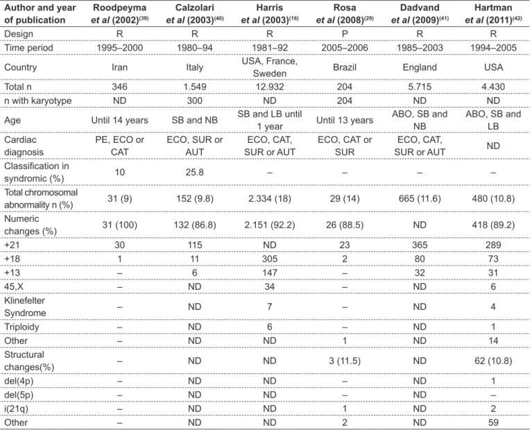
According to Table 1(16,19,29,33-42), there was no study that
assessed all patients of the same way. They were characterized by being, most of them, retrospective, developed in Eu-rope, and not all patients of the samples studied underwent
Author and year of publication
Ferencz et al
(1989)(33)
Stoll et al
(1989)(19)
Pradat (1992)(34)
Hanna et al
(1994)(35)
Goodship
et al (1998)(36)
Grech e Gatt (1999)(37)
Meberg et al
(2000)(38)
Design R R R R P R R
Time period 1981–86 1979–86 1981–86 1974–78 1994–95 1990–94 1982–96 Country United States France Sweden Northern
Ireland England Malta Norway Total n 2.102 801 1.605 388 207 231 360 n with karyotype ND 153 ND ND 173 ND ND Age Until 1 year ABO, LB and
SB
SB and LB
until 6 months Until 7 years Children Until 1 year Until 18 years Cardiac
diagnosis
ECO, CAT, SUR or AUT
ECO, CAT, SUR or AUT
ECO, CAT, SUR or AUT
CAT, SUR or
AUT ND
ECO, CAT, SUR or AUT
ECO, CAT, SUR or AUT
Classiication in
syndromic (%) – – – – – 11 –
Total chromosomal
abnormality n (%) 271 (12.9) 72 (9) 202 (13) 12 (3) 21 (12.1) 21 (9) 24 (6.7) Numeric
changes (%) 259 (95.6) 69 (95.8) ND ND 19 (90.5) 20 (95.2) 20 (83.3)
+21 ND ND ND ND 15 ND 15
+18 17 15 15 ND 1 1 3
+13 16 7 6 ND – – –
45,X 7 1 ND ND 2 – 1
Klinefelter
Syndrome – – ND ND – – –
Triploidy – – – ND – – –
Other 1 46 ND ND 1 19 1
Structural
changes(%) 12 (4.4) 3 (4.2) ND ND 2 (9.5) 1 (4.8) 4 (16.7)
del(4p) ND 1 ND ND – – 2
del(5p) ND 1 ND ND – – 1
i(21q) ND – ND ND – – –
Other ND 1 ND ND 2 1 1
karyotype examination (most of the studies does not describe how many patients were assessed through this exam). We obtained only one study conducted in Latin America, in Brazil(29). The sample sizes of the studies are also variable,
being smaller on those developed prospectively(29,36).
The age of analyzed patients also varied greatly. Some studies included spontaneous abortions and stillbirths. The top age limit observed was 18 years(38). As for cardiac
evalu-ation of patients, in most studies, there was a report of echo-cardiography, cardiac catheterization, surgery, and autopsy. Despite the classiication of syndromic or not observed in
some studies, there was no data describing the performance of dysmorphologic physical examination by a clinical geneti-cist. The classiication of patients in syndromic ranged from 10 to 25.8% of the samples analyzed(16,19,29,33-42) (Table 1).
The frequency of chromosome abnormalities detected by karyotype in patients with congenital heart disease ranged from 3 to 23% (usually around 9%)(16,19,29,33-42) (Table 1).
Thus, they are present in about one in every 10 patients with congenital heart disease, i.e., their frequency is about 12 times greater among individuals with congenital heart disease than in the general population, for which the rate Author and year
of publication
Roodpeyma
et al (2002)(39)
Calzolari
et al (2003)(40)
Harris
et al (2003)(16)
Rosa
et al (2008)(29)
Dadvand
et al (2009)(41)
Hartman
et al (2011)(42)
Design R R R P R R
Time period 1995–2000 1980–94 1981–92 2005–2006 1985–2003 1994–2005 Country Iran Italy USA, France,
Sweden Brazil England USA Total n 346 1.549 12.932 204 5.715 4.430
n with karyotype ND 300 ND 204 ND ND
Age Until 14 years SB and NB SB and LB until
1 year Until 13 years
ABO, SB and NB
ABO, SB and LB Cardiac
diagnosis
PE, ECO or CAT
ECO, SUR or AUT
ECO, CAT, SUR or AUT
ECO, CAT or SUR
ECO, CAT,
SUR or AUT ND
Classiication in
syndromic (%) 10 25.8 – – – –
Total chromosomal
abnormality n (%) 31 (9) 152 (9.8) 2.334 (18) 29 (14) 665 (11.6) 480 (10.8) Numeric
changes (%) 31 (100) 132 (86.8) 2.151 (92.2) 26 (88.5) ND 418 (89.2)
+21 30 115 ND 23 365 289
+18 1 11 305 2 80 73
+13 – 6 147 – 32 31
45,X – ND 34 – ND 6
Klinefelter
Syndrome – ND 7 – ND 4
Triploidy – ND 6 – ND 1
Other – ND ND 1 ND 14
Structural
changes(%) – ND ND 3 (11.5) ND 62 (10.8)
del(4p) – ND ND – ND 1
del(5p) – ND ND – ND –
i(21q) – ND ND 1 ND 2
Other – ND ND 2 ND 59
R: retrospective; P: prospective; ND: not described; ABO: abortions; LB: live births; SB: stillborn; NB: newborn; PE: physical examination; ECO: echocardiography; CAT: catheterism; SUR: surgery; AUT: autopsy; -: absent; +21: full trisomy of chromosome 21; +18: full trisomy of chromosome 18; +13: full trisomy of chromosome 13; 45,X: chromosome X monosomy; del(4p): deletion of the short arm of chromosome 4; del(5p): deletion of the short arm of chromosome 5; i(21q): Down syndrome secondary to isochromosome of the long arm of chromosome 21.
is one in every 120 newborns(18). The major chromosomal
changes observed are numeric and correspond to the ad-ditional presence or lack of a chromosome. These were the irst genetic abnormalities to be described in patients with congenital heart disease(17) and usually account for over 80%
of the abnormalities observed(16,19,29,33-42). Among them,
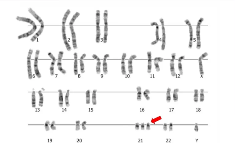
stands out the full trisomy of chromosome 21 (+21), the main chromosome constitution observed in individuals with Down syndrome (Figure 1). Another relatively common change was the full trisomy of chromosome 18 (+18), respon-sible for Edwards syndrome. Recurrent, but less frequent abnormalities consisted of full trisomy 13 (Patau syndrome), chromosome X monosomy (Turner syndrome), Klinefelter syndrome, and the triploidy syndrome(16,19,29,33-42) (Table 1).
A smaller percentage of chromosomal abnormalities ob-served in patients with congenital heart disease consisted of structural abnormalities. The main abnormalities correspond to those with loss (deletion) or gain (duplication) of part of a chromosome. Among them, stand out the deletion of the short arm of chromosome 4 (Wolf-Hirschhorn syndrome)
and of chromosome 5 (Cri-du-Chat syndrome). The isochro-mosome of the long arm (chroisochro-mosome with loss of short arm and duplication of the long arm) of chromosome 21, less common cause of Down syndrome, was also frequently described (16,19,29,33-42) (Table 1 and Figure 2).
The chromosomal abnormalities most frequently observed and cited are characterized by having a high percentage of cardiac involvement. For instance, the frequency of con-genital heart disease among individuals with Edwards and Patau syndrome ranges from 80 to 100%(43-46). Furthermore,
about 40 to 50% of patients with Down’s syndrome have this defect(16,17,20) (Table 2). Another important feature is
the relationship of certain chromosomal abnormalities with speciic heart defects. Down syndrome, for instance, shows association with atrioventricular septal defects(47,48); and
Edwards and Patau syndromes, with septal defects, such as interventricular and atrial communication. The polyvalvular disease is also common among these individuals(43-46).
Pa-tients with Turner syndrome have more often bicuspid aortic valve and coarctation of the aorta(49). The 22q11 deletion has
Figure 2 - Partial karyotype by GTG-banding (trypsin-Giemsa G-band) and ideograms showing, respectively, a Robertsonian translocation between chromosomes 14 and 21 [der(14;21)], one isochromosome of the long arm (q) of chromosome 21 [i(21q)]
and one partial interstitial deletion of long arm of chromosome 5 [del(5p)]. The irst two forms represent changes associated with
Down syndrome, and the third, to the Cri-du-Chat syndrome
great association with defects involving the outlow tract of the heart (conotruncal heart defects), such as interrupted aortic arch type B, truncus arteriosus, and the tetralogy of Fallot(16,17,20,29-32) (Table 2).
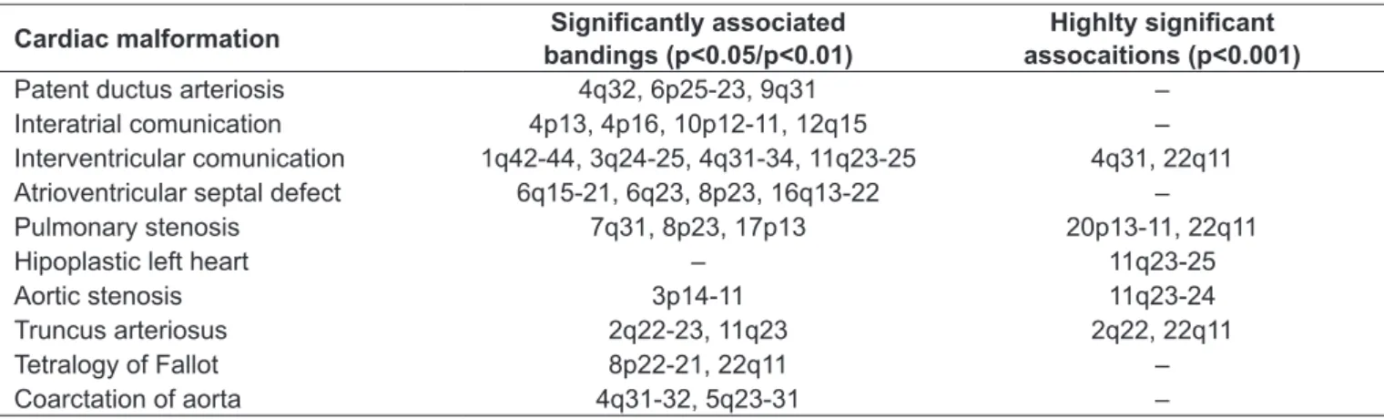
On the other hand, some types of heart defects showed a greater association with chromosomal abnormalities (Table 3)
(50). Among them, we highlight the atrioventricular septal defect
(frequency greater than 50%, mainly due to Down syndrome), as well as interrupted aortic arch type B, truncus arteriosus, and tetralogy of Fallot (as already mentioned, they are rather associ-ated with 22q11 deletion) (Table 3)(50). The involvement of some
chromosomal regions due to deletions and duplications is well reported in the literature, according to Table 4(51).
Importance of identifying chromosomal abnormalities in patients with congenital heart disease
As already mentioned, around 15 to 20% of patients with congenital heart disease present known etiology, and chromosomal abnormalities identiied by karyotype stand
out(5,18). These are common in individuals with congenital
heart disease, with a frequency of 3 to 23%, which highlights the importance of karyotyping for this population(16,19,29,33-42).
Individuals with chromosomal disorders usually have an aspect that is considered syndromic, i.e., they present a scenario of dysmorphias, both major and minor, associated with other disabilities (such as intellectual) and behav-ioral changes. These dysmorphic features can be identiied through a physical examination (dysmorphologic examina-tion) conducted by a trained professional, such as the clinical geneticist or even a pediatrician with experience. Therefore, this professional is vital for the best choice of individuals to undergo genetic evaluation by examining the karyotype. Patients with chromosomal abnormalities often have associated extracardiac malformations, and are therefore at a higher risk of morbidity and mortality, which makes the cardiac surgery even riskier(20,35,52,53). Moreover, such
*Described percentage of congenital heart defects in patients with the respective chromossolam abnormality; ♀: females; ?: unknown; IVC: inter
-ventricular communication; AVSD: atrio-ventricular septal defect; PDA: patent ductus arteriosus; IAC: interatrial communication; TOF: tetralogy of Fallot; CoAo: Coarctation of the aorta; PS: pulmonary stenosis; DORV: double outlet right ventricle; BAV: bicuspid aortic valve; LV: left ventricle; MVP: mitral valve prolapse; AS: aortic stenosis; LSVC: left superior vena cava; TGA: transposition of the great arteries; TAPVR: total anomalous pulmonary venous return; TrA: tricuspid atresia; IVC: inferior vena cava; IAA: interruption of aortic arch; TA: truncus arteriosus; PA: pulmonary atresia
Syndrome Chromosome alterations Incidence at birth
% with heart
defects* Associated heart defects
Down Trisomy of chromosome 21 40–50 CIV, DSAV, PCA, CIA e TOF
Edwards Trisomy of chromosome 18 1:3.600–8.500 80–100
CIV, CIA, PCA, CoAo, EP, DVSVD, TOF, doença
polivalvular e DSAV
Patau Trisomy of chromosome 13 1:5.000–12.000 80–100 CIA, CIV, PCA, doença
polivalvular e dextrocardia
Ullrich-Turner Partial or total monosomy of
Chromosome X 1:2.000–3.000 (♀) 17–60
CoAo, VAB, hipoplasia de VE, PVM e EA
Mosaic trisomy of chromosome 9
Mosaicism for chromosome 9
trisomy ±30 cases described 60
CIA, CIV, PCA, DVSVD e persistência VCSE
Triploidy Triploidy ? 50 CIV, PCA e CIA
Wolf-Hirschorn Partial deiciency of 4p 1:50.000 30–60 CIA, CIV e EP
Cri-du-chat (cat’s cry) Partial deiciency of 5p 1:50.000 10–55 CIV, CIA, PCA e TOF
Deletion 8p Partial deiciency of 8p ±40 cases described 65–75 CIV, EP, CIA e TGA
Deletion 9p Partial deiciency of 9p ±100 cases described 30–65 CIV, PCA e EP
Jacobsen Partial deiciency of 11q ±75 cases described 65 CIV, Ventrículo único,
hipoplasia de VE e CIA
Duplication11q Partial duplication of 11q ? 60 Variável
Cat-eye syndrome Tetrasomy or partial trisomy in
chromosome 2222 1:50.000–150.000 30–40
Persistência VCSE, DVAPT, TOF, CIV, AT e
ausência de VCI
Pallister-Killian Mosaic tetrasomy for the short
arm in chromosome 12 ? 25
CIV, EA, PCA e cardiomiopatia hipertróica
Deletion 22q11.2 Deletion 22q11.2 1:2.000 75–100
TOF, IAA do tipo B,
TA, CIV, AP, EP e
arco aórtico à direita Table 2 - Main chromosomal abnormalities associated to cardiac malformations potentially detected through karyotype examination. Based on Marino and Digilio (2000)(20), Harris et al (2003)(16), and Fahed et al (2013)(17)
is usually need for multidisciplinary assessment and moni-toring, involving not only areas of Cardiology and Medical Genetics. It is also worth noting that some chromosomal abnormalities such as trisomy 13 (Patau syndrome) and trisomy 18 (Edwards syndrome), are associated with a very poor prognosis, and the literature discusses whether patients might actually beneit from heart surgery(44,45). Therefore, all
this information is critical for the patient’s proper manage-ment and risk/prognosis assessmanage-ment.
The importance of establishing an accurate diagnosis of the etiology of congenital heart disease also lies in the fact that families need genetic counseling with accurate information about the risks of recurrence(21). Older studies on the
recur-rence of congenital heart disease suggested multifactorial
inheritance(20), because they simply measured familial
Cardiac Malformation Associated risk (%) Cromossome abnormality
Tetralogy of Fallot
6–10 +21
10–15 +13; +18
10–19 deletion 22q11
Tetralogy of Fallot with pulmonary atresia 26 deletion 22q11
Interrupted aortic arch 25–50 deletion 22q11
Truncus arteriosus 40 deletion 22q11
Tetralogy of Fallot with absent pulmonary valve >60 deletion 22q11
Double outlet right ventricle 5 +13; +18
Coarctation of aorta 10 Turner Syndrome
Atrioventricular septal defect (complete or partial) >50 +21; +13; +18
Pulmonary atresia with interventricular communcation 22 deletion 22q11
Pulmonary atresia with ventricular septal defect and major
aortopulmonary communicating arteries 35 deletion 22q11
Interventricular communcation outlet 20 +21
Perimembranosa IVC 20 +21; +18
Doubly-committed interventricular communication 20 deletion 22q11
Table 3 - Cardiac malformations and their association with chromosomal abnormalities. Adapted from Manning et al(50)
+21: chromosome 21 full trisomy; +13: chromosome 13 full trisomy; +18: chromosome 18 full trisomy
Cardiac malformation Signiicantly associated bandings (p<0.05/p<0.01)
Highlty signiicant assocaitions (p<0.001)
Patent ductus arteriosis 4q32, 6p25-23, 9q31 –
Interatrial comunication 4p13, 4p16, 10p12-11, 12q15 –
Interventricular comunication 1q42-44, 3q24-25, 4q31-34, 11q23-25 4q31, 22q11
Atrioventricular septal defect 6q15-21, 6q23, 8p23, 16q13-22 –
Pulmonary stenosis 7q31, 8p23, 17p13 20p13-11, 22q11
Hipoplastic left heart – 11q23-25
Aortic stenosis 3p14-11 11q23-24
Truncus arteriosus 2q22-23, 11q23 2q22, 22q11
Tetralogy of Fallot 8p22-21, 22q11 –
Coarctation of aorta 4q31-32, 5q23-31 –
Table 4 - Regions of chromosome deletion statistically signiicant associated to speciic heart malformations. Adapted from Brewer et al(51)
q: log arm of chromosome; p: short arm of chromosome
hypothesis of one of them carrying a balanced chromosomal abnormality related to that observed in the child(18).
It is worth noting, however, that the result of a traditional karyotype test does not exclude the fact that the patient might still present a syndrome. As shown previously, micro-scopic changes (such as microdeletions or microduplications) or gene mutations are not detected by this test. In such cases, clinical evaluation of the patient, especially by the geneticist, is essential to generate hypotheses and therefore choose appropriate tests for diagnosis.
Based on this review, authors believe that an accurate dysmorphologic examination, performed by an experienced
pediatrician or by a geneticist, is rather important to indi-cate the karyotype in patients with congenital heart disease. This would help both to save costs with the exam and to the early identiication of patients with chromosomal ab-normalities, which might relect in better supervision and genetic counseling.
Acknowledgements
References
1. Robinson A, Linden MG. Clinical genetics handbook. 2nd ed. Boston: Blackwell
Science Inc; 1994.
2. Amorim LF, Pires CA, Lana AM, Campos AS, Aguiar RA, Tibúrcio JD et al. Presentation of congenital heart disease diagnosed at birth: analysis of 29,770 newborn infants. J Pediatr (Rio J) 2008;84:83-90.
3. Kramer HH, Majewski F, Trampisch HJ, Rammos S, Bourgeois M. Malformation patterns in children with congenital heart disease. Am J Dis Child 1987;141:789-95.
4. Hoyme HE. Minor anomalies: diagnostic clues to aberrant human morphogenesis. Genetica 1993;89:307-15.
5. Van der Bom T, Zomer AC, Zwinderman AH, Meijboom FJ, Bouma BJ, Mulder BJ. The changing epidemiology of congenital heart disease. Nat Rev Cardiol 2011;8:50-60.
6. Hoffman JI, Kaplan S. The incidence of congenital heart disease. J Am Coll Cardiol 2002;39:1890-900.
7. Pinto Junior VC, Daher CV, Sallum FS, Jatene MB, Croti UA. The situation of congenital heart surgeries in Brazil. Braz J Cardiovasc Surg 2004;19:3-6. 8. Ransom J, Srivastava D. The genetics of cardiac birth defects. Semin Cell Dev Biol
2007;18:132-9.
9. Guitti JC. Epidemiological characteristics of congenital heart diseases in Londrina, Paraná south Brazil. Arq Bras Cardiol 2000;74:395-404.
10. Hagemann LL, Zielinsky P. Rastreamento populacional de anormalidades cardíacas fetais por ecocardiograia pré-natal em gestações de baixo risco no município de Porto Alegre. Arq Bras Cardiol 2004;82:313-9.
11. Acharya G, Sitras V, Maltau JM, Dahl LB, Kaaresen PI, Hanssen TA et al. Major congenital heart disease in Northern Norway: shortcomings of pre- and postnatal diagnosis. Acta Obstet Gynecol Scand 2004;83:1124-9.
12. Jenkins KJ, Correa A, Feinstein JA, Botto L, Britt AE, Daniels SR et al. Noninherited risk factors and congenital cardiovascular defects: current knowledge: a scientiic statement from the American Heart Association Council on Cardiovascular Disease in the Young: endorsed by the American Academy of Pediatrics. Circulation 2007;115:2995-3014.
13. Pedra CA, Haddad J, Pedra SF, Peirone A, Pilla CB, Marin-Neto JA. Paediatric and congenital heart disease in South America: an overview. Heart 2009;95:1385-92. 14. Dolk H, Loane M, Garne E; European Surveillance of Congenital Anomalies
(EUROCAT) Working Group. Congenital heart defects in Europe: prevalence and perinatal mortality, 2000 to 2005. Circulation 2011;123:841-9.
15. Thulstrup AM, Bonde JP. Maternal occupational exposure and risk of speciic birth defects. Occup Med (Lond) 2006;56:532-43.
16. Harris JA, Francannet C, Pradat P, Robert E. The epidemiology of cardiovascular defects, part 2: a study based on data from three large registries of congenital malformations. Pediatr Cardiol 2003;24:222-35.
17. Fahed AC, Gelb BD, Seidman JG, Seidman CE. Genetics of congenital heart disease: the glass half empty. Circ Res 2013;112:707-20.
18. Blue GM, Kirk EP, Sholler GF, Harvey RP, Winlaw DS. Congenital heart disease: current knowledge about causes and inheritance. Med J Aust 2012;197:155-9. 19. Stoll C, Alembik Y, Roth MP, Dott B, De Geeter B. Risk factors in congenital heart
disease. Eur J Epidemiol 1989;5:382-91.
20. Marino B, Digilio MC. Congenital heart disease and genetic syndromes: speciic correlation between cardiac phenotype and genotype. Cardiovasc Pathol 2000;9:303-15.
21. Prasad C, Chudley AE. Genetics and cardiac anomalies: the heart of the matter. Indian J Pediatr 2002;69:321-32.
22. 22. Tjio JH, Levan A. The chromosome number of man. Hereditas 1956;42:1-6. 23. Lejeune J, Gautier M, Turpin R. Étude des chromosomes somatiques de neuf
enfants mongoliens. C R Hebd Seances Acad Sci 1959;248:1721-2.
24. Caspersson T, Zech L, Johansson C. Differential binding of alkylating luorochromes in human chromosomes. Exp Cell Res 1970;60:315-9.
25. Yunis JJ. New chromosomes techniques in the study of human neoplasia. Hum Pathol 1981;12:540-9.
26. Smeets DF. Historical prospective of human cytogenetics: from microscope to microarray. Clin Biochem 2004;37:439-46.
27. Jauch A, Daumer C, Lichter P, Murken J, Schroeder-Kurth T, Cremer T. Chromosomal in situ suppression hybridization of human gonosomes and autosomes and its use
28. Bejjani BA, Shaffer LG. Clinical utility of contemporary molecular cytogenetics. Annu Rev Genomics Hum Genet 2008;9:71-86.
29. Rosa RF, Pilla CB, Pereira VL, Flores JA, Golendziner E, Koshiyama DB et al. 22q11.2 deletion syndrome in patients admitted to a cardiac pediatric intensive care unit in Brazil. Am J Med Genet A 2008;146A:1655-61.
30. Rosa RF, Zen PR, Roman T, Graziadio C, Paskulin GA. 22q11.2 deletion syndrome: catching the CATCH22. Rev Paul Pediatr 2009;27:211-20.
31. Rosa RF, Trevisan P, Koshiyama DB, Pilla CB, Zen PR, Varella-Garcia M et al. 22q11.2 deletion syndrome and complex congenital heart defects. Rev Assoc Med Bras 2011;57:62-5.
32. Rosa RFM, Zen PRG, Graziadio C, Paskulin GA. Síndrome de deleção 22q11.2 e cardiopatias congênitas. Rev Paul Pediatr 2011;29:251-60.
33. Ferencz C, Neill CA, Boughman JA, Rubin JD, Brenner JI, Perry LW. Congenital cardiovascular malformations associated with chromosome abnormalities: an epidemiologic study. J Pediatr 1989;114:79-86.
34. Pradat P. Epidemiology of major congenital heart defects in Sweden, 1981-1986. J Epidemiol Community Health 1992;46:211-5.
35. Hanna EF, Nevin NC, Nelson J. Genetic study of congenital heart defects in Northern Ireland (1974-1978). J Med Genet 1994;31:858-63.
36. Goodship J, Cross I, Liling J, Wren C. A population study of chromosome 22q11 deletions in infancy. Arch Dis Child 1998;79:348-51.
37. Grech V, Gatt M. Syndromes and malformations associated with congenital heart disease in a population-based study. Int J Cardiol 1999;68:151-6.
38. Meberg A, Otterstad JE, Frøland G, Lindberg H, Sørland SJ. Outcome of congenital heart defects — a population-based study. Acta Paediatr 2000;89:1344-51. 39. Roodpeyma S, Kamali Z, Afshar F, Naraghi S. Risk factors in congenital heart
disease. Clin Pediatr 2002;41:653-8.
40. Calzolari E, Garani G, Cocchi G, Magnani C, Rivieri F, Neville A et al. Congenital heart defects: 15 years of experience of the Emilia-Romagna Registry (Italy). Eur J Epidemiol 2003;18:773-80.
41. Dadvand P, Rankin J, Shirley MD, Rushton S, Pless-Mulloli T. Descriptive epidemiology of congenital heart disease in Northern England. Paediatr Perinat Epidemiol 2009;23:58-65.
42. Hartman RJ, Rasmussen SA, Botto LD, Riehle-Colarusso T, Martin CL, Cragan JD et al. The contribution of chromosomal abnormalities to congenital heart defects: a population-based study. Pediatr Cardiol 2011;32:1147-57.
43. Rosa RF, Rosa RC, Lorenzen MB, de Moraes FN, Graziadio C, Zen PR et al. Trisomy 18: experience of a reference hospital from the south of Brazil. Am J Med Genet A. 2011;155A:1529-35.
44. Rosa RF, Rosa RC, Lorenzen MB, de Oliveira CA, Graziadio C, Zen PR et al. Trisomy 18: frequency, types, and prognosis of congenital heart defects in a Brazilian cohort. Am J Med Genet A 2012;158A:2358-61.
45. Petry P, Polli JB, Mattos VF, Rosa RC, Zen PR, Graziadio C et al. Clinical features and prognosis of a sample of patients with trisomy 13 (Patau syndrome) from Brazil. Am J Med Genet A 2013;161:1278-83.
46. Rosa RF, Rosa RC, Zen PR, Graziadio C, Paskulin GA. Trisomy 18: review of the clinical, etiologic, prognostic, and ethical aspects. Rev Paul Pediatr 2013;31:111-20. 47. Nisli K. Prevalence of congenital heart defects in patients with Down’s syndrome.
J Pediatr (Rio J) 2009;85:377-8.
48. Vilas Boas LT, Albernaz EP, Costa RG. Prevalence of congenital heart defects in patients with Down syndrome in the municipality of Pelotas, Brazil. J Pediatr (Rio J) 2009;85:403-7.
49. Carvalho AB, Guerra Júnior G, Baptista MT, de Faria AP, Marini SH, Guerra AT. Cardiovascular and renal anomalies in Turner syndrome. Rev Assoc Med Bras 2010;56:655-9.
50. Manning N, Kaufman L, Roberts P. Genetics of cardiological disorders. Semin Fetal Neonatal Med 2005;10:259-69.
51. Brewer C, Holloway S, Zawalnyski P, Schinzel A, FitzPatrick D. A chromosomal deletion map of human malformations. Am J Hum Genet 1998;63:1153-9. 52. Begić H, Tahirović H, Mesihović-Dinarević S, Ferković V, Atić N, Latifagić A.
Epidemiological and clinical aspects of congenital heart disease in children in Tuzla Canton, Bosnia-Herzegovina. Eur J Pediatr 2003;162:191-3.

![Figure 2 - Partial karyotype by GTG-banding (trypsin-Giemsa G-band) and ideograms showing, respectively, a Robertsonian translocation between chromosomes 14 and 21 [der(14;21)], one isochromosome of the long arm (q) of chromosome 21 [i(21q)]](https://thumb-eu.123doks.com/thumbv2/123dok_br/19058523.485521/7.892.62.831.561.1007/karyotype-ideograms-respectively-robertsonian-translocation-chromosomes-isochromosome-chromosome.webp)