Cop
yright
© ABE&M t
odos os dir
eit
os r
eser
vados
.
Arq Bras Endocrinol Metab. 2011;55/8 628
original article
1 Pediatric Endocrinology Unit,
Department of Pediatrics, Irmandade da Santa Casa de Misericórdia de São Paulo (ISCMSP), São Paulo, SP, Brazil
2 Department of Physiology,
Faculdade de Ciências Médicas da Santa Casa de São Paulo (FCMSC-SP), São Paulo, SP, Brazil
Correspondence to: Cláudia Dutra Costantin Faria Faculdade de Ciências Médicas da Santa Casa de São Paulo Rua Dr. Cesário Mota Junior, 112 01221-020 – São Paulo, SP, Brazil ceclau@terra.com.br
Received on 4/Oct/2011 Accepted on 10/Nov/2011
TSH neurosecretory dysfunction
(TSH-nd) in Down syndrome
(DS): low risk of progression
to Hashimoto’s thyroiditis
Disfunção neurossecretora de TSH na síndrome de Down: baixo risco de progressão para a tireoidite de Hashimoto
Claudia Dutra Costantin Faria1, Simone Ribeiro1, Cristiane Kochi1,2, Aryane Pereira Neves da Silva2, Bruna Natalia Freire Ribeiro2, Lilian Teixeira Marçal2, Felipe Henrique Yyazawa Santos2, Calliari Procópio Luis Eduardo1, Osmar Monte, Carlos Alberto Longui1,2
ABSTRACT
Introduction: Patients with Down syndrome (DS) often have elevated TSH (hypothala-mic origin), which is called TSH neurosecretory dysfunction (TSH-nd). In these cases, there is slight elevation in TSH (5-15 μUI/mL), with normal free T4 and negative thyroid antibodies (AB). Objective: To recognize the risk of progression to Hashimoto’s thyroiditis (HT). Sub-jects and methods: We retrospectively analyzed 40 DS patients (mean age = 4.5 years), followed up for 6.8 years. Results: HT was diagnosed in 9/40 patients, three early in moni-toring, and six during evolution. In 31/40 patients, TSH-nd diagnosis remained unchan-ged over the years, with maximum TSH values ranging from 5 to 15 μUI/mL. In this group, free T4 also remained normal and AB were negative. There was a signiicant TSH reduction (p = 0.017), and normal TSH concentrations (< 5.0 μUI/mL) were observed in 29/31 patients, in at least one moment. No patient had TSH > 15 μUI/mL. Conclusion: DS patients with TSH-nd pre-sent low risk of progression to HT (10% for females and 6% for males). Arq Bras Endocrinol Metab. 2011;55(8):628-31
Keywords
Down syndrome; Hashimoto’s thyroiditis; TSH neurosecretory dysfunction; isolated TSH elevation
RESUMO
Introdução: Pacientes com síndrome de Down (SD) geralmente apresentam TSH elevado (de origem hipotalâmica), uma desordem chamada de disfunção neurossecretora de TSH (TSH--nd). Nesses casos, há uma leve elevação do TSH (5-15 μUI/mL), com T4 livre normal e anticor-pos antitireoide (AB) negativos. Objetivo: Reconhecer o risco de progressão para a tireoidite de Hashimoto (HT). Sujeitos e métodos: Analisamos retrospectivamente 40 pacientes com SD (idade média = 4,5 anos), acompanhados por 6,8 anos. Resultados: A HT foi diagnosticada em 9/40 pacientes, três logo no início da avaliação e seis durante a evolução. Em 31/40 dos pacientes, o diagnóstico de TSH-nd permaneceu estável durante os anos, com valores máxi-mos de TSH variando de 5 a 15 μUI/mL. Neste grupo, o T4 livre também permaneceu normal e os AB foram negativos. Houve uma redução signiicativa do TSH (p = 0,017), e concentrações normais de TSH (< 5,0 μUI/mL) foram observadas em 29/31 pacientes, em pelo menos um mo-mento. Nenhum paciente apresentou TSH > 15 μUI/mL. Conclusão: Pacientes com SD e TSH-nd apresentam baixo risco de progressão para a HT (10% para o sexo feminino e 6% para o sexo masculino). Arq Bras Endocrinol Metab. 2011;55(8):628-31
Descritores
Cop
yright
© ABE&M t
odos os dir
eit
os r
eser
vados
.
629 Arq Bras Endocrinol Metab. 2011;55/8
Elevated TSH in Down syndrome
INTRODUCTION
D
own syndrome (DS) is a common chromosomal abnormality and the most common genetic cause of mental retardation (1). Autoimmune diseases, es-pecially Hashimoto’s thyroiditis (HT), are frequently observed in patients with DS. In these situations, auto-antibodies (AB) directed against the thyroid are found in 13% to 34% of patients (2). Therefore, it is suggested that thyroid function, including measurement of TSH, free T4 and AB, is performed as part of routine moni-toring of DS patients (3-4). Thyroid hormones have im-portant functions in the central nervous system (CNS). They are involved in neuronal migration and differentia-tion, neurotransmitter synthesis and secredifferentia-tion, myelina-tion, and regulation of gene expression in neuronal cells (5). Hypothyroidism is a potentially aggravating factor of neurological abnormalities of DS patients (5).In addition to primary thyroid disease, patients with DS may present inadequate dopaminergic regulation of pituitary TSH secretion (6). These changes determi-ne an abnormal secretion pattern, resulting in isolated TSH elevation without changes in thyroid hormones (7). In TSH neurosecretory dysfunction (TSH-nd), TSH concentrations are slight elevated (5-10 μUI/ mL), free T4 values are normal, and AB are negative. In most cases, there is no detectable anatomic abnor-mality, and the etiology is not identiied. Frequency presentation is similar in males and females (7).
Despite the absence of alteration in thyroid hormones, it is questionable whether TSH-nd may reduce growth velocity in these children (8). In these cases, there is no consensus on levothyroxine replacement therapy (9).
Cutler and cols. suggested that isolated TSH eleva-tion could be a sign of risk of progression to primary autoimmune hypothyroidism, even in the absence of detectable anti-thyroid AB (10).
The aim of this study was to identify a risk of pro-gression to HT, by means of a follow up of DS patients with TSH-nd.
SUBJECTS AND METHODS
This study was based on the retrospective analysis of 40 patients with DS (mean age = 4.5 years), both females and males, followed up at the Pediatric Endocrinology Department Unit of ISCMSP, referred to us because of previous elevated TSH and/or positive anti-thyroid AB. The study was approved by the Ethics Committee in Human Research of the ISCMSP.
The studied variables were: weight (kg), height (cm), pubertal characteristics (Tanner score); T4, free T4, TSH, anti-thyroperoxidase and anti-thyroglobulin concentrations and, for some patients, thyroid ultrasound. Weight was measured on an analog scale (Filizola®), and
height, on a wall-mounted stadiometer (Gomes & Tonelli®).
Height and BMI were expressed as standard devia-tion z scores (SDS), calculated from reference values of SD patients (11).
T4, free T4 and TSH were measured by luorome-tric assays (AutoDELFIA automatic immunoassay sys-tem). Anti-thyroperoxidase and anti-thyroglobulin AB were quantiied by an immunometric assay (IMMULI-TE 2000). Negative results were below 35 IU/mL and 40 IU/mL, respectively.
TSH-nd diagnosis was established when TSH values were slightly elevated (5-15 μUI/mL), T4 and free T4 concentrations were normal, and thyroid AB were negative.
Statistical analysis used SigmaStat for Windows, v.2.03 (SPSS, Chicago, Il, USA). Comparisons of ini-tial and inal values for height SDS, BMI SDS, T4, free T4 and TSH of the same patient were performed by Paired T test. Statistical signiicance was determined by p < 0.05.
RESULTS
Forty DS patients (mean age = 4.5 years) were con-sidered for retrospective evaluation. Three patients (3/40) were initially excluded because they had symp-toms compatible with primary hypothyroidism already at diagnosis.
During follow-up, 6/37 patients (4F: 2M) initially considered as having TSH-nd presented progression to HT diagnosis, without hypothyroidism. Anti-thyroid AB were positive after a follow-up period ranging from 8 to 28 months.
TSH-nd diagnosis remained unchanged in thirty one individuals (12F: 19M). Median follow-up period of this subgroup was 6.8 years (1.0 - 20.2). Mean age at irst visit was 3.4 years (SDS = 3.7), ranging from 0.1 to 14.3 years. Table 1 summarizes initial and inal des-criptive data, including anthropometric measurements and laboratory results.
Cop
yright
© ABE&M t
odos os dir
eit
os r
eser
vados
.
630 Arq Bras Endocrinol Metab. 2011;55/8
Elevated TSH in Down syndrome
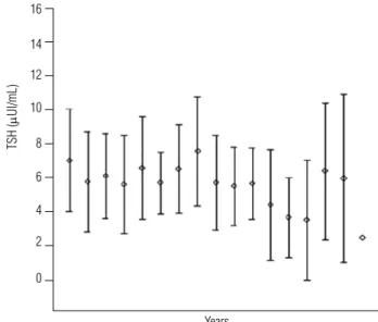
When analyzed individually, TSH peak values were sli-ghtly elevated (mean = 9.7 μUI/mL, ranging from 5.4 to 15.0). There was a wide variation in TSH, with 29/31 patients (93.5%) showing normal values in at least once during follow-up. Eleven patients (11/31) had at least one TSH measurement between 10 and 15 μUI/mL. Only one patient had TSH = 15 μUI/mL, although this value was observed only once during the follow-up period of 5.4 years. Figure 1 represents TSH evolution in the subgroup of DS patients with TSH-nd during follow-up.
Table 1. Initial and final assessment of anthropometric measurements and laboratory results of patients with DS and TSH-nd
Initial values (N = 31) Mean (SDS)
Range Median
Final values (N = 31) Mean (SDS)
Range Median
P value
Height SDS -0.6 (1.2)
-2.7 – 3.0 -0.7
-0.9 (0.9) -3.0 – 0.9 -0.8
0.13
BMI SDS -0.3 (1.4)
-3.3 – 2.2 0.02
0.6 (1.2) -1.4 – 3.5
0.5
0.003a
T4 (µd/dL) 9.9 (1.9)
5.3 – 14.0 9.5
9.6 (1.3) 7.4 – 11.7
9.8
0.49
Free T4 (ng/dL) 1.4 (0.2)
0.7 – 1.9 1.4
1.4 (0.3) 0.9 – 2.5 1.3
0.42
TSH (µU/mL) 7.1 (3.0)
0.8 – 13.7 6.8
5.5 (2.6) 1.7 – 11.5
5.0
0.017b
Height SDS: z score standard deviation of height; BMI SDS: z score standard deviation of body
mass index; a: BMI SDS final values vs. BMI SDS initial values (Paired T test); b: TSH final mean
values vs. TSH initial mean values (Paired T test).
Figure 1. TSH evolution during follow-up of DS patients with TSH-nd.
TSH (
m
Ul/mL)
16
14
12
10
8
6
4
2
0
Years
Also in the TSH-nd subgroup, no patient developed hypothyroidism. By analyzing T4 and free T4 concentra-tions, the lowest mean individual values were respective-ly: 7.6 mg/dL (5.3 to 10.9) and 1.1 ng/dL (0.7 to 1.7).
Thyroid ultrasound was performed in 16/40 patients. In 5/9 patients diagnosed with HT, ultrasound results showed only heterogeneous texture. In 11/31 patients with TSH-nd, all results were considered normal.
DISCUSSION
The close relationship between thyroid disorders and DS is well-documented (12). Considering the total universe of patients with DS, the frequency of autoim-mune thyroid disease is described to be between 20% and 66%, depending on the type of study, sample size, region, age group and the inclusion of hypo-or hyper-thyroidism cases (13-14). It is possible that previous studies also included DS patients with TSH-nd, wi-thout TH and wiwi-thout hypothyroidism, producing a large number of false positive results.
In this study, long-term monitoring showed positi-ve AB in only 6/37 patients (16%) in a period ranging from 8 to 28 months of observation. From this total, about 10% were represented by females and 6% by ma-les. These results corroborate literature data, which show a higher frequency of HT in girls than in pre--pubertal boys (2:1) (15).
For most patients (31/37) analyzed in this study, there were only intermittent and isolated elevations of TSH without thyroid disease progression. For all these patients, anti-thyroid AB were not detected, values of free T4 remained normal, and there was no reduction in longitudinal growth over a mean period of 6.8 years. These long-term observations suggest that isolated ele-vation of TSH in DS does not seem to be a condition that predisposes to the development of thyroid disease, since in these cases, normal TSH values were often ob-served at some point of monitoring.
Cop
yright
© ABE&M t
odos os dir
eit
os r
eser
vados
.
631 Arq Bras Endocrinol Metab. 2011;55/8
was detected in only 4/169 of the DS patients. Thus, they concluded that isolated TSH elevation in DS chil-dren may be a transitional condition in these patients.
Other studies also observed transient elevation of TSH in DS patients. Selikowitz (17) suggested a less active form of TSH. However, Konings and cols. (18) showed normal TSH bioactivity in DS children who had subclinical hypothyroidism.
Other studies have suggested that the genesis of isolated TSH elevation in DS patients may be related to reduced dopaminergic tonus, determining increased secretion of TSH, which is related to down-regulation of thyroid TSH receptors and may maintain normal free T4 values (6).
Dopamine is a catecholamine that acts both on the hypothalamus and on D2 receptors of pituitary thyro-trophs, inhibiting the secretion of TSH. Therefore, dysfunctions of the Hypothalamic-Pituitary-Thyroid axis in patients with DS may be associated with prima-ry thyroid diseases or with disorders of TSH secretion dependent on insuficient dopaminergic control of pi-tuitary secretion (19).
Previous studies conducted on patients with DS rein-force the hypothesis of reduced dopamine secretion in the CNS, and describe atrophy and reduction in the number of dopamine-producing cells in the substantia nigra at the base of the brain, and ventral tegmental area. These in-dings are common in individuals with DS who are over 40 years old, and occur as dementia similar to Alzheimer’s disease. Just like the substantia nigra and the ventral teg-mental area (important dopamine-producing areas in the brain), the arcuate nuclei of the hypothalamus (dopamine producers that act as inhibitory hormone upon TSH se-cretion) could also be compromised (19-21).
We concluded that patients with DS may have eleva-ted TSH, even when thyroid hormone values are nor-mal. Thus, caution is recommended in relation to the indication of levothyroxine replacement therapy (22), since monitoring of these patients showed long-term, low risk of progression to HT. Repeated clinical and laboratory evaluation is necessary for correct indication of treatment in DS patients who have TSH-nd.
Disclosure: no potential conlict of interest relevant to this article was reported.
REFERENCES
1. Brodtmann A. Hashimoto encephalopathy and Down syndrome. Arch Neurol. 2009;66(5):663-6.
2. Regueras L, Prieto P, Muñoz-Calvo MT, Pozo J, Arguinzoniz L, Ar-gente J. Endocrinological abnormalities in 1,105 children and ado-lescents with Down syndrome. Med Clin (Barc). 2011;136(9):376-81. 3. Van Vliet G. How often should we screen children with Down’s syndrome for hypothyroidism? Arch Dis Child. 2005;90(6):557-8. 4. Carroll KN, Arbogast PG, Dudley JA, Cooper WO. Increase in inci-dence of medically treated thyroid disease in children with Down syndrome after rerelease of American Academy of Pediatrics Heal-th Supervision guidelines. Pediatrics. 2008;122(2):e493-8. 5. Prasher VP, Corbett JA. Thyroid function in fetuses with
chromo-somal abnormalities. BMJ. 1991;302(6785):1151-2.
6. Reichlin S. Neuroendocrinology of the pituitary gland. Toxicol Pa-thol. 1989;17(2):250-5.
7. Oliveira AT, Longui CA, Calliari EP, Ferone Ede A, Kawaguti FS, Mon-te O. [Evaluation of the hypothalamic-pituitary-thyroid axis in chil-dren with Down syndrome]. J Pediatr (Rio J). 2002;78(4):295-300. 8. O’Grady MJ, Cody D. Subclinical hypothyroidism in childhood.
Arch Dis Child. 2011;96(3):280-4.
9. Trbojevi B. [Subclinical thyroid disease--should we treat, should we screen for it?]. Srp Arh Celok Lek. 2003;131(11-12):467-73. 10. Cutler AT, Benezra-Obeiter R, Brink SJ. Thyroid function in young
children with Down syndrome. Am J Dis Child. 1986;140(5):479-83. 11. Cremers MJ, van der Tweel I, Boersma B, Wit JM, Zonderland M.
Growth curves of Dutch children with Down’s syndrome. J Intel-lect Disabil Res. 1996t;40:412-20.
12. Gibson PA, Newton RW, Selby K, Price DA, Leyland K, Addison GM. Longitudinal study of thyroid function in Down’s syndrome in the irst two decades. Arch Dis Child. 2005;90(6):574-8. 13. Posner EB, Colver AF. Thyroid dysfunction in Down’s
syndro-me: relation to age and thyroid autoimmunity. Arch Dis Child. 1999;81(3):283.
14. Nisihara RM, Kotze LM, Utiyama SR, Oliveira NP, Fiedler PT, Mes-sias-Reason IT. Celiac disease in children and adolescents with Down syndrome. J Pediatr (Rio J). 2005;81(5):373-6.
15. Zak T, Noczy ska A, Wasikowa R, Zaleska-Dorobisz U, Golenko A. Chronic autoimmune thyroid disease in children and adoles-cents in the years 1999-2004 in Lower Silesia, Poland. Hormones (Athens). 2005;4(1):45-8.
16. Dias VM, Nunes JC, Araújo SS, Goulart EM. [Etiological assess-ment of hyperthyrotropinemia in children with Down’s syndro-me]. J Pediatr (Rio J). 2005;81(1):79-84.
17. Selikowitz M. A ive-year longitudinal study of thyroid func-tion in children with Down syndrome. Dev Med Child Neurol. 1993;35(5):396-401.
18. Konings CH, van Trotsenburg AS, Ris-Stalpers C, Vulsma T, Wie-dijk BM, de Vijlder JJ. Plasma thyrotropin bioactivity in Down’s syndrome children with subclinical hypothyroidism. Eur J Endo-crinol. 2001;144(1):1-4.
19. Krulich L. Neurotransmitter control of thyrotropin secretion. Neu-roendocrinology. 1982;35(2):139-47.
20. Männistö PT. Central regulation of thyrotropin secretion in rats: methodological aspects, problems and some progress. Med Biol. 1983;61(2):92-100.
21. Zimmermann RC, Krahn LE, Klee GG, Ditkoff EC, Ory SJ, Sauer MV. Prolonged inhibition of presynaptic catecholamine synthesis with alpha-methyl-para-tyrosine attenuates the circadian rhythm of human TSH secretion. J Soc Gynecol Investig. 2001;8(3):174-8. 22. Szeliga DVM, Setian N, Passos L, Lima TMR, Kuperman H, Della
Manna T, et al. Tireoidite de Hashimoto na infância e na adoles-cência: estudo retrospectivo de 43 casos. Arq Bras Endocrinol Metab. 2002;46/2:150-4.
