Cop
yright
© ABE&M t
odos os dir
eit
os r
eser
vados
.
1 Centro de Biologia Molecular e
Engenharia Genética (CBMEG), Universidade Estadual de Campinas (Unicamp), Campinas, SP, Brazil
2 Departamento de Genética
Médica, Faculdade de Ciências Médicas (FCM), Unicamp, Campinas, SP, Brazil
3 Departamento de Pediatria,
FCM-Unicamp, Campinas, SP, Brazil
Correspondence to:
Maricilda Palandi de Mello Universidade Estadual de Campinas, CBMEG
Av. Cândido Rondon, 400 13083-875 – Campinas, SP, Brazil [email protected]
Received on 6/Oct/2011 Accepted on 19/Oct/2011
Multifunctional role of
steroidogenic factor 1 and
disorders of sex development
Papel multifuncional do fator esteroidogênico 1 e as doenças do desenvolvimento sexual
Maricilda Palandi de Mello1, Emerson Salvador de Souza França1, Helena Campos Fabbri1, Andréa Trevas Maciel-Guerra2, Gil Guerra-Júnior3
SUMMARY
Disorders of sex development (DSD) involve several conditions that result from abnormalities during gonadal determination and differentiation. Some of these disorders may manifest at birth by ambiguous genitalia; others are diagnosed only at puberty, by the delayed onset of se-condary sexual characteristics. Sex determination and differentiation in humans are processes that involve the interaction of several genes such as WT1, NR5A1, NR0B1, SOX9, among others,
in the testicular pathway, and WNT4, DAX1, FOXL2 and RSPO1, in the ovarian pathway. One of
the major proteins in mammalian gonadal differentiation is the steroidogenic nuclear receptor factor 1 (SF1). This review will cover some of the most recent data on SF1 functional roles and findings related to mutations in its coding gene, NR5A1. Arq Bras Endocrinol Metab. 2011;55(8):607-12
Keywords
Steroidogenic factor 1; NR5A1 gene; disorders of sex development
SUMÁRIO
Os distúrbios do desenvolvimento sexual (DDS) envolvem várias condições que resultam de anormalidades que podem acontecer tanto na determinação como durante a diferenciação gonadal. Algumas dessas doenças podem se manifestar ao nascimento principalmente por ge-nitália ambígua, outras são diagnosticadas apenas na puberdade por atraso no aparecimento de características sexuais secundárias. A determinação e a diferenciação do sexo em seres hu-manos são processos que envolvem interações entre vários genes nas vias testicular, tais como
NR5A1, NR0B1, WT1, SOX9, entre outros, e ovariana, tais como WNT4, DAX1, FOXL2 e RSPO1.
Uma das principais proteínas na diferenciação gonadal de mamíferos é o fator esteroidogênico e receptor nuclear 1 (SF1). Esta revisão cobrirá alguns dos dados mais recentes sobre os papéis funcionais de SF1 e as últimas descobertas relacionadas a mutações em seu gene, NR5A1. Arq Bras Endocrinol Metab. 2011;55(8):607-12
Descritores
Fator esteroidogênico 1; gene NR5A1; doenças do desenvolvimento sexual
T
he presence or absence of the Y chromosome in the karyotype of most mammals, including hu mans, is linked, respectively, to the processes of male and female sex determination and differentiation (1,2). Sex development is classically divided into three stages: determination, which is chromosomally established at fertilization; differentiation of the gonads from undifCop
yright
© ABE&M t
odos os dir
eit
os r
eser
vados
.
manifest at birth by ambiguous genitalia; others are di agnosed only at puberty by delayed onset of secondary sexual characteristics (4,5). In cases of newborns with ambiguous genitalia, the deinition of appropriate sex is urgent, because it generally requires a series of surgical procedures, hormone therapy and counseling (5). The understanding of the intricate processes that involve sex determination and differentiation has been a challenge for physicians and scientists in general.
DSD are characterized by incomplete or disordered gonadal or genital development, leading to divergen ces between genetic sex, gonadal sex and phenotypic sex, specially in individuals with 46,XY karyotype (6,7). These cases present female or ambiguous genitalia that may vary from mild to severe hypospadias, with or wi thout penoscrotal chordee, dysgenetic testes, reduced or null sperm production, and Müllerian structures that may be absent or present as fully developed uterus and fallopian tubes (8). In cases of 46,XY DSD, gonadal dysgenesis refers to a set of abnormalities characterized by dysgenetic gonads as a result of failures in the ex pression or function of genes involved in testicular de velopment (4,9). Dysgenetic gonads are mainly formed by ibrous tissue without hormonal function that is not able to produce gametes; whereas dysgenetic testes are associated with abnormalities in the differentiation of the Wolfian ducts, in external genitalia virilization, and in the regression of Müllerian ducts (10).
Gonadal dysgenesis may be classiied as pure (com plete), partial (incomplete) or mixed dysgenesis (11). Complete gonadal dysgenesis (CGD) is characterized by phenotypically female individuals without genital ambiguity, and presence of dysgenetic gonads, and nor mal Müllerian derivatives. It may occur in individuals with normal karyotypes, 46,XX or 46,XY (12). Indivi duals with XY CGD are prone to developing gonadal tumors (13). Conversely, incomplete gonadal dysgene sis (DGI) is characterized by individuals 46,XY without mosaicism, with partial testicular differentiation, deri vatives of Müllerian ducts and genital ambiguity. Usu ally, seminiferous tubules are present and associated with areas similar to ovarian stroma. Internal genitalia consists of derivatives of both Wolfian and Müllerian ducts (12). The presence of a second cell lineage pre senting 45,X in the karyotype characterizes mixed go nadal dysgenesis (14).
After two decades of the identiication of SRY gene as the sexdetermining region on the Y chromosome responsible for initiating male development, it is well
known that sex determination and differentiation in humans are processes that involve the interaction of several genes, such as WT1, NR5A1, NR0B1, SOX9, among others, in the testicular pathway; and WNT4,
DAX1, FOXL2 and RSPO1, in the ovarian pathway (8,15). Figure 1 illustrates the main steps and genes involved in gonadal differentiation that result in male and female characteristics.
One of the major proteins in mammalian gonadal differentiation is the steroidogenic nuclear receptor fac tor 1 (SF1) now known as nuclear receptor subfamily 5 group A member 1 (NR5A1 [MIM 184757]). The hu man SF1 protein is formed by 461 amino acids and is a member of the orphan nuclear receptor family, conside red the main regulator of enzymes involved in adrenal and gonadal steroidogenesis, and is expressed in undi fferentiated gonads before SRY (16). The expression of
NR5A1 gene, which encodes SF1 protein, is necessary at three stages throughout testis determination and di fferentiation: in the formation of bipotential gonads; in the Sertoli cells, to regulate the expression of anti Müllerian hormone gene (AMH); and in the Leydig cells, to regulate the expression a number of steroid hormones (17,18). Therefore, it plays an important role in the expression of male speciic genes. In female development, it is also actively present and participates in different steps of ovarian development and function (19). The wide range of action of this protein became evident after observations in mice, showing that the gene is expressed very early in the urogenital ridge, and acts later on in the development of the adrenals, gona ds, pituitary, and ventromedial hypothalamus (20,21).
Cop
yright
© ABE&M t
odos os dir
eit
os r
eser
vados
.
Figure 1. Sexual development cascade [adapted from Swain (1) and Biason-Lauber (2)]. The most important proteins in male and female pathways are shown in the figure. WT1 and SF1 are expressed in the bipotential gonad that develops into an ovary under the action of LIM1, FOXL2, RSPO1 and WNT4, whereas in the presence of SRY, a testis develops with additional action of SOX9, DHH and DMRT1, among others. In the testis, germ cells, Sertoli cells, and Leydig cells differentiate. SF1-regulated production of AMH, Insl3, and testosterone induce the differentiation of the male-specific internal and external genitalia. In the ovaries, beside germ cells, there are follicle cells and theca cells, in which SF1-regulation is also important.
cells, early in folliculogenesis (26). In addition, SF1 is the main regulator of cholesterol metabolism in steroi dogenic cells, stimulating the expression of almost all factors involved in the mobilization of cholesterol and steroid hormone biosynthesis (27).
NR5A1 is an autosomal gene located at 9q33 (OMIM 184757). It expands over 30 kb of genomic DNA divided into one noncoding exon followed by six coding exons (28). The structure of human SF1 pro tein includes: a DNAbinding domain (DBD) contai ning two zinc ingers, a ligandbinding domain (LBD), two functional activation domains (AF1 and AF2); an accessory region, and a hinge region. The irst zinc in ger contains a proximal (P box) region, that is involved in nuclear receptor speciic recognition of DNA target sequences (29). The region contains an accessory box that stabilizes DNA binding. The hinge region is im portant for SF1 transcriptional activity (30). Moreover, unlike most nuclear receptors, SF1 binds DNA as a monomer with high afinity for the region 5’YCAA GGYCR’3 (where Y = T / C, R = G / A) (31). As a member of the NR5A subfamily, it has the DBD ex tended by a FTZF1 box, which is important for DNA anchoring. This box contains a Tbox that supports an Abox that, in turn, interacts cooperatively with the P
box in the irst Zninger of the DBD, and deines the speciicity of the monomeric DNA binding (20).
Similar to the great majority of transcription factors, the activation of SF1 transcriptional activity requires the interaction with other proteins through its protein activation domains (20). Furthermore, transcriptional activity of SF1 is modiied by posttranslational modi ications such as phosphorylation/dephosphorylation, acetylation and SUMOylation (20,21). Its expression is precisely regulated in a time and tissuedependent manner by promoters formed through alternative non coding exons 1 (32,33), upstream stimulatory factors 1 and 2 (USF1, USF2) (34), interactions at the ba sal promoter region with different regulatory elements (20), and methylation at the basal promoter region, as well as at intronic enhancers (35).
Several studies have shown that the NR5A1 gene is highly conserved among species, and the overall ami no acid similarity between mice and humans is 95%. Homozygous mutations that inactivate SF1 in mice are manifested by the absence of adrenal and gonadal de velopment, the absence of pituitary gonadotropins and structural changes in the ventral and median hipota lamus regions. Nr5A1 gene deletions in such animals cause complete adrenal and gonadal agenesis, defects in virilization and retention of Müllerian ducts in XY ani mals (36,37). Abnormalities in the pituitary and in the brain, speciically in the ventromedial hypothalamus, are also attributed to the Nr5A1 deletion (21).
Sequencing NR5A1 gene of an XY female reve aled the heterozygosity for the missense mutation p.Gly35Glu (38). This was the irst mutation described in humans, and the reported patient presented primary adrenal insuficiency, complete gonadal dysgenesis, and Müllerian duct persistence. It was a de novo heterozygous mutation, leading to an amino acid substitution in the irst zinc inger Pbox DNAbinding region, severely affecting SF1 function (38,39). A homozygous patient with similar phenotype had been subsequently described as carrying the p.Arg92Gln mutation located in the A box region. This inherited mutation caused partial loss of SF1 function in vitro, what explained that hetero zygous carriers were normal (40). Several studies sho wed an haploinsuficiency effect caused by inactivating mutations in the NR5A1 gene in 46,XY heterozygous individuals with gonadal dysgenesis and without adrenal failure (4143). Actually, mutations in the NR5A1 gene may be more frequent in patients with gonadal dysge nesis without adrenal insuficiency than in patients with
Epididymus vas deferens seminal
vesicles
Wolffian duct
Testis descent
Penis Genital
tubercle DHT
Prostate Urogenital
sinus
Testoterone Steroidogenic
enzymes
Insl3
SF1 Leydig
cells
Müllerian duct regression Sertoli cells
Germ cells in mitotic arrest Somatic cells from
mesonephros
Germ cells from yolk sac
Genital ridge Bipotential gonad Ovary
Theca cells
Steroidogenic enzymes
Germ cells in meiosis SF1
FOXL2 RSPO1
AMH SF1
SF1
SF1 SF1 WNT4 LIM1
WT1 DAX1
DMRT1 DHH SRY SOX9
Cop
yright
© ABE&M t
odos os dir
eit
os r
eser
vados
.
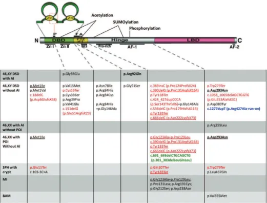
both gonadal dysgenesis and adrenal insuficiency (43). Considering the type of each mutation already identiied in NR5A1, it is dificult to establish a direct phenotype genotype correlation (Figure 2). If we consider that there is a dosage effect, it is dificult to understand why all patients heterozygous for severe nonsense mutations occurring in the irst and second coding exons, as well as other frameshift mutations that are considered to under go mRNA nonsensemediated decay, do not present the severe phenotype, including adrenal insuficiency. Ne vertheless, heterozygous mutations such as p.Glu11Ter, p.Cis16Ter and p.Glu51ArgfsX23 were identiied in pa tients with, respectively, XY DSD and severe penoscrotal hypospadia, and cryptorchidism without adrenal insu ficiency. Most mutations were found in heterozygous patients either as de novo mutations or as inherited from a nonaffected parent, indicating a dominant inheritan
ce with different degree of penetrance. It is interesting to note that from the nine mutations identiied in XY DSD and in XX primary ovarian insuficiency, eight are nonsense or frameshift mutations (Figure 2), and most of them had been inherited. Conversely, there are two cases of homozygosity for inherited mutations enabling the conclusion that, in those cases, the mutation was transmitted in a recessive manner. The p.Arg92Gln described above is one, and the other is p.Asp293Asn, which was found segregating in a family, and resulted in three different phenotypes: XX primary ovarian in suficiency, XY incomplete gonadal dysgenesis, and XY complete gonadal dysgenesis (44,45).
Many studies have shown that variations in the NR5A1
may be associated not only with gonadal dysgenesis and adrenal failure, but also with hypospadias, anorchia, male infertility, and premature ovarian failure, affecting both
Figure 2.Schematic overview of SF-1 and locations of each NR5A1 mutation[adapted from Hoivik and cols. (20) and Ferraz-de-Souza and cols. (46)]. The
Cop
yright
© ABE&M t
odos os dir
eit
os r
eser
vados
.
46,XY and 46,XX individuals. In some cases, it may be also associated with adrenal tumors and endometriosis (46).
The identiication of NR5A1 nucleotide changes in women with primary ovarian failure clearly indica tes that SF1 is indeed a key factor in the development and function of ovaries in humans (44), although it was considered before that NR5A1 mutations might not interfere with ovarian development (47). SF1 fai lure may affect the ovary at several levels: reducing the number of germ cells, damaging stroma integrity, and causing abnormal folliculogenesis. Phenotypic varia bility is veriied for either 46,XY or 46,XX individuals carrying NR5A1 mutations within families, proba bly as a result of multifunctional roles of SF1 protein (8,15,44,45,48). Another example of a mutated SF1 allele affecting both male and female development is the allele carrying the mutations p.Gly123Ala and p.Pro129Leu within the SF1 hinge region, which has been associated to either ovarian insuficiency or male infertility (44,49). In addition, p.Gly146Ala, also loca ted in the hinge region, has been described as a missen se associated with micropenis and cryptorchidism (50). Finally, all the molecular studies reported so far in dicate different clinical manifestations for different nu cleotide changes in the NR5A1 gene. Bilateral anorchia or testicular regression, in addition to all other manifes tations, was reported as a result of p.Val355Met muta tion in one out of 24 children evaluated (51). Howe ver, a recent study involving 26 patients with the same condition did not reveal any mutation (52), indicating that NR5A1 mutation might be not a frequent cause of anorchia. It can be inferred from all data reported so far that mutations in the NR5A1 gene are more fre quent than in the SRY gene in cases of 46,XY DSD (53). However, more research on mutation screenin gs is necessary to indicate NR5A1 gene mutations as frequent causes of either primary or premature ovarian failure without adrenal insuficiency.
The indings reviewed here indicate a complex ex pressivity of the phenotype, penetrance, and modes of inheritance of NR5A1 mutations, and suggest that more research has to be done to igure out how NR5A1
mutations correlate with different phenotypes, whether and how other genes modulate their expressivity, and to identify these genes for each affected phenotype.
Acknowledgements: the authors would like to thank Fundação de Amparo à Pesquisa do Estado de São Paulo and Conselho Nacional de Desenvolvimento Cientíico e Tecnológico (CNPq Brasil) for their inancial support.
Disclosure: no potential conlict of interest relevant to this article was reported.
REFERENCES
1. Swain A. Sex determination and differentiation. In: Neill JD, edi-tor. Knobil and Neill’s Physiology of Reproduction. 3rd ed. New York: Elsevier; 2006. p. 245-60.
2. Biason-Lauber A. Control of sex development. Best Pract Res Clin Endocrinol Metab. 2010;24(2):163-86.
3. Goodfellow PN, Darling SM. Genetics of sex determination. De-velopment. 1988;102(2):251-8.
4. De-Mello MP, Soardi FC. Genes envolvidos na determinação se-xual. In: Maciel-Guerra AT, Guerra-Júnior G, editores. Menino ou menina? Os distúrbios da diferenciação do sexo. 2. ed. São Paulo: Rubio; 2010. p. 3-14.
5. Guerra-Júnior G, Maciel-Guerra AT. The role of the pediatrician in the management of children with genital ambiguities. J Pediatr (Rio J). 2007;83(5 Suppl):S184-91.
6. Hughes IA, Houk C, Ahmed SF, Lee PA, Group LC, Group EC. Con-sensus statement on management of intersex disorders. Arch Dis Child. 2006;91(7):554-63.
7. Nabhan ZM, Lee PA. Disorders of sex development. Curr Opin Obstet Gynecol. 2007;19(5):440-5.
8. Kousta E, Papathanasiou A, Skordis N. Sex determination and disorders of the sex development according to the revised no-menclature and classification in 46,XX individuals. Hormones. 2010;9(3):218-31.
9. Mello MP, Assumpção JG, Hackel C. Genes involved in sex de-termination and differentiation. Arq Bras Endocrinol Metabol. 2005;49(1):14-25.
10. Ribeiro Scolfaro M, Aparecida Cardinalli I, Gabas Stuchi-Perez E, Palandi de Mello M, de Godoy Assumpção J, Matias Baptista MT, et al. Morphometry and histology of gonads from 13 children with dysgenetic male pseudohermaphroditism. Arch Pathol Lab Med. 2001;125(5):652-6.
11. Lipay MV, Bianco B, Verreschi IT. Disgenesias gonadais e tumo-res: aspectos genéticos e clínicos. Arq Bras Endocrinol Metabol. 2005;49(1):60-70.
12. Andrade JG, Guerra-Júnior G, Maciel-Guerra AT. 46,XY and 45,X/46,XY testicular dysgenesis: similar gonadal and genital phenotype, different prognosis. Arq Bras Endocrinol Metabol. 2010;54(3):331-4.
13. Gourlay WA, Johnson HW, Pantzar JT, McGillivray R, Crawford R, Nielsen W. Gonadal tumors in disorders of sexual differentiation. Urology. 1994;43(4):537-40.
14. Alvarez-Nava F, Puerta H, Soto M, Pineda L, Temponi A. High incidence of Y- chromosome microdeletions in gonadal tissues from patients with 45,X/46,XY gonadal dysgenesis Fertil Steril. 2008;89(2):458-60.
15. Biason-Lauber A. Control of sex development. Best Pract Res Clin Endocrinol Metab. 2010;24(2):163-86.
16. El-Khairi R, Martinez-Aguayo A, Ferraz-de-Souza B, Lin L, Acher-mann JC. Role of DAX-1 (NR0B1) and steroidogenic factor-1 (NR5A1) in human adrenal function. Endocr Dev. 2011;20:38-46. 17. Hanley NA, Ball SG, Clement-Jones M, Hagan DM, Strachan T,
Lindsay S, et al. Expression of steroidogenic factor 1 and Wilms’ tumour 1 during early human gonadal development and sex de-termination. Mech Dev. 1999;87(1-2):175-80.
18. Shen WH, Moore CCD, Ikeda Y, Parker KL, Holly AI. Nuclear re-ceptor steroidogenic factor 1 regulates the Mullerian inhibiting substance gene: a link to the sex determination cascade. Cell. 1994;77(5):651-61.
Cop
yright
© ABE&M t
odos os dir
eit
os r
eser
vados
.
20. Hoivik EA, Lewis AE, Aumo L, Bakke M. Molecular aspects of ste-roidogenic factor 1 (SF-1). Mol Cell Endocrinol. 2010;315(1-2):27-39. 21. Büdefeld T, Tobet SA, Majdic G. Steroidogenic factor 1 and the
central nervous system. J Neuroendocrinol. 2011 Jun 11. doi: 10.1111/j.1365-2826.2011.02174.x. [Epub ahead of print].
22. Lala DS, Rice DA, Parker KL. Steroidogenic factor I, a key regula-tor of steroidogenic enzyme expression, is the mouse homolog of fushi tarazu-factor I. Mol Endocrinol. 1992;6(8):1249-58. 23. Parker KL, Rice DA, Lala DS, Ikeda Y, Luo X, Wong M, et al.
Ste-roidogenic factor 1: an essential mediator of endocrine develop-ment. Recent Prog Horm Res. 2002;57:19-36.
24. Zimmermann S, Schwarzler A, Buth S, Engel W, Adham IM. Trans-cription of the Leydig insulin-like gene is mediated by steroidoge-nic factor-1. Mol Endocrinol. 1998;12(5):706-13.
25. Lin L, Achermann JC. Steroidogenic factor-1 (SF-1, Ad4BP, NR5A1) and disorders of testis development. Sex Dev. 2008;2(4-5):200-9. 26. Takayama K, Sasano H, Fukaya T, Morohashi K, Suzuki T,
Tamu-ra M, et al. Immunohistochemical localization of Ad4 binding protein with correlation to steroidogenic enzyme expression in cycling human ovaries and sex cord stromal tumors. J Clin Endocrinol Metab. 1995;80(9):2815-21.
27. Parker KL, Schimmer BP. Steroidogenic factor 1: a key deter-minant of endocrine development and function. Endocr Rev. 1997;18(3):361-77.
28. Wong M, Ramayya MS, Chrousos GP, Driggers PH, Parker KL. Clo-ning and sequence analysis of the human gene encoding steroi-dogenic factor 1. J Mol Endocrinol. 1996;17(2):139-47.
29. Oba K, Yanase, T, Nomura M, Morohashi K, Takayanagi R, Nawata H. Structural characterization of human Ad4bp (SF-1) gene. Bio-chem Biophys Res Commun. 1996;226(1):261-7.
30. Little TH, Zhang Y, Matulis CK, Weck J, Zhang Z, Ramachandran A, et al. Sequence-specific deoxyribonucleic acid (DNA) recognition by steroidogenic factor 1: a helix at the carboxy terminus of the DNA binding domain is necessary for complex stability. Mol En-docrinol. 2006;20(4):831-43.
31. Wilson TE, Fahrner TJ, Milbrandt J. The orphan receptors NGFI--B and steroidogenic factor 1 establish monomer binding as a third paradigm of nuclear receptor-DNA interaction. Mol Cell Biol. 1993;13(9):5794-804.
32. Kimura R, Yoshii H, Nomura M, Kotomura N, Mukai T, Ishihara S, et al. Identification of novel first exons in Ad4BP/SF-1 (NR5A1) gene and their tissue- and species-specific usage. Biochem Bio-phys Res Commun. 2000;278(1):63-71.
33. Demura M, Wang F, Yoneda T, Karashima S, Mori S, Oe M, et al. Multiple noncoding exons 1 of nuclear receptors NR4A family (nerve growth factor-induced clone B, Nur-related factor 1 and neuron-derived orphan receptor 1) and NR5A1 (steroidogenic factor 1) in human cardiovascular and adrenal tissues. J Hyper-tens. 2011;29(6):1185-95.
34. Wood MA, Mukherjee P, Toocheck CA, Walker WH. Upstream stimulatory factor induces Nr5a1 and Shbg gene expression during the onset of rat sertoli cell differentiation. Biol Reprod. 2011;85(5):965-76. Epub 2011 Jul 6.
35. Hoivik EA, Bjanesoy TE, Mai O, Okamoto S, Minokoshi Y, Shima Y, et al. DNA methylation of intronic enhancers directs tissue-speci-fic expression of steroidogenic factor 1/adrenal 4 binding protein (SF-1/Ad4BP). Endocrinology. 2011;152(5):2100-12.
36. Luo X, Ikeda Y, Parker KL. A cell-specific nuclear receptor is essen-tial for adrenal and gonadal development and sexual differentia-tion. Cell. 1994;77:481-90.
37. Sadovsky Y, Crawford PA, Woodson KG, Polish JA, Clements MA, Tourtellotte LM, et al. Mice deficient in the orphan receptor ste-roidogenic factor 1 lack adrenal glands and gonads but express P450 side-chain-cleavage enzyme in the placenta and have
nor-mal embryonic serum levels of corticosteroids. Proc Natl Acad Sci USA. 1995;92(24):10939-43.
38. Achermann JC, Ito M, Hindmarsh PC, Jameson JL. A mutation in the gene encoding steroidogenic factor-1 causes XY sex reversal and adrenal failure in humans. Nature Genetics. 1999;22(2):125-6. 39. Ito M, Achermann JC, Jameson JL. A naturally occurring steroi-dogenic factor-1 mutation exhibits differential binding and activa-tion of target genes. J Biol Chem. 2000;275(41):31708-14. 40. Achermann JC, Ozisik G, Ito M, Orun UA, Harmanci K, Gurakan
B, et al. Gonadal determination and adrenal development are regulated by the orphan nuclear receptor steroidogenic fac-tor-1, in a dose-dependent manner. J Clin Endocrinol Metab. 2002;87(4):1829-33.
41. Correa RV, Domenice S, Bingham NC, Billerbeck AEC, Rainey WE, Parker KL, et al. A microdeletion in the ligand binding domain of human steroidogenic factor 1 causes XY sex reversal without adrenal insufficiency. J Clin Endocrinol Metab. 2004;89(4):1767-72. 42. Mallet D, Bretones P, Michel-Calemard L, Dijoud F, David M, Morel
Y. Gonadal dysgenesis without adrenal insufficiency in a 46,XY patient heterozygous for the nonsense C16X mutation: a case of SF1 haploinsufficiency. J Clin Endocrinol Metab. 2004;89:4829-32. 43. Tajima T, Fujiwara F, Fujieda K. A novel heterozygous mutation
of steroidogenic factor-1 (SF-1/Ad4BP) gene (NR5A1) in a 46, XY disorders of sex development (DSD) patient without adrenal fai-lure. Endocr J. 2009;56(4):619-24.
44. Lourenço D, Brauner R, Lin L, De Perdigo A, Weryha G, Muresan M, et al. Mutations in the NR5A1 associated with ovarian insuffi-ciency. New Eng J Med. 2009;360(12):1200-10.
45. Soardi FC, Coeli FB, Maciel-Guerra AT, Guerra-Júnior G, De-Mello MP. Complete XY gonadal dysgenesis due to p.D293N homo-zygous mutation in the NR5A1 gene: a case study. J Appl Genet. 2010;51(2):223-4.
46. Ferraz-de-Souza B, Lin L, Achermann JC. Steroidogenic fac-tor-1 (SF-1, NR5A1) and human disease. Mol Cell Endocrinol. 2011;336(1-2):198-205.
47. Biason-Lauber A, Schoenle EJ. Apparently normal ovarian di-fferentiation in a prepubertal girl with transcriptionally inactive steroidogenic factor 1 (NR5A1/SF-1) and adrenocortical insuffi-ciency. Am J Hum Genet. 2000;67(6):1563-8.
48. Warman DM, Costanzo M, Marino R, Berensztein E, Galeano J, Ramirez PC, et al. Three new SF-1 (NR5A1) gene mutations in two unrelated families with multiple affected members: within-family variability in 46,XY subjects and low ovarian reserve in fertile 46,XX subjects. Horm Res Paediatr. 2011;75(1):70-7.
49. Bashamboo A, Ferraz-de-Souza B, Lourenço D, Lin L, Sebire NJ, Montjean D, et al. Human male infertility associated with muta-tions in NR5A1 encoding steroidogenic factor 1. Am J Hum Ge-net. 2010;87(4):505-12.
50. Wuqiang F, Yanase T, Wei L, Oba K, Nomura M, Okabe T, et al. Functional characterization of a new human Ad4BP/SF-1 varia-tion, G146A. Biochem Biophys Res Commun. 2003;311(4):987-94. 51. Philibert P, Zenaty D, Lin L, Soskin S, Audran F, Léger J, et al. Mu-tational analysis of steroidogenic factor 1 (NR5a1) in 24 boys with bilateral anorchia: a French collaborative study. Hum Reprod. 2007;22(12):3255-61.
52. Brauner R, Neve M, Allali S, Trivin C, Lottmann H, Bashamboo A, et al. Clinical, biological and genetic analysis of anorchia in 26 boys. PLoS One. 2011;6(8):e23292. Epub 2011 Aug 10.
![Figure 1. Sexual development cascade [adapted from Swain (1) and Biason-Lauber (2)]. The most important proteins in male and female pathways are shown in the figure](https://thumb-eu.123doks.com/thumbv2/123dok_br/19017667.470161/3.918.110.442.121.382/figure-sexual-development-cascade-adapted-important-proteins-pathways.webp)