Rev Saúde Pública 2004;38(5) www.fsp.usp.br/rsp
Lead level, enamel defects and dental caries
in deciduous teeth
Viviane Elisângela Gomesa, Ronaldo S Wadab, Jaime Aparecido Curyc and M aria da Luz
Rosário de Sousab
aDepartamento de Odontologia Social.Faculdade de Odontologia de Piracicaba da Universidade
Estadual de Campinas (Unicamp). Piracicaba, SP, Brasil. bDepartamento de Odontologia Social da Faculdade de Odontologia de Piracicaba. Unicamp. Piracicaba, SP, Brasil. cDepartamento de Ciências Fisiológicas da Faculdade de Odontologia de Piracicaba. Unicamp. Piracicaba, SP, Brasil
Correspondence to: Maria da Luz Rosário de Sousa
Departamento de Odontologia Social - FOP/ Unicamp
Av. Limeira, 901
13414-900 Piracicaba, SP, Brasil E-mail: [email protected]
Funded by Fundação de Amparo à Pesquisa do Estado de São Paulo (Fapesp – Process n. 00/07353-6). Work based on master’s dissertation presented to the Dentistry School of Piracicaba, Universidade Estadual de Campinas, 2002.
Received on 8/10/2003. Approved on 16/4/2004.
Keywords
Lead. Dental enamel. Dental caries, epidemiology. Dental enamel hypoplasia.
Abstract
Objective
To verify the relationship between lead concentration in the enamel of deciduous teeth and the presence of enamel defects and, consequently, with dental caries among preschool children.
Methods
The sample consisted of 329 preschool children in Piracicaba, State of São Paulo: 132 attending municipal kindergartens close to industrial plants and 197 attending kindergartens in non-industrial areas. This sample belonged to an initial study made between 2000 and 2001, in which the lead concentration was obtained by means of enamel biopsy. Oral clinical examination of the children from both regions was performed to verify the prevalence of enamel defects, using the Developmental Defects of Enamel (DDE) Index of the World Dental Federation (FDI), and of dental caries, using the decayed, missing and filled surfaces (dmfs) index of the World Health Organization. The chi-squared test and relative risk calculation were utilized in relation to a significance level of 5%, considering each region separately.
Results
Among the children from the non-industrial area, there was a higher proportion with dental caries among those with higher lead concentrations in deciduous teeth (p=0.02). This was not, however, observed among the children from the industrial area (p=0.89). There was an increased relative risk (RR) of caries among the children from the non-industrial area, but this was not seen among the children from the non-industrial area. No relationship was observed between the presence of lead and enamel defects.
Conclusions
No data was found that would give evidence of a relationship between lead concentration and enamel defects in either of the areas studied. No relationship was found between lead and dental caries in the industrial area, thus emphasizing that more studies of such relationships are needed.
INTRODUCTION
In an earlier study,10 it was found that preschool children living in an industrial region of the city of Piracicaba, State of São Paulo, presented greater lead (Pb) concentrations in their dental enamel than did those living in a non-industrial region. There are re-ports in the literature indicating that the presence of
lead in the chemical composition of dental enamel may alter dental ultrastructure, thereby giving rise to defective enamel. It was therefore considered impor-tant to investigate such relationships among this same population of preschool children.
Within this context, in vitro studies have shown that
Rev Saúde Pública 2004;38(5) www.fsp.usp.br/rsp
Chumbo, cárie e defeitos em dentes decíduos Gomes VE et al
regions (the industrial and non-industrial regions), through data collected during a survey in 1999.5
The schools health sector of the Piracicaba munici-pal authorities furnished listings of the children en-rolled in the ten kindergartens in the two regions se-lected for the study. There was a total of 421 children: 209 in the industrial region and 212 in the non-in-dustrial region.
Because of losses, the sample consisted of 329 pre-school children of both sexes, aged four and five years old. The adapted Frias Matrix16 was utilized for the age calculations. In the sample, there were 197 chil-dren from six kindergartens in the non-industrial re-gion and 132 children from four kindergartens in the industrial region.
The delineation of the study was single-blind. The scheduling of the children for them to participate in the study was done by another researcher. It was thus avoided that the examiner would have prior knowl-edge about the children.
The participants were attended at consultation of-fices in the university dental laboratory.
Some modifications to the original biopsy tech-nique3 were made so as to adapt it to the present sam-ple. These modifications were made after performing a pilot study.10
The teeth were submitted to professional prophy-laxis, using a Robinson-type brush, rubber cup and pumice, followed by washing and drying using air and gauze and relative isolation using cotton wool rolls. These procedures were accomplished by an as-sistant so that the researcher would not touch any metallic surface before performing the biopsy.
Adhesive tape with a central perforation of 1.6 mm in diameter (±0.03) was firmly stuck onto the ves-tibular surface of one of the upper central deciduous incisors (51 or 61) of each participant in the study, thereby demarcating the area of the biopsy. 5 µl of 1.6 mol/l HCl prepared in 70% glycerol (v/v) was deposited onto this area of exposed enamel. The drop of acid was agitated for 20 seconds, using the end of the pointer, and was then aspirated and transferred to a collecting tube containing 200 µl of MILLI-Q ul-trapurified water. To recover the residual acid and dis-solved enamel, 5 µl of ultrapurified water was placed on the enamel, agitated for 10 seconds and added to the biopsy solution in the collecting tube. The lead and phosphorus concentrations were determined from this solution, in the laboratory. The phosphorus con-alterations to the ultrastructure of the enamel,8 given
that ameloblasts are cells that are extremely sensitive to environmental alterations. Such alterations may, in turn, be associated with modifications in the physico-chemical behavior of the enamel,15 thereby making it more susceptible to demineralization.
Thus, some studies have indicated that there is an association between the presence of lead in dental tissue and clinical alterations in the enamel. Such variations may relate to coloration9 or hypoplasia.1 However, these studies have not been sufficient for establishing a clear relationship between the pres-ence of lead and such enamel defects.
A relationship between the presence of lead in the dental tissue and increased dental caries, albeit indi-rect, has been reported by some researchers.2,9,17 None-theless, the mechanism through which this takes place has not been well established yet.
Because of the divergences in the literature and scar-city of data on this subject among Brazilian communi-ties, an epidemiological investigation was made in order to determine the relationship between the pres-ence of lead in dental enamel, enamel defects and car-ies, in Brazilian children’s deciduous teeth. Thus, the objective of the present work was to verify the rela-tionship between the concentration of lead present in the enamel of preschool children’s deciduous teeth and the prevalence of enamel defects and caries.
M ETH O D S
In order to determine which regions of the city of Piracicaba, State of São Paulo, to be studied, the following bodies were firstly consulted to obtain information on lead pollution in the city: Piracicaba Municipal Department of the Environment, the En-vironmental Sanitation Technology Company (Companhia de Tecnologia de Saneamento Ambiental - Cetesb) and the Center for Nuclear En-ergy in Agriculture of the University of São Paulo (CENA/USP). These bodies reported the existence of a car battery factory in an industrial district lo-cated in the Dois Córregos region, which was taken as the industrial region for the study. The kindergar-tens located in this industrial region were then found by using a map of the city. Kindergartens in another region known as Campestre, which does not have industrial plants and was taken as the non-indus-trial region, were also localized.
!
Rev Saúde Pública 2004;38(5) www.fsp.usp.br/rsp
Chumbo, cárie e defeitos em dentes decíduos Gomes VE et al
centration was used for estimating the mass of the enamel removed by the biopsy. After concluding the biopsy, the tape was removed and the tooth was washed with water for 30 seconds, dried using air jets and again isolated, in order to receive the application of fluoride gel.
Control biopsies were performed on the bench sur-face or on the caps of the pointer supports, so as to check the lead contamination in the working envi-ronment during the procedures. All the material uti-lized and the bench on which the instruments and pipettes were laid out were cleaned using a 10% so-lution of nitric acid (HNO3), in order to remove possi-ble prior lead contamination. The collecting tubes were also previously washed using 10% nitric acid: immersed in the solution for 24 hours and then rinsed 20 times using ultrapurified water and dried under a hood that had been decontaminated from metals.
The examinations were conducted under artificial light, with drying of the surface using air and gauze, using a clinical mirror and a CPI (Community Peri-odontal Index) probe. The prevalence of caries was described using the dmfs index, which consists of summing the decayed, missing and filled surfaces of the deciduous teeth, according to the criteria of the World Health Organization (WHO),19 and the preva-lence of enamel defects using the DDE (developmen-tal defects of enamel) index, which describes the dif-ferent types of defects, from opacity to hypoplasia.6 The examinations were conducted by a single cali-brated examiner, and the intra-examiner error was calculated. Around 10% of the total sample was ran-domly selected for reexamination, with calculation of the intra-examiner error during data collection.
To avoid possible evaporation and loss of sample volume, it was decided to dehydrate the samples by placing them in a desiccator with anhydrous calcium chloride (CaCl2) for 36 hours. This procedure was tested previously to verify that no alterations would occur. At the time when the lead and phosphorus assays were performed, the samples were rehydrated using 210 µl of ultrapurified water, agitated and divided into two parts, for lead and phosphorus assaying.
The lead determination was performed using a graphite furnace atomic absorption spectrometer (GFAAS), calibrated using standard solutions con-taining 0 to 100 ppb of lead. The sample with 490 µl of a solution of 0.2% (w/v) NH4H2PO4 with 0.5% (v/ v) Triton X-100 and 0.2% HNO3.
The analysis of the phosphorus concentration was done in order to determine the quantity of enamel
removed from the tooth surface and the depth of the biopsy, so that the quantity of lead could be calcu-lated in µg per g of enamel removed (ppm). The phos-phorus was assayed via the Fiske & Subbarow colori-metric method.7 Thus, 105 µl of sample, 295 µl of MILLI-Q ultrapurified water and 83 µl of molybdic acid were pipetted together and agitated. After 10 minutes, 33 µl of reducer were added and the mixture was again agitated. After 20 minutes, the color inten-sity was measured in a spectrophotometer (Beckman DU-70) at 660 nm. The apparatus was calibrated us-ing samples that presented the followus-ing known phos-phorus concentrations: 0.58 µg/ml, 1.16 µg/ml, 2.32 µg/ml, 4.65 µg/mland 9.30 µg/ml.
Considering that human enamel contains 17% phosphorus by weight, this was the starting point for determining the mass of enamel removed. To de-termine the thickness of enamel removed by the bi-opsy, it was assumed that the enamel had a density of 2.95 g/cm3.
Information on the socioeconomic level of the children studied was collected by applying a ques-tionnaire to each mother or the person responsible for the child, with questions relating to family in-come and the mothers’ education level. This infor-mation was collected by the health agents of the kindergartens.
The statistical analysis was done using the SAS (Statistical Analysis System) and Epi Info version 5.0 statistical software. Correlation analysis, the chi-squared test and calculations of relative risk (RR) in relation to a significance level of 5% were utilized, and each region was considered separately.
The present research was approved by the Research Ethics Committee of the Dentistry School of Piraci-caba, Universidade Estadual de Campinas (FOP/ Unicamp) (protocol no. 29/99), in accordance with Resolution 196/96 of the National Commission for Research Ethics (CONEP).
RESU LTS
" Rev Saúde Pública 2004;38(5) www.fsp.usp.br/rsp
Chumbo, cárie e defeitos em dentes decíduos Gomes VE et al
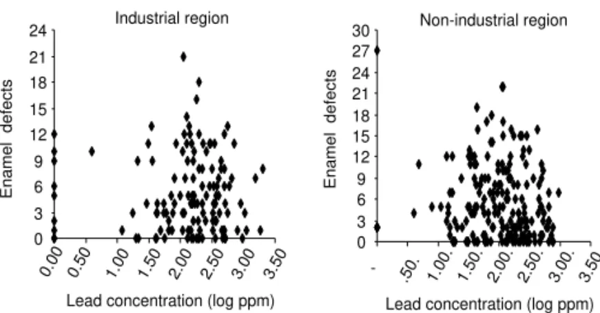
Figure - D i stri buti on of dental enamel defects accordi ng to l ead concentration in the enamel of children in the industrial and non-industrial regions. Piracicaba, SP, 2001.
0 3 6 9 12 15 18 21 24
0.00 0.50 1.00 1.50 2.00 2.50 3.00 3.50
Lead concentration (log ppm)
Enamel defects
0 3 6 9 12 15 18 21 24 27 30
-.50. 1.00. 1-.50. 2.00. 2-.50. 3.00. 3-.50. Lead concentration (log ppm)
Enamel defects
Industrial region Non-industrial region
Kappa of k=0.9, and 90% concordance for the defects.
The sample loss was 36.8% in the indus-trial region and 7.1% in the non-indusindus-trial region.
The average value (± standard deviation, SD) of phosphorus found in the enamel bi-opsy samples from the exposed group (indus-trial region) was 4.1 µg/ml (±1.5) and 4.4 µg/ ml (±1.6) from the non-exposed group (non-industrial region). The average masses of enamel removed (± SD) were 0.24 µg (±0.095) and 0.25 µg (±0.097), respectively. There was
no statistical difference in the depths of the biopsies (± SD) between the groups from the two regions (p>0.05): these depths were 4.1 µm (±1.5) for the exposed group and 4.4 µm (±1.6) for the non-exposed group.
A negative correlation between lead concentration and the total number of enamel defects was found in the non-industrial region (r2=-0.1497 and p=0.0357), while such a correlation was not observed in the in-dustrial region (p>0.05). When the total number of defects was discriminated, there were no correlations (p>0.05) between lead concentration and hypopla-sia, lead concentration and demarcated opacity and lead concentration and diffuse opacity, either in the industrial or non-industrial region.
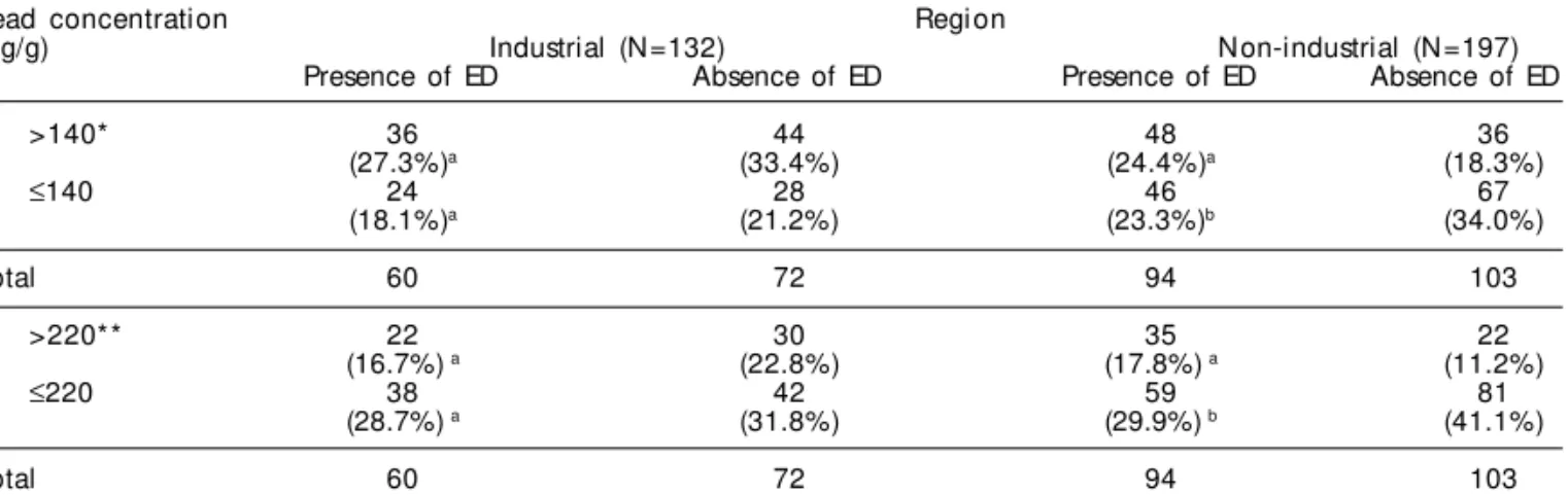
The results for the relationship between lead con-centration and enamel defects (presence/absence) in the industrial and non-industrial regions are ex-pressed in Table 1.
According to Table 1, no statistically significant dif-ference was found between the regions (p>0.05) with regard to the presence/absence of enamel defects.
The relative risks between lead concentration (140 and 220 ppm) and enamel defects (presence/absence) were 1.01 (0.87<RR<1.16) and 1.03 (0.89<RR<1.19), respectively, for the industrial region, and 1.03 (0.91<RR<1.16) and 0.99 (0.86<RR<1.14), respec-tively, for the non-industrial region, thus showing that there was no risk (p>0.05).
The distribution of enamel defects according to the lead concentration found in the enamel of deciduous teeth of the children from the industrial and non-in-dustrial regions is represented in the Figure.
There was no correlation between lead concentra-tion and caries in either region (p>0.05).
The relationship between lead concentration and car-ies (presence/absence) in the samples from the indus-trial and non-indusindus-trial regions is shown in Table 2.
There was a statistically significant difference for the proportions of individuals with presence of car-ies and lead concentrations of more than 140 and 220 ppm, in relation to those with presence of caries and lead concentrations of less than or equal to 140
Table 1 - Lead concentration in the enamel of preschool children’s deciduous teeth, according to region and the presence or absence of enamel defects (ED). Piracicaba, 2001.
Lead concentration Regi on
(µg/g) Industrial (N=132) Non-industrial (N=197)
Presence of ED Absence of ED Presence of ED Absence of ED
>140* 68 12 71 13
(51.6%)a (9.0%) (36.0%)a (6.6%)
≤140 44 8 93 20
(33.4%)a (6.0%) (47.2%)a (10.2%)
Total 112 20 164 33
>220** 45 7 47 10
(34.0%) a (5.3%) (23.9%) a (5.0%)
≤220 67 13 117 23
(50.8%) a (9.9%) (59.4%) a (11.7%)
Total 112 20 164 33
Values followed by the same letter, in the same column, indicate that there was no statistically significant difference according to the chi-squared test (p<0.05).
#
Rev Saúde Pública 2004;38(5) www.fsp.usp.br/rsp
Chumbo, cárie e defeitos em dentes decíduos Gomes VE et al
and 220 ppm, only for the non-industrial region (p=0.0223 and p=0.0141, respectively).
There was an increased risk of caries among chil-dren from the non-industrial region who had lead con-centrations of more than the median and average: 1.40 (1.05<RR<1.88) and 1.46 (1.10<RR<1.93), re-spectively. This was not seen among the children from the industrial region: 0.98 (0.67<RR<1.43) and 0.89 (0.60<RR<1.32).
There was a statistically significant difference in family income (p<0.0001) and education level (p<0.0001) between the groups from the industrial and non-industrial regions: these levels were greater in the industrial region.
D ISCU SSIO N
The lead concentrations in the deciduous teeth were analyzed in two ways (median and average), because no defined reference values exist in the literature for the concentrations in dental enamel that would re-flect a condition of lead poisoning or, furthermore, would relate to enamel defects and caries. The studies in the Brazilian literature have established reference values only for blood and have reported the impor-tance of verifying lead levels as a routine examina-tion.14 However, blood measurements indicate acute lead contamination, thus differing from dental enamel, which indicates past contamination.
Nonetheless, no relationship was found between any of the lead concentrations analyzed in the present study and enamel defects in the deciduous teeth. The results from the correlations and the relative risks between lead concentrations and enamel defects, when these existed, were weak. For the non-industrial region, for which the correlation was negative, this relationship ought to be
better investigated, considering that this result diverges from some reports in the literature.1,10 However, the latter reports did not produce conclusive results and reported the relationship between lead in dental tissue and enamel defects in an indirect manner. Moreover, in examining the Figure of the present study, it can be seen that, in the industrial region, there were more cases of children with enamel defects even in the absence of lead concentra-tion, thereby indicating that other factors must be con-tributing towards the presence of these defects. Besides this, the visualization of the industrial and non-indus-trial regions in the Figure allows the observation that, where there were greater lead concentrations, there were greater numbers of enamel defects in the industrial re-gion than in the non-industrial rere-gion.
With regard to the relationship between lead and caries, surprisingly, a greater proportion of individu-als with the presence of caries at lead concentrations of more than the median and average was observed in the non-industrial region. However, it was seen that, in the industrial region, both the mother’s education level and the family income were greater than those in the non-industrial region. These may have acted as confounding factors that could have exercised a role that perhaps was more important than the environ-mental lead contamination in the industrial region. Although the characterization of socioeconomic lev-els from the education network has been utilized by other researchers,12 in the present work this appears not to have been a variable that homogenized the groups in relation to similarity of socioeconomic level. Another matter that may have interfered was the greater sample loss in the industrial region.
One important epidemiological finding was that the relative risk of having caries was greater among the children living in the non-industrial region who presented lead concentrations of more than 140 and
Table 2 - Lead concentration in the enamel of preschool children’s deciduous teeth, according to region and the presence or absence of dental caries. Piracicaba, 2001.
Lead concentration Regi on
(µg/g) Industrial (N=132) Non-industrial (N=197)
Presence of ED Absence of ED Presence of ED Absence of ED
>140* 36 44 48 36
(27.3%)a (33.4%) (24.4%)a (18.3%)
≤140 24 28 46 67
(18.1%)a (21.2%) (23.3%)b (34.0%)
Total 60 72 94 103
>220** 22 30 35 22
(16.7%) a (22.8%) (17.8%) a (11.2%)
≤220 38 42 59 81
(28.7%) a (31.8%) (29.9%) b (41.1%)
Total 60 72 94 103
Values followed by different letters, in the same column, indicate that there was a statistically significant difference, according to the chi-squared test (p<0.05).
$ Rev Saúde Pública 2004;38(5) www.fsp.usp.br/rsp
Chumbo, cárie e defeitos em dentes decíduos Gomes VE et al
220 ppm in their deciduous teeth. This result reaf-firms the tendency reported in the literature towards a relationship between presence of lead in dental tis-sue and increased prevalence of caries.2,9,17
No data were found that would give evidence of a relationship between the lead concentration in the enamel of deciduous teeth and enamel defects, in ei-ther of the regions studied, However, a relationship was found between lead and caries in the non-indus-trial region, thus emphasizing the need for more stud-ies on such relationships, taking into consideration the importance of socioeconomic factors in caries.
REFEREN CES
1. Brook AH, Fearne JM, Smith JM . Environmental causes of enamel defects. In: Symposium on Dental Enamel, 1996, Ciba Foundation. Proceedings. New York: John Wiley & Sons, 1997. Ciba Foundation Symposium, v. 205.
2. Brudevold F, Aasenden R, Srinivasian BN, Bakhos Y. Lead in enamel and saliva, dental caries and the use of enamel biopsies for measuring past exposure to lead. J Dent Res 1977;56:1165-71.
3. Brudevold F, Reda A, Aasenden R, Bakhos Y. Determination of trace elements in surface enamel of human teeth by a new biopsy procedure. Arch Oral Biol 1975;20:667-73.
4. Bulman JS, Osborn JF. Measuring diagnostic consistency. Br Dent J 1989;166:377-81.
5. Cypriano S, Sousa MLR, Rihs LB, Wada RS. Saúde bucal dos pré-escolares em Piracicaba, Brasil, 1999. Rev Saúde Pública 2003;37:247-53.
6. Fédération Dentaire Internationale. Comission on Oral Health, Research and Epidemilogy. A review of the developmental defects of enamel index (DDE Index). Int Dent J 1992;42:411-26.
7. Fiske CH, Subbarow Y. The colorimetric determination of phosphorus. J Biol Chem 1925;66:375-400.
8 Gerlach RF, Souza AP, Cury JA, Line SRP. Effect of lead, cadmium and zinc on the activity of enamel matrix proteinases in vitro. Eur J Oral Sci 2000;108:327-34.
9. Gil F, Facio A, Villanueva E, Pérez ML, Tojo R, Gil A. The association of tooth content with dental health factors. Sci Total Environ 1996;192:183-91.
10. Gomes VE, Gerlach RF, Sousa MLR, Krug FJ. Concen-tração de chumbo em dentes decíduos de pré-escolares de Piracicaba, SP, Brasil – Estudo Piloto. Rev Odonto Ciência 2003;18:3-7.
11. Gomes VE, Sousa MLR, Barbosa FJr, Krug FJ, Saraiva MCP, Cury JA, Gerlach RF. In vivo studies on lead content of deciduos teeth superficial enamel of preschool children. Sci Total Environ 2004;320:25-35.
12. Maltz M, Silva BB. Relação entre cárie, gengivite e fluorose e nível socioeconômico em escolares. Rev Saúde Pública 2001;35:170-6.
13. Oliveira, AGR. Levantamentos epidemiológicos em saúde bucal: análise metodológica proposta pela Organização Mundial da Saúde. Rev Bras Epidemiol 1998;1:180-4.
14. Paoliello MMB, Gutierrez PR, Turini CA, Matsuo T, Mezzaroba L, Barbosa DS et al. Valores de referência para plumbemia em uma população urbana do Sul do Brasil. Rev Panam Salud Publica 2001;9:315-9.
15. Simons TJ. Cellular interactions between lead and calcium. Br Med Bull 1986;42:431-4.
16. Universidade de São Paulo. Faculdade de Saúde Pública. Secretaria de Estado da Saúde de São Paulo. Levantamento Epidemiológico em Saúde Bucal – Estado de São Paulo, 1998. São Paulo: Núcleo de Estudos e Pesquisas de Sistemas de Saúde; 1998. [Relatório apresentado à Secretaria de Estado da Saúde de São Paulo como conclusão do projeto realizado com o Núcleo de Estudos e Pesquisas em Serviços de Saúde, 1999].
17 Watson GE, Davis BA, Raubertas RF, Pearson SK, Bowen WH. Influence of maternal lead ingestion on caries in rat pups. Nature Med 1997;3:1024-5.
18. World Health Organization. Oral health surveys: basic methods. 3rd ed. Geneva: WHO; 1987.
19. World Health Organization. Oral health surveys: basic methods. 4th ed. Geneva: WHO; 1997.
ACKN O W LED GEM EN TS