J of Evidence Based Med & Hlthcare, pISSN- 2349-2562, eISSN- 2349-2570/ Vol. 2/Issue 48/Nov. 16, 2015 Page 8403
AGE WISE HISTOMORPHOLOGICAL CHANGES IN HUMAN LIVER
Tribeni Medhi1, Jayasri Devi2
1Assistant Professor, Department of Anatomy, Fakhruddin Ali Ahmed Medical college, Barpeta, Assam. 2Professor, Department of Anatomy, Fakhruddin Ali Ahmed Medical college, Barpeta, Assam.
ABSTRACT: CONTEXT: Hepato cellular carcinoma (HCC) results in between 2.5 lakhs to 1million deaths globally per annum. Liver transplantation nowadays is a well accepted treatment option for end-stage liver disease and acute liver failure.
AIMS: Keeping this concept in view, a study was conducted in the Guwahati Zone of Northeast India, to compare the histomorphological features of the human liver in different age groups.
SETTING AND DESIGN: Apparently healthy livers were obtained from 21 subjects on whom medicolegal post-mortems had
been performed. Their ages varied from newborn to 90 years. Subjects were divided into 3 groups. 7 specimens were taken from each group. (1) Pediatric (2) Adult (3) Old age.
METHODS AND MATERIALS: In all the above age groups, immediately after removal of the livers, they were washed in normal saline, dried with blotting paper and weighed in an electronic weighing machine. Sections of liver were fixed, processed, cut and stained with Harris Haematoxylin and Eosin stain.
RESULTS: The liver loses weight from 50 years onwards. There appears to be racial and environmental differences in the change in liver weight in old age. Autopsy studies show a diminution of nearly 46% in liver weight between the 3rd and 10th
decades of life. The liver decreases in size with age. The hepatocytes are radially disposed in the liver lobule. They are piled up, forming a layer one cell thick (except in young children) in a fashion similar to the bricks of a wall. These plates are directed from the periphery of the lobule to its centre and anastomose freely forming a complex labyrinthine and sponge-like structure.
CONCLUSIONS: From the findings in the present study it can be concluded that: 1. Nowadays, the measurement of liver volume has gained practical use in relation to liver transplantation. 2. We have compared the histomorphology of adult liver with a child. The findings in both the groups are very similar. This feature is important, since in pediatric liver transplantation, a portion of an adult liver can be used for an infant or small child. 3. The present study has open scopes for further research with more advanced techniques.
HOW TO CITE THIS ARTICLE: Tribeni Medhi, Jayasri Devi. “Age Wise Histomorphological Changes in Human Liver”. Journal of Evidence based Medicine and Healthcare; Volume 2, Issue 48, November 16, 2015; Page: 8403-8408,
DOI: 10.18410/jebmh/2015/1145
INTRODUCTION: The size of the liver varies a little in health, its average transverse diameter is about 22-28 cms. Its antero posterior diameter 17-20 cms and thickness from above downwards 7 to 9 cms. In the adult the right lobe is about 4 times the bulk of the left. In infant the lobes approximate in size, the right lobe being about twice the size of the left and the liver as a whole larger in proportion to the size of the body. The capsule is thin, smooth,shining,translucent,permitting the fine lobular mottling to be seen.
A few studies on liver weights of Indians have also been reported. At birth, the liver constitutes 4-4.5% of the body weight, its relative size decreases with growth so that after puberty it comprises between 2.5-3% of the weight.1
The liver is the largest gland in the body and is reddish brown in colour, very vascular, soft and friable, it weighs
about 3 pounds but relative to the body weight, is twice as heavy at birth as in the adult, hence the prominence of a
child’s abdomen.2
In the newborn infant, the liver occupies nearly 2/5th of the
abdominal cavity, the average weight of the organ in newborn, 1 year, 6 year and puberty are 150g, 300g, 550g and 1500g respectively.3
HISTOLOGY OF HUMAN LIVER: The hepatic cells are arranged as plates which form the lacunae.4 The
hepatocytes(liver cells) form plates(cellae murales) one cell thick(except in young children) in whom two-cell thick plates may be seen.5
MATERIALS AND METHODS: The study was conducted
on human liver in the department of Anatomy. The specimens of livers were divided into three groups according to different age groups which are shown as follows.
Groups Age
(Years)
Number of specimens
Pediatric group 0 to 14 7
Adult group 15-50 7
Elderly group More
than 50 7
Submission 05-11-2015, Peer Review 06-11-2015, Acceptance 07-11-2015, Published 14-11-2015. Corresponding Author:
Dr. Tribeni Medhi, Department of Anatomy,
Fakhruddin Ali Ahmed Medical College, Barpeta, Assam. E-mail: [email protected]
J of Evidence Based Med & Hlthcare, pISSN- 2349-2562, eISSN- 2349-2570/ Vol. 2/Issue 48/Nov. 16, 2015 Page 8404
The livers were collected from the following sources:
1. Cadavers in the department of Forensic Medicine, within stipulated time limit after fulfilling the formalities. The livers were collected after excluding all possible history of abnormalities of the livers.
2. From the department of obstetrics and gynaecology, cases of neonatal death were collected and autopsies were done in Forensic Medicine to take out the neonatal liver.
In all the above age groups, immediately after the removal of the livers, they were washed in normal saline, dried with blotting paper and weighed in an electronic weighing machine. The length, breath and thickness were measured by means of graph paper, scale, pin to locate the maximum length/breath and vernier calliper mainly for thickness measured at the level of inferiorvenacavae.
Biometrical values of different age groups were statistically analyzed and significant difference of length, breath and thickness were noted.
Immediately after biometry, depending on the size of liver, slices were made by cutting the specimen with sharp scalpel in planes passing through hilum to capsule. The sizes of slices were about 3-5mm thick and 4-5mm in dimension. The fixation of the slides were done by keeping them in 10% formal saline(10% formal saline=100ml formalin+8.5g sodium chloride+900ml tap water) for 24-48 hours.
The tissues were subjected to dehydration by immersing them into ascending strength of alcohol- 50%, 70%, 90%, and absolute alcohol for specified time. The slices were immersed into clearing agent xylol for half to one hour.
Then the wax impregnation was done by passing the tissue through liquid paraffin bath, maintained at around 60 degree centigrade temperatures. Wax impregnation removes the clearing agent from the tissue.
STAINING OF THE SLIDES: The sections of the tissues were stained by routine haematoxylin and eosin according to standard method.6
RESULTS AND OBSERVATION: In the present study,
the human livers were grouped into three as given below.
GROUP I–pediatric age group–0 to 14 years.
GROUP II–adult age group–15 to 50 years.
GROUP III–old age group–more than 50 years.
MORPHOLOGY: The liver is found to be reddish brown in colour in pediatric and adult age group. In the old age group, the liver is dark brown in colour. The right lobe constitutes about 2/3rd of the whole liver and contributes to
form all its surfaces. At birth, the right lobe is found to be twice as large as the left lobe. In infancy, the size of the right lobe is found to be proportionately greater than in an adult. In young children and adolescents, the right lobe is three times as large as the left lobe. In the adult, the right lobe is found to be about four times the bulk of the left.
BIOMETRY: Comparison between pediatric and adult age group was done by statistical analysis. Similarly comparison was done between old age and adult age group.
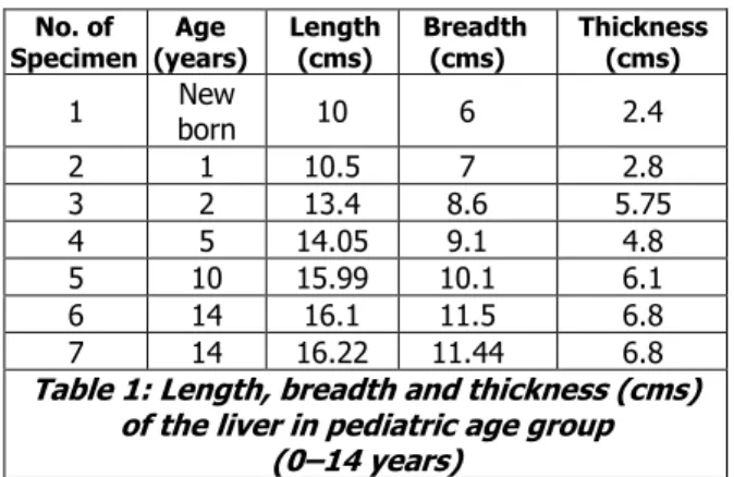
No. of Specimen
Age (years)
Length (cms)
Breadth (cms)
Thickness (cms)
1 New
born 10 6 2.4
2 1 10.5 7 2.8
3 2 13.4 8.6 5.75
4 5 14.05 9.1 4.8
5 10 15.99 10.1 6.1
6 14 16.1 11.5 6.8
7 14 16.22 11.44 6.8
Table 1: Length, breadth and thickness (cms) of the liver in pediatric age group
(0–14 years)
Average: 13.751cms 9.105cms 5.064cms
No. of specimen
Age (years)
Length (cms)
Breadth (cms)
Thickness (cms)
1 15 17.01 12.05 7.11
2 20 18.42 16.10 9.51
3 31 18.10 16.25 10.01
4 40 17.99 16.80 9.83
5 44 18.91 16.66 9.65
6 45 18.65 16.25 9.51
7 50 22.9 13.75 9.84
Table 2: Length,Breadth and Thickness (cms) of the liver in adult age group (15–50 years)
Average: 18.85cms 15.408cms 9.351cms
No. of specimen
Age (years)
Length (cms)
Breadth (cms)
Thickness (cms)
1 51 23 13.5 9.9
2 60 22.67 12.05 8.01
3 65 22.42 12.42 7.69
4 76 23 11.5 7.65
5 78 20.18 11.01 7.11
6 84 20.02 10.75 7.05
7 90 19.05 10.05 6.95
Table 3:Length,Breadth And Thickness (Cms) Of The Liver In Old Age Group (>50 Years)
Average: 21.47 cms 11.61 cms 7.76 cms
Parameters Group
I
Group
II SE
Value of
‘t’
Length
13.751 18.85 0.651 7.832(S)
Breadth
9.105 15.408 0.557 0.0113(NS)
Thickness
5.064 9.351 0.418 10.255(S)
Table 4: Mean Values For Length, Breadth and Thickness (Cms) Of Liver Between Group I and
Group II
J of Evidence Based Med & Hlthcare, pISSN- 2349-2562, eISSN- 2349-2570/ Vol. 2/Issue 48/Nov. 16, 2015 Page 8405
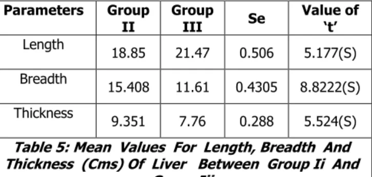
Parameters Group
II
Group
III Se
Value of
‘t’
Length
18.85 21.47 0.506 5.177(S)
Breadth
15.408 11.61 0.4305 8.8222(S)
Thickness
9.351 7.76 0.288 5.524(S)
Table 5: Mean Values For Length, Breadth And Thickness (Cms) Of Liver Between Group Ii And
Group Iii GROUP II – Adult S=Significant GROUP III – Old age
The average liver weight in the three age groups are recorded in the table. From the table it is seen that the average liver weight in pediatric age group is 568.07g. The weight increased gradually but erratically in the adult age group to 1556.99g. Subsequently, it decreased erratically in the old age group to 1276.67g.
The weight of apparently healthy livers were obtained from 7 specimens from each age group. Age related variations in measurements were also noted.
Means and standard deviations of the liver weight were
obtained from various age groups. Unpaired students ‘t’
test was applied to obtain the differences in the parameters. Statistically the difference between pediatric and adult age group was highly significant ( p<0.05). There was also some difference between the old age group and adult age group. The tables showing the weight of liver in different age groups are shown below
Number of
specimen Age (years) Weight (g)
1 Newborn 160.65
2 1 343.05
3 2 366.07
4 3 477.33
5 5 543.75
6 9 812.04
7 14 1273.65
TABLE 6: WEIGHT (g) OF THE LIVER IN PEDIATRIC AGE GROUP (0–14 years)
Average – 568.077 g
Number of
specimen Age (years) Weight (g)
1 18 1425.26
2 21 1513.11
3 25 1512.21
4 37 1685.91
5 41 1585.09
6 45 1583.75
7 50 1593.60
TABLE 7: WEIGHT (g) OF THE LIVER IN ADULT AGE GROUP (15–50 years)
Average – 1556.99 g
Number of specimen
Age
(years) Weight (g)
1 53 1485.31
2 56 1491.90
3 60 1489.05
4 65 1468.46
5 71 1101.24
6 80 952.08
7 90 948.70
Table 8: Weight (G) Of The Liver In Old Age Group (>50years)
Average – 1276.67 g
Parameter Group
I
Group
II SE
Value of
‘t’
Weight 568.077 1556.99 76.681 12.896 (S)
Table 9: Mean values for weight (g) of liver between group I and group II
Parameter Group
II
Group
III SE
Value of
‘t’
Weight 1556.99 1276.677 55.629 5.0389 (S)
Table 10: Mean values for weight (g) of liver between group II and group III
GROUP I – Pediatric, S=Significant GROUP II – Adult
GROUP III – Old age
MICROMETRY: HISTOLOGY OF HEPATIC LOBULES:
As observed in the slides of liver in different age groups, the liver is organised into lobules, which take the shape of irregular polygonal prisms. It is delimited by interlobular connective tissue which is very little, if any visible in humans. Along the central axis of each lobule, runs a central vein, which is a branch of the hepatic vein. The lobular organisation of the human liver is not immediately evident under the microscope. Lobules do not have distinct boundaries and they are seldom cut neatly in cross section. At the central axis of each lobule, runs a central vein. The central veins are conspicuous spaces with no associated connective tissue, located roughly midway between the portal areas. These central veins mark the centers of the lobules.
Number of specimen
Age (years)
Lobule size(mm)
1 Newborn 1.11
2 1 1.10
3 2 1.12
4 3 2.1
5 5 2.11
6 10 2.13
7 14 2.0
Table 11: Liver Lobule Size (Mm) In The Pediatric Age Group (0 To 14 Years)
J of Evidence Based Med & Hlthcare, pISSN- 2349-2562, eISSN- 2349-2570/ Vol. 2/Issue 48/Nov. 16, 2015 Page 8406
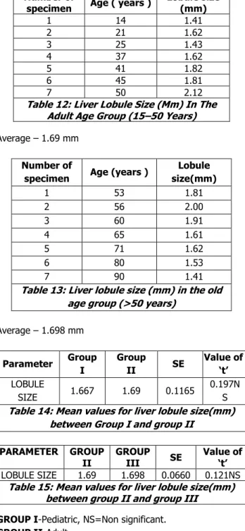
Number of
specimen Age ( years )
Lobule size (mm)
1 14 1.41
2 21 1.62
3 25 1.43
4 37 1.62
5 41 1.82
6 45 1.81
7 50 2.12
Table 12: Liver Lobule Size (Mm) In The Adult Age Group (15–50 Years)
Average – 1.69 mm
Number of
specimen Age (years )
Lobule size(mm)
1 53 1.81
2 56 2.00
3 60 1.91
4 65 1.61
5 71 1.62
6 80 1.53
7 90 1.41
Table 13: Liver lobule size (mm) in the old age group (>50 years)
Average – 1.698 mm
Parameter Group
I
Group
II SE
Value of
‘t’
LOBULE
SIZE 1.667 1.69 0.1165
0.197N S Table 14: Mean values for liver lobule size(mm)
between Group I and group II
PARAMETER GROUP
II
GROUP
III SE
Value of
‘t’
LOBULE SIZE 1.69 1.698 0.0660 0.121NS
Table 15: Mean values for liver lobule size(mm) between group II and group III
GROUP I-Pediatric, NS=Non significant.
GROUP II-Adult.
GROUPIII-Old age.
CYTOARCHITECTURE: In the pediatric age group, the lobules are made up of hepatocytes which extend radially from the central vein to the periphery.
The liver cell plates are originally 3 to 5 cell thick. These are gradually split into less cell thick plates by ramifications and widening of sinusoids. In the newborn, most of the plates are two cells thick.
At 2 years single cell plates are seen in our present study. In young children two cell thick plates may be seen. Anastomosing plates of hepatocytes, two cell thick is found prior to the age of 7 years. One cell thick plate is found after that age.
In the adult liver the parenchyma cells are arranged in elongated, anastomosing cords made up of single rows of cells. In sections of the liver of man, the cord appears to
be either one cell thick or two cell thick, depending on the plane of the section.
It is seen that the hepatocytes are radially disposed in the liver lobule.
They are piled up, forming a layer one cell thick in a fashion similar to the bricks of a wall. These plates are directed from the periphery of the lobule to its centre and anastomose freely forming a complex sponge like structure. In adults the hepatocytes form plates one cell thick.
In the old age group, the hepatocytes are found to be enlarged. Hepatocytes with age are enlarged in size and more frequently contain multiple nuclei. Further, the nuclei are larger in size, are more often polyploidy and chromosomal abnormalities are common.
In our present study it is seen that in old age the mean volume of hepatocytes increases
DISCUSSION: Results and observations obtained in the present study which have been described in the preceeding chapters revealed several points of interest having marked importance in medical science. Hence, these have been considered worthy of discussion in conjunction with the findings of the other workers to draw a definite conclusion in respect of morphology, biometry and histomorphological differences of the liver among different age groups which may help in investigation and management of both benign and malignant disease of the liver.
Morphology: In the present study it was found that, in infancy the size of the right lobe is proportionately greater than in an adult. The right lobe constitutes about 2/3rd of
the whole liver and contributes to form all its surfaces in parts. The reduction in size of the left lobe is considered to be due to proportionate reduction in its blood supply.7 At
birth, the right lobe is found to be twice as large as the left lobe; in young children and adolescents it is found to be three times as large.8
The liver is reddish brown in colour in young age and adults.7 In old age it was observed that the liver is dark
brown in colour which might be due to accumulation of lipofuschin granules in lysosomes of hepatocytes.9
Weight of the liver in different age groups is compared
statistically by unpaired ‘t’ test. Significant difference of
weight is found between pediatric and adult age group. The variation in weight is also found to significant between adult age group and old age group. If we compare the liver weight in the different age groups we can see that the liver is twice as heavy at birth as in the adult.The liver at puberty(14 years) is found to be ten times the birth weight.2
SUMMARY AND CONCLUSION: The present study was
J of Evidence Based Med & Hlthcare, pISSN- 2349-2562, eISSN- 2349-2570/ Vol. 2/Issue 48/Nov. 16, 2015 Page 8407 different age groups amongst each other and to compare
these findings with previous research works. The histomorphological study of the liver was done and the salient findings of the present study is summarised below.
The size and shape of the human liver in different age groups were noted.Their length, breadth and thickness were recorded. The weight of the liver was also noted in the three age groups. Statistical analysis was also done.
The liver of human is found to be triangular in shape and divided into distinct right and left lobes by the falciform ligament, and the right and left triangular ligament. It also presents the caudate and quadrate lobes.
At birth, the right lobe is twice as large as the left lobe. In infancy, the size of the right lobe is found to be proportionately greater than in an adult.
The reduction in size of the left lobe is considered to be due to proportionate reduction in its blood supply.
The liver is found to be reddish brown in colour in pediatric and adult age group. In the old age, the liver is dark brown in colour, which may be due to accumulation of lipofuschin granules in lysosomes of hepatocytes.
Variations of thickness was noted between pediatric and adult age groups. Difference was also noted between the adult and the old age groups.
Statistically the difference between pediatric and adult age group was highly significant. There was also some difference between the old age group and adult age group. If we compare the liver weight in the different age groups we can see that the liver is twice as heavy at birth as in the adult.The liver at puberty(14 years) is found to be ten times the birth weight.
The liver was studied in each age group under light microscope in low and high magnification.
As observed in the slides of liver in different age groups, the liver is organised into lobules, which take the shape of irregular polygonal prisms. It is delimited by interlobular connective tissue which is very little, if any visible in humans.
The size of the lobule is measured under low power microscope. The lobules in different age groups are statistically analysed. There is no significant difference in the lobule size between the pediatric and the adult age groups. There is also no significant dufference in the lobule size between the adult and the old age groups.
The liver cell plates are originally 3 to 5 cell thick. These are gradually split into less cell thick plates by ramifications and widening of sinusoids. In the newborn, most of the plates are two cells thick.In young children two cell thick plates may be seen. Anastomosing plates of hepatocytes, two cell thick is found prior to the age of 7 years. One cell thick plate is found after that age.
CONCLUSION: From the findings of the present study it may be concluded that histomorphological features of the liver in the three age groups significantly differ from each other.The liver is a major early hematopoietic organ of the embryo. Despite being an organ whose cells are renewed at a slow rate, the liver has an extra-ordinary capacity for regeneration. Liver transplantation nowadays is a well accepted treatment option for end-stage liver disease and acute liver failure. A major advance in pediatric liver transplantation was the development of reduced size liver transplantation in which a portion of an adult liver is used for an infant or small child. Unfortunately, the supply of liver allografts from non-living donors is far short of the number of potential recipients, a reality that has spurred the development of living donor liver transplantation. The present study has opened scopes for investigation and treatment of liver diseases and further research with more advanced techniques needs to be undertaken to know more about histomorphological architectures of the liver with respect to different ages.
BIBLIOGRAPHY:
1. L. Emmett Holt, Rustin Mc Intosh(1953), “Holt
Pediatrics”, 12th edition, Appleton-Century–Crafts, Inc.
New York
2. R.D.Lockhart, G.F.Hamilton(1959), “Anatomy of the
Human body”, pg 529-532, Faber and Faber limited. 3. Ernest H.Watson, George H.Lowrey(1962), “Growth and
Development of children”, Year book Medical
publishers. INC
4. Julian A. Sterling(1955), “The Biliary Tract”, The Williams and Wilkins Company.
5. Ivan Damjanov et al. (1996), “Anderson’s Pathology”, 10th edition, vol 2, Mosby Yearbook, INC, 1996.
6. H.M.Carleton(1956), Schaffer’s Essentials of Histology”, 16th edition,
7. Dr S.N.Sahana(1985), “Human Anatomy”, 4th edition,
vol-2, New Central Book Agency Calcutta.
8. Victor C.Vaughan, R.James Mc Kay(1975), “Nelson
Textbook of Pediatrics”, 10th edition, W. B. Saunders
Company IGASU SHOIN ltd.
9. O.P Sharma(1999), “Geriatric Care in India,A Textbook