A rare non-Robertsonian translocation
involving chromosomes 15 and 21
Uma rara translocação não robertsoniana envolvendo os cromossomos 15 e 21
Marcelo Razera Barui
I, Deise Helena Souza
II, Rosana Aparecida Bicudo Silva
III, Ester Silveira Ramos
IV, Danilo Moretti-Ferreira
VDepartment of Genetics, Instituto de Biociências (IBB), Universidade Estadual Paulista (Unesp), Botucatu, São Paulo, Brazil
ABSTRACT
CONTEXT: Robertsonian translocations (RT) are among the most common balanced structural rearrange-ments in humans and comprise complete chromatin fusion of the long arm of two acrocentric chromo-somes. Nevertheless, non-Robertsonian translocation involving these chromosomes is a rare event. CASE REPORT: We report a de novo unbalanced translocation involving chromosomes 15 and 21. The newborn was the daughter of a 29-year-old mother and a 42-year-old father. The couple was non-con-sanguineous. Clinical indings led to the diagnosis of Down syndrome (DS) with severe congenital heart defects (persistent arterial duct, and complete atrioventricular septal defect), as well as low birth length and weight (< 5th and < 10th percentile, respectively, based on speciic measurement curves for DS).
Con-ventional cytogenetic analysis revealed the karyotype 46,XX,der(15)(15pter→15q26.2::21q11.2→21qter). The translocation was conirmed by means of luorescence in situ hybridization. The parents had normal karyotypes.
CONCLUSIONS: Diferently from RT, in our case a rare event occurred involving the distal segment of 15q and the proximal segment of 21q. Only two reports of this translocation, involving chromosomes 15 and 21 but diferent breakpoints, have been described so far. The association between 21q duplication and 15q deletion makes it diicult to separate the efect of each chromosome, but might also be respon-sible for increasing the growth retardation, as detected in our case. Cytogenetic analysis on DS patients is mandatory not only to conirm the diagnosis, but also to assess the risk of recurrence at genetic counsel-ing, as well as to evaluate the contribution of other chromosome aberrations in the inal phenotype.
RESUMO
CONTEXTO: Translocações robertsonianas (TR) estão entre os rearranjos estruturais balanceados mais comuns em humanos e compreendem a fusão da cromatina completa do braço longo de dois cromossomos acrocêntricos. No entanto, são raras as translocações não Robertsonianas envolvendo esses cromossomos.
RELATO DE CASO: Nós descrevemos uma translocação não balanceada de novo envolvendo os cro-mossomos 15 e 21. A recém-nascida era ilha de uma mãe de 29 anos e de um pai de 42 anos, casal não consanguíneo. Os achados clínicos levaram ao diagnóstico de síndrome de Down (SD) com de-feitos cardíacos congênitos graves (persistência do canal arterial e defeito do septo atrioventricular completo), além de baixos comprimento e peso ao nascimento (< 5o e < 10o percentil em curvas de
medidas especíicas para SD, respectivamente). A análise citogenética convencional revelou o cariótipo 46,XX,der(15)(15pter→15q26.2::21q11.2→21qter). A translocação foi conirmada por hibridação in situ
luorescente. Os pais apresentavam cariótipo normal.
CONCLUSÕES: Diferentemente das TR, nesse caso ocorreu evento raro envolvendo o segmento distal de 15q e o proximal de 21q. Apenas dois relatos dessa translocação, envolvendo os cromossomos 15 e 21 e diferentes pontos de quebra, já foram descritos. A associação entre duplicação 21q e deleção 15q diiculta a distinção dos efeitos de cada cromossomo, mas poderia também ser responsável pelo acentuado retar-do de crescimento. A análise citogenética é obrigatória em pacientes com SD não apenas para conirmar o diagnóstico, mas também para avaliar o risco de recorrência no aconselhamento genético, bem como avaliar a contribuição de alterações de outros cromossomos no fenótipo inal.
IBSc, PhD. Assistant Professor, Department
of Genetics, Instituto de Biociências (IBB), Universidade Estadual Paulista (Unesp), Botucatu, São Paulo, Brazil.
IIBSc, MSc. Biomedic, Department of Genetics,
Instituto de Biociências (IBB), Universidade Estadual Paulista (Unesp), Botucatu, São Paulo, Brazil.
IIIBSc. Technician, Department of Genetics,
Instituto de Biociências (IBB), Universidade Estadual Paulista (Unesp), Botucatu, São Paulo, Brazil.
IVMD, PhD. Assistant Professor, Department of
Genetics, Faculdade de Medicina de Ribeirão Preto (FMRP), Universidade de São Paulo (USP), Ribeirão Preto, São Paulo, Brazil
VBSc, PhD. Associate Professor, Department
of Genetics, Instituto de Biociências (IBB), Universidade Estadual Paulista (Unesp), Botucatu, São Paulo, Brazil.
KEY WORDS: Translocation, genetic. Down syndrome.
Chromosomes, human, pair 15. Chromosomes, human, pair 21. Chromosome deletion.
PALAVRAS-CHAVE: Translocação genética. Síndrome de Down.
INTRODUCTION
he majority (approximately 95%) of Down syndrome cases are caused by simple trisomy of chromosome 21. Translocations involving this chromosome account for approximately only 1-3%
of all Down syndrome cases.1-3 Almost all of these translocations
involve a Robertsonian translocation, which comprises the long-arm elements of two acrocentric chromosomes (chromosomes
13, 14, 15, 21 and 22).3
Less common distinct forms of Down syndrome result from structural changes in chromosome 21. For example, reciprocal translocations are extraordinarily rare and are the cause of less than 0.1% of Down syndrome cases. hese translocations with partial trisomy have been used to deine a Down syndrome
criti-cal region or Down syndrome loci, consisting of a segment of the
long arm of chromosome 21.1,2
Recent studies have attempted to delineate an association between rare terminal deletions of the long arm of chromosome 15 and a speciic phenotype, in particular short stature caused
especially by the loss of one copy of the IGF1R (insulin-like
growth factor receptor) gene.4,5
he main aim of this study was to report a very rare de novo
non-Robertsonian translocation involving chromosomes 15 and 21, and to show the importance of cytogenetic investigation in all cases of clinical diagnosis of Down syndrome.
CASE REPORT
A female newborn was referred to the Genetic Counseling Service of Universidade Estadual Paulista (Unesp), Botucatu, São Paulo, with a clinical diagnosis of Down syndrome. She was the only child of non-consanguineous parents. Her 29-year-old mother was normal and her 42-year-old healthy father had a normal daughter from a previous marriage. Ultrasound exami-nation during pregnancy detected the presence of oligohydram-nios, but no renal malformations. She was born at term by means
of vaginal delivery with a birth length of 41 cm (< 5th percentile),
weight of 2000 g (< 10th percentile) and head circumference of
33 cm (50th percentile, all measurement based on a standard chart
for Down syndrome).6
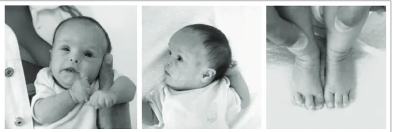
Clinical examination revealed hypotonia, brachycephaly, frontal bossing, lat face, upslanting palpebral issures, protrud-ing tongue, short neck with excess skin, brachydactyly, clinod-actyly of the ith ingers with a single interdigital crease on the
right side and overlapping toes (Figure 1). At the age of three
months, her length (51.5 cm) and weight (3,350 g) were still < 5th
and < 10th percentiles, respectively, and her head circumference
(36 cm) was normal, based on speciic standard measurement
charts for Down syndrome.6 Echocardiography showed a
persis-tent arterial duct, and a complete atrioventricular septal defect, which included a ventricular septal defect.
Metaphase chromosome spreads were obtained from tempo-rary lymphocyte cultures and the slides were subjected to Giemsa
trypsin G-banding (GTG-banding). Fluorescence in situ
hybrid-ization (FISH) was performed using commercial probes. For chromosome 15, we used a digoxigenin-labeled chromo-some 15 probe (Coatchromo-some; Cat. No. P5216; ONCOR, Gaithersburg,
MD, USA) and chromosome 15 α-satellite probe D15Z (Cat. No.
P5033; ONCOR, Gaithersburg, MD, USA). Hybridization was performed in accordance with the manufacturer’s instructions, followed by detection by means of luorescein isothiocyanate (FITC)-labeled anti-digoxigenin and counting using propidium
iodide staining (inal concentration: 0.3 μg/ml in antifade).
For chromosome 21, we used the Chromoprobe Multiprobe System Octochrome (Cytocell). FISH was performed to investi-gate the centromere regions of chromosomes 15 and 21, using
a Cytocell probe for the centromeres (α-satellites) of
chromo-some 13/21 (Cat. No. LPE 013G) and chromochromo-some 15 (Cat. No. LPE 015G). Hybridization was carried out in accordance with the manufacturer’s instructions.
B
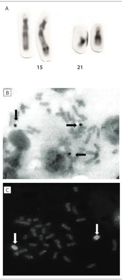
Cytogenetic analysis (Figure 2) revealed the karyotype
46,XX,der(15)(15pter→15q26.2::21q11.2→21qter). he parents presented normal karyotypes.
Unfortunately, the patient did not return for a follow-up and our service was unable to contact this speciic patient/family due to a change in their address and phone number. Consequently, it was impossible to have more information about this case and to collect more blood samples.
DISCUSSION
A translocation involving chromosome 21 has the potential to
produce duplication of the Down syndrome critical region.1,2
Duplication of a long segment of 21q could explain the predomi-nant Down syndrome phenotype, as detected in our case. Unlike in Down syndrome caused by trisomy of chromosome 21, termi-nal deletions of the long arm of chromosome 15 have rarely been
described.4,7-13
A large number of genes have been mapped within the
termi-nal 15q region, and among them, IGF1R seems to play the main
role in the phenotype of 15qter syndrome. Copy number varia-tions of this gene result in prenatal and postnatal growth
restric-tion. Additionally, IGF1R contributes towards development of
the central nervous and cardiovascular systems.4,5,12
Our study had some limitations due to the loss of contact with the patient. For example, we were unable to observe the IGF1 lev-els, which might have shown evidence of the IGF1R deiciency.
From reviewing the literature, we observed that the major-ity of the patients who were known to present terminal dele-tions of 15q displayed prenatal and postnatal growth retarda-tion, cardiac defects, delayed development, ear abnormalities
and clinodactyly.4,5,7-13 All these features and the Down
syn-drome indings overlap. For this reason, it was diicult to sepa-rate the efects of 21q duplication from those of 15q deletion, in our case. he association between these two chromosomal aber-rations may have been responsible for increasing the growth retardation in our case.
To the best of our knowledge (Table 1), only two reports on
non-Robertsonian translocation, involving chromosomes 15 and 21, have been published so far. Abeliovich et al. described the
karyotype t(15;21)(q15;q22.1)pat in two siblings.14 One of them
had Prader-Willi syndrome. An interesting case of a patient with typical Down syndrome phenotype and apparently
nor-mal karyotype was studied by Nadal et al.15 Using FISH, these
authors found the unbalanced karyotype t(15;21)(q26;q22.1).14
he father and other members of the family carried a balanced translocation between chromosomes 15 and 21. Although these cases have some similarities to our patient, these three transloca-tions present diferent breakpoints. A deeper search with more complex and more expensive methods, such as array comparative
21
Figure 2. Cytogenetic analysis. (A) GTG-banded partial metaphase. Chromosomes are (from left to right): normal chromosome 15, der(15) and two normal chromosomes 21. (B) luorescence hybridization in situ (FISH) using chromosome 21 probe. (C) FISH using chromosome 15 probe.
15
C
A
B
genomic hybridization (CGH), might have clariied some points such as the precise breakpoints. Unfortunately, we were unable to obtain further blood samples from this patient.
On the other hand, the techniques carried out in our study were suicient to show the rare non-Robertsonian translocation, the involvement of chromosomes 21 and 15 and the chromosome 21 trisomy. he results obtained provide evidence for the occur-rence of this atypical chromosome aberration, and for the impor-tance of cytogenetic analysis.
CONCLUSION
An association between these two chromosomal aberrations could be responsible for increasing the growth retardation, as detected in our case.
Cytogenetic analysis on Down syndrome patients is manda-tory, not only to conirm the diagnosis, but also to assess the risk of recurrence at genetic counseling, in particular when translo-cations are involved. Moreover, this makes it possible to evaluate the contribution of other chromosome aberrations to the inal phenotype.
REFERENCES
1. Gardner RJM, Sutherland, GR. Down syndrome, other full
aneuploidies, and polyploidy. In: Gardner RJM, Sutherland, GR,
editors. Chromosome abnormalities and genetic counseling. 3rd ed.
New York: Oxford University Press; 2004. p. 249-63.
2. Bornstein E, Lenchner E, Donnenfeld A, et al. Complete trisomy
21 vs translocation Down syndrome: a comparison of modes of
ascertainment. Am J Obstet Gynecol. 2010;203(4):391.e1-5.
3. Gardner RJM, Sutherland, GR. Robertsonian translocations. In:
Gardner RJM, Sutherland GR, editors. Chromosome abnormalities
and genetic counseling. 3rd ed. New York: Oxford University Press;
2004. p. 122-37.
4. Rudaks LI, Nicholl JK, Bratkovic D, Barnett CP. Short stature due to
15q26 microdeletion involving IGF1R: report of an additional case
Table 1. Results from our review of the medical databases (with and without using the words “case report” as a ilter). Date of search: January 9, 2013
Database Search strategy Results
PubMed
Chromosome 21 AND Chromosome 15 AND Translocation AND Robertsonian 15* Chromosome 21 AND Deletion chromosome 15 AND translocation AND syndrome 06†
Chromosome 21 AND deletion chromosome 15 AND translocation AND ‘Non Robertsonian’ 01*
Embase
Chromosome and 15 and 21 and translocation and Robertsonian 214*
Translocation non-Robertsonian 46*
Translocation and non Robertsonian and acrocentric 02*
15 AND chromosome AND 21 AND ‘Non-Robertsonian’ AND Translocation 04*
Lilacs
Translocation AND chromosome 21 04*
Translocation AND chromosome 15 05*
Translocation AND chromosome 15 05*
*None of them involves chromosome 15 and 21 at the same time and/or Down syndrome; †Only one with a t(15;21).
and review of the literature. Am J Med Genet A. 2011;155A(12):
3139-43.
5. Jezela-Stanek A, Kucharczyk M, Pelc M, Chrzanowska KH,
Krajewska-Walasek M. Minimal clinical indings in a patient with 15qter
microdeletion syndrome: delineation of the associated phenotype.
Am J Med Genet A. 2012;158A(4):922-6.
6. Hall J, Allanson J, Gripp K, et al. Measurements for speciic syndromes.
In: Hall J, Allanson J, Gripp K, Slavotinek A, editors. Handbook of
physical measurement. 2nd ed. New York: Oxford University Press;
2006. p. 424-37.
7. Pinson L, Perrin A, Plouzennec C, et al. Detection of an unexpected
subtelomeric 15q26.2 --> qter deletion in a little girl: clinical and
cytogenetic studies. Am J Med Genet A. 2005;138A(2):160-5.
8. Rump P, Dijkhuizen T, Sikkema-Raddatz B, et al. Drayer’s syndrome of
mental retardation, microcephaly, short stature and absent phalanges
is caused by a recurrent deletion of chromosome 15(q26.2-->qter).
Clin Genet. 2008;74(5):455-62.
9. Drayer NM, Kamps WA, ten Kate LP, et al. Microcephaly, ocular
hypertelorism, low-set ears, hand and feet anomalies, short stature
and mental retardation. Syndrome Identiication. 1977;5(1):9-11.
10. Houlston RS, Renshaw RM, James RS, Ironton R, Temple IK. Duplication
of 16q22-->qter conirmed by luorescence in situ hybridisation and
molecular analysis. J Med Genet. 1994;31(11):884-7.
11. Rosenberg C, Blakemore KJ, Kearns WG, et al. Analysis of reciprocal
translocations by chromosome painting: applications and limitations
of the technique. Am J Hum Genet. 1992;50(4):700-5.
12. Roback EW, Barakat AJ, Dev VG, et al. An infant with deletion of the
distal long arm of chromosome 15 (q26.1----qter) and loss of
insulin-like growth factor 1 receptor gene. Am J Med Genet. 1991;38(1):74-9.
13. Dateki S, Fukami M, Tanaka Y, et al. Identiication of chromosome
15q26 terminal deletion with telomere sequences and its bearing on
genotype-phenotype analysis. Endocr J. 2011;58(3):155-9.
14. Abeliovich D, Dagan J, Lerer I, et al. t(15;21)(q15;q22.1)pat resulting in
partial trisomy and partial monosomy of chromosomes 15 and 21 in
15. Nadal M, Moreno S, Pritchard M, et al. Down syndrome:
characterisation of a case with partial trisomy of chromosome 21
owing to a paternal balanced translocation (15;21) (q26;q22.1) by
FISH. J Med Genet. 1997;34(1):50-4.
Acknowledgements: We thank Bruna Paes Barros for technical assistance
Sources of funding: Fundação de Amparo à Pesquisa do Estado de São
Paulo – Fapesp (procedural no. 2008/51903-2)
Conlict of interest: None
Date of irst submission: June 18, 2012 Last received: May 20, 2013
Accepted: July 5, 2013
Address for correspondence:
Marcelo Razera Barui
Departamento de Genética
Instituto de Biociências (IBB), Universidade Estadual Paulista (Unesp)
Campus de Rubião Jr.
Distrito de Rubião Jr., s/no
Botucatu (SP)
CEP 18618-970
Tel. (+55 14) 3815-3747