Trends Psychiatry Psychother. 2016;38(1) – 50-55
T
APRS
Transcranial magnetic stimulation for posttraumatic stress
disorder: an updated systematic review and meta-analysis
Estimulação magnética transcraniana para transtorno de estresse
pós-traumático: revisão sistemática de literatura e metanálise
Alisson Paulino Trevizol,1 Mirna Duarte Barros,1 Paula Oliveira Silva,1 Elizabeth Osuch,2 Quirino Cordeiro,1 Pedro Shiozawa1
Abstract
Introduction: Transcranial magnetic stimulation (TMS) is a promising non-pharmacological intervention for posttraumatic stress disorder (PTSD). However, randomized controlled trials (RCTs) and meta-analyses have reported mixed results.
Objective: To review articles that assess the eicacy of TMS in PTSD treatment.
Methods: A systematic review using MEDLINE and other da-tabases to identify studies from the irst RCT available up to September 2015. The primary outcome was based on PTSD sco-res (continuous variable). The main outcome was Hedges’ g. We used a random-efects model using the statistical packages for meta-analysis available in Stata 13 for Mac OSX. Heterogenei-ty was evaluated with I2 (> 35% for heterogeneity) and the χ2
test (p < 0.10 for heterogeneity). Publication bias was evaluated using a funnel plot. Meta-regression was performed using the random-efects model.
Results: Five RCTs (n = 118) were included. Active TMS was signiicantly superior to sham TMS for PTSD symptoms (Hedges’ g = 0.74; 95% conidence interval = 0.06-1.42). Heterogeneity was signiicant in our analysis (I2 = 71.4% and p = 0.01 for the
χ2 test). The funnel plot shows that studies were evenly
distri-buted, with just one study located marginally at the edge of the funnel and one study located out of the funnel. We found that exclusion of either study did not have a signiicant impact on the results. Meta-regression found no particular inluence of any variable on the results.
Conclusion: Active TMS was superior to sham stimulation for ame-lioration of PTSD symptoms. Further RCTs with larger sample sizes are fundamental to clarify the precise impact of TMS in PTSD.
Keywords: Meta-analysis, posttraumatic stress disorder, trans-cranial magnetic stimulation, non-pharmacological therapies, systematic review.
Resumo
Introdução: A estimulação magnética transcraniana (EMT) é uma intervenção não farmacológica promissora no tratamento de transtorno de estresse pós-traumático (TEPT). No entanto, estudos controlados e metanálises apresentaram resultados conlitantes até o momento.
Objetivo: Revisar os artigos sobre a eicácia da EMT para o tra-tamento de TEPT.
Métodos: Conduzimos uma revisão sistemática da literatura no MEDLINE para identiicar estudos controlados e randomizados pu-blicados até setembro de 2015. O desfecho primeiro foi baseado nas escalas de gravidade de TEPT como variáreis contínuas. O des-fecho principal foi o g de Hedges. Utilizamos o modelo de efeito randômico com as análises estatísticas para metanálise do Stata 13 para Mac OSX. A heterogeneidade foi avaliada com o I2 (> 35%
para heterogeneidade) e o teste do χ2 (p < 0,01 para
heterogenei-dade). Viés de publicação foi avaliado utilizando-se o gráico do fu-nil. Realizamos metarregressões com modelo de efeito randômico.
Resultados: Cinco estudos foram incluídos. A EMT ativa foi supe-rior ao placebo para o tratamento de TEPT (g de Hedges = 0,74; intervalo de coniança 95% = 0,06-1,42). A heterogeneidade en-tre os estudos foi signiicativa em nossa análise (I2 = 71,4% e p
= 0,01 para o teste do χ2). O gráico do funil nos mostrou estudos
simetricamente distribuídos, com apenas um estudo localizado marginalmente ao gráico e um estudo localizado fora do funil. Encontramos que a exclusão de cada estudo não alterou signi-icativamente o resultado inal. A metarregressão não mostrou inluência de nenhuma variável no resultado.
Conclusões: A estimulação ativa de EMT foi superior à estimula-ção simulada para melhora dos sintomas de TEPT. Novos estudos randomizados e controlados por simulação são necessários para esclarecer com melhor precisão o impacto da EMT no TEPT.
Descritores: Metanálise, transtorno de estresse pós-traumáti-co, estimulação magnética transcraniana, terapias não farmaco-lógicas, revisão sistemática.
1 Centro Interdisciplinar de Neuromodulação Clínica, Faculdade de Ciências Médicas da Santa Casa de São Paulo, São Paulo, SP, Brazil. 2 Department of
Psychiatry, University of Western Ontario, Schulich School of Medicine and Dentistry, London, Ontario, Canada. Financial support: none.
Submitted Oct 15 2015, accepted for publication Nov 29 2015. No conlicts of interest declared concerning the publication of this article.
Introduction
Posttraumatic stress disorder (PTSD) is a trauma and stress-related disorder, characterized by intrusive, avoidance and hyperarousal core symptoms that may
result in signiicant social or occupational dysfunction. It
is estimated that 7.8% of the United States population experience PTSD in their lifetime and it is estimated that it
causes impaired ability to work that costs in excess of $3
billion per year in lost productivity in the United States.1
There is no deinitive pharmacotherapy for core PTSD
symptoms. Although medications and psychotherapy have been shown to help reduce symptoms and treat comorbid anxiety and depressive symptoms, in one third of patients there is no improvement in symptoms.2
Brain mechanisms related to PTSD (such as, for instance, threatening processing and fear-inducing stimuli) have been traced to particular pathways related to the amygdala, the frontal lobe, and the hippocampus.3
Working from the hypothesis that dysfunctional
brain structures underlie PTSD symptoms, the use of transcranial magnetic stimulation (TMS) was proposed with the objective of modulating target areas. Several
diferent protocols have emerged focusing on the left
and right dorsolateral prefrontal cortex (DLPFC).4 We
hereby present a literature review and meta-analysis
of the eicacy of active vs. sham TMS for treatment of
PTSD. It is relevant to point out that a previous meta-analysis has already been published.4 However, in the
present study we aim to enlarge the pooled sample and improve the quality of data analysis.
Method
A systematic review and meta-analysis was conducted in accordance with the recommendations of the Cochrane group and the PRISMA guidelines.5 Two
authors (PS and APT) performed independent systematic reviews and data extraction, and any discrepancies were resolved by consensus.
Literature review
We reviewed the following references and databases: a) MEDLINE and EMBASE databases using the
key words: (1) transcranial stimulation; (2) TMS; (3)
transcranial magnetic stimulation; (4) non-invasive brain stimulation; (5) NIBS; (6) post-traumatic stress disorder; (7) PTSD; and (8) anxiety disorder. Boolean terms were used as follows: [(1) OR (2) OR (3) OR (4)
OR (5)] AND [(6) OR (7) OR (8)]. We searched for work
published up to September 30, 2015.
b) The references listed in articles found by a) above and review articles, particularly those included in the meta-analyses by Karsen et al.4
We also attempted to identify controlled trials by
contacting specialists in the ield and by searching the
website clinicaltrials.gov for additional unpublished/ ongoing trials.
Eligibility criteria
We adopted the following inclusion criteria: 1) manuscript written in English, Spanish or Portuguese (in fact all articles retrieved were written in English); 2) describing randomized, sham-controlled trials; and 3) providing data (in the manuscript or upon request) needed to estimate the main outcomes, i.e., mean (standard deviation [SD]) values and response and remission rates. We excluded case reports and case series, uncontrolled trials and trials assessing conditions other than PTSD or interventions other than TMS.
Data extraction
The following variables were extracted according
to a structured checklist developed by the authors in
advance: 1) metadata (authorship, publication date, etc.); 2) demographics (sample size in each group, age, gender); 3) PTSD characteristics (baseline PTSD scores; use of medication; psychometric scales,
interviews and checklists used for PTSD diagnosis
and assessment of avoidance, hyperarousal and reexperiencing); 4) characteristics of the TMS technique (frequency; motor threshold; time period of stimulation; train; inter-train interval; number of sessions; side of brain); 5) research methods (randomization protocol; sham technique; blinding assessment; number of dropouts).
The primary outcome was based on PTSD scores (continuous variable). Although categorical variables might be more readily interpretable than continuous ones (despite the fact that the odds ratio is often
misinterpreted as a risk ratio), our choice was based on
the fact that the primary outcome of all studies included was based on continuous variables and so we considered
that a continuous efect size would better synthesize the
studies chosen for review.
For continuous outcomes, the meta-analysis was performed on endpoint PTSD scores. Since studies used more than one PTSD scale, we extracted data
corresponding to the study’s deinitions of the primary
For studies in which three groups were compared, two separate datasets were compared in each of two
diferent analyses. For example, in the study by Boggio
et al.,6 high frequency TMS for left and right DLPFC were
compared with sham stimulation. We therefore compared active left DLPFC stimulation vs. sham stimulation in one analysis and we compared active right DLPFC stimulation
with sham stimulation in another analysis. Likewise, in the
study by Cohen et al.,7 low frequency and high frequency
stimulation of the right DLPFC were compared with sham stimulation. We therefore compared high frequency right DLPFC stimulation with sham stimulation in one analysis and in another analysis we compared low frequency right DLPFC stimulation with sham stimulation.
The study by Isserles et al.8 requires further
explanation, since patients were randomly allocated into three treatment groups, combining deep transcranial magnetic stimulation (DTMS) and brief exposure of script-driven imagery of the traumatic event. The groups
were conigured as follows: in group a) EXP-STIM,
patients were given DTMS after script-driven imagery of the traumatic experience immediately followed by script-driven imagery of a neutral event; in group b) NOEXP-STIM, patients were given DTMS after script-driven imagery of a positive experience immediately followed by script-driven imagery of a neutral event; and in group c) EXP-SHAM patients were given sham-DTMS after script-driven imagery of the traumatic experience immediately followed by script-driven imagery of a neutral event.
Even though all groups received some kind of treatment,
we considered group c) to be the sham group since TMS was not applied. In a crossover study conducted by Osuch et al.,9 patients underwent active or sham TMS
combined with exposure therapy.
Quality assessment
We assessed the methodological quality of each trial by assessing the following: 1) methods of randomization – whether the study was correctly randomized and/ or the authors reported the randomization method; 2) sham TMS – how sham TMS was performed.
Quantitative analysis
Main outcomes
All analyses were performed using the statistical
packages for meta-analysis available in Stata 13 for
Mac OSX. For the main outcome (PTSD scores) we
initially calculated the standardized mean diference and
the pooled SD of each comparison. This procedure is
convenient when handling diferent scales (such as PTSD scales) since it standardizes the efect sizes across all
studies based on the SD of each study. For the study by Boggio et al.,6 PTSD scale scores were assessed by
graphic evaluation. For the study by Osuch et al.,9 data
were provided by the authors. Hedges’ g was used as the
measure of efect size, which is appropriate for studies with small sample sizes. The pooled efect size was
weighted by the inverse variance method and measured
using the random-efects model. Studies that failed to
provide crucial data such as SD or scale assessment
were excluded from the inal analysis.
Quantitative assessment of heterogeneity and bias
Heterogeneity was evaluated with the I2 statistic (>
35% for heterogeneity) and the χ2 test (p < 0.10 for
heterogeneity). Publication bias was evaluated using a
funnel plot, which displays conidence interval boundaries
to assist in visualizing whether the studies are within the funnel, thus providing an estimate of publication bias (e.g., whether the studies are distributed asymmetrically and/or fall outside the funnel). A sensitivity analysis was also performed, assessing the impact of each study on the overall results by excluding one study at a time.
Meta-regression
Meta-regression was performed using the
random-efects model as modiied by Knapp & Hartung,10 using
only one variable at a time.
Results
Overview
Our systematic review yielded 54 studies after duplicates were removed. Of these, 49 articles did not meet the eligibility criteria. Five studies6-9,11 (n = 118 patients) were
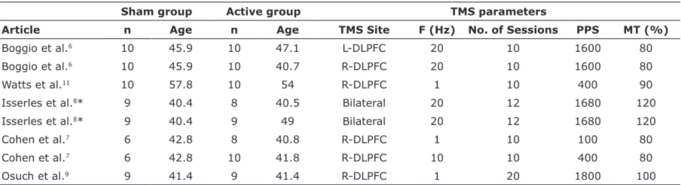
selected for the quantitative analysis. Mean age was 51.5 (SD = 2.5) years and 44% of the participants were women. No washout of drugs was performed. Demographics and stimulation protocols are summarized in Table 1.
The quality assessment revealed that all studies were
randomized. Sham TMS was performed in four diferent
ways: 1) a sham coil that produced a similar acoustic artifact and scalp sensation as the active coil; 2) a sham
magnetic coil that looked and sounded identical to the active coil, but did not provoke scalp sensation; 3) the
coil was held at 90° vertical over the stimulated head
area (no signiicant magnetic ield was evoked, just
that of the previous review by 59% with the inclusion of two further trials. We also improved the quality of data analysis. The previous investigation did not analyze heterogeneity or publication bias and did not conduct meta-regression to
detect the inluence of variables or exclude each study one
by one in the meta-analysis to evaluate their impact on the
inal result. The funnel plot assessment conducted in the present study showed that the risk of publication bias was
also low, further strengthening our results. Importantly, we found that between-study heterogeneity was high. New clinical trials with uniform intervention protocols could clarify results in future analyses.
Another characteristic of our meta-analysis is that we analyzed separately two datasets for each of three studies, Boggio et al.,6 Cohen et al.7 and Isserles et al.8
This is because they used a triple-arm design. Indeed, using this approach we were able to increase the sample
size and narrow the conidence interval further.
Our meta-regressions did not identify clinical and/ or methodological predictors of TMS responsiveness. In our meta-analysis we included intervention protocols that either stimulated or inhibited left or right DLPFC. Meta-regressions were performed in order to identify the
possibility of diferent results if protocols were evaluated
separately. None of the protocols (stimulation of the left DLPFC; stimulation of the left DLPFC and inhibition of right DLPFC; stimulation of right DLPFC; inhibition of
right DLPFC; bilateral DTMS) were identiied as predictive
of TMS non-responsiveness. Moreover, in order to verify
the inluence of each study on the overall efect, subgroup
analysis was performed using the metaninf command in
Stata. No single study inluenced the overall efect by itself. The neurobiological hypothesis for the eicacy of TMS
in PTSD treatment is based on dysfunctions of brain regions so far associated with processing threatening and fear-inducing stimuli, including the amygdala, the frontal lobe, and the hippocampus. The ventral prefrontal areas are richly connected to lateral prefrontal areas and amygdala. The right ventromedial frontal area provides access to
Primary outcome
We calculated the efect size for the endpoint. We found that active TMS was signiicantly superior to sham
TMS (Hedges’ g = 0.74; 95%CI 0.06-1.42) (Figure 1).
Quantitative assessment of heterogeneity and bias
Heterogeneity was signiicant in our analysis (I2 =
71.4% and p = 0.01 for the χ2 test). The funnel plot
shows that studies were evenly distributed, with just one study located marginally at the edge of the funnel and one study located out of the funnel. We found that exclusion of each of these studies in turn did not have
a signiicant impact on the results, with resulting efect sizes close to the overall efect size. Therefore, no single
study in particular was driving the results of our analysis.
Meta-regression
The following variables were assessed: baseline depression severity scores, session duration, whether a crossover study, use of brief exposure, frequency, number of sessions, duration of sessions, number of pulses per session, total number of pulses, motor threshold, type of blinding, whether deep TMS was used, number of days of stimulation, side of brain stimulation, and subject’s age. Meta-regression showed no particular
inluence of any variable on the results.
Discussion
In this systematic review of ive randomized clinical trials (n = 118), we found that active TMS was signiicantly superior
to sham TMS for treatment of core PTSD symptoms. Our results are in line with those of a previous meta-analysis.4
However, we were able to enlarge the pooled sample over
Table 1 - Overview of demographics and stimulation parameters
Sham group Active group TMS parameters
Article n Age n Age TMS Site F (Hz) No. of Sessions PPS MT (%)
Boggio et al.6 10 45.9 10 47.1 L-DLPFC 20 10 1600 80
Boggio et al.6 10 45.9 10 40.7 R-DLPFC 20 10 1600 80
Watts et al.11 10 57.8 10 54 R-DLPFC 1 10 400 90
Isserles et al.8* 9 40.4 8 40.5 Bilateral 20 12 1680 120
Isserles et al.8* 9 40.4 9 49 Bilateral 20 12 1680 120
Cohen et al.7 6 42.8 8 40.8 R-DLPFC 1 10 100 80
Cohen et al.7 6 42.8 10 41.8 R-DLPFC 10 10 400 80
Osuch et al.9 9 41.4 9 41.4 R-DLPFC 1 20 1800 100
* Deep transcranial magnetic stimulation.
PTSD treatment, since it is a safe, non-invasive treatment
that uses an electromagnetic ield to modulate the activity
of cortical areas based on a high-intensity current through a magnetic coil placed on the scalp, generating a
time-varying pulsed magnetic ield that penetrates the cranium
approximately 2-cm from the scalp surface to cortical tissue. Low-frequency TMS (1 Hz) is inhibitory, and high-frequency TMS (high-frequency above 10 Hz) is excitatory to underlying neural tissue.
In the present systematic review and meta-analysis we found that active TMS was clinically and statistically superior to sham TMS for treatment of PTSD. Notwithstanding, given the relatively small number of trials published to date and the heterogeneity of those studies, further phase III studies assessing broader samples are fundamental for clarifying the potential impact of TMS for treatment of PTSD in daily clinical practice.
References
1. Kessler RC, Sonnega A, Bromet E, Hughes M, Nelson CB. Posttraumatic stress disorder in the National Comorbidity Survey. Arch Gen Psychiatry. 1995;52:1048-60.
object-recognition systems to the amygdala, where the fear or threat response is mediated.12 Electroencephalographic
(EEG) studies have shown alpha power decreases in the right hemisphere in PTSD patients compared to control groups while they are exposed to trauma-related pictures.13
These indings have been corroborated by single photon
emission computed tomography (SPECT) studies that
have shown cerebral blood low to the right hemisphere is
increased in PTSD patients when they hear trauma-related sounds.3 Morey et al.,14 compared trauma-related stimuli
during a visual working-memory task in combat veterans
without PTSD and combat veterans with PTSD. These authors showed greater activation in the right ventrolateral prefrontal cortex (VLPFC) compared to the control group,
suggesting that PTSD patients may require more efort
to ignore emotionally distracting stimuli. The increased activation in the VLPFC was associated with decrease in the dorsolateral prefrontal cortex and with increased activation of the amygdala. The interpretation was that amygdala activation signaled the emotional distraction, which was
inhibited by the VLPFC, taking attention away from tasks,
as indicated by activation of the DLPFC. Modulating the
DLPFC using TMS was proposed to assess its eicacy in
Figure 1 - Forest plot of effect sizes (Hedges’ g). The forest plot was used to graphically illustrate the relative strength of treatment effects for each study reviewed. The vertical line represents the overall effect. 95%CI = 95% confidence interval;
SMD = standardized mean difference. Study
ID SMD (95%CI) Weight
%
Boggio et al.6
Boggio et al.6
Watts et al.11
Isserles et al.8
Isserles et al.8
Cohen et al.7
Cohen et al.7
Osuch et al.9
Overall (l-squared = 71.4%, p = 0.001)
NOTE: Weights are from random effects analysus
0.21 (-0.67, 1.09)
3.08 (1.70, 4.46)
0.74 (-0.17, 1.66)
1.44 (0.34, 2.55)
0.00 (-0.92, 0.92)
-0.12 (-1.18, 0.94)
1.50 (0.32, 2.67)
-0.26 (-1.19, 0.67)
0.74 (0.06, 1.42)
13.31 11.57 12.38 13.35 12.08 13.42 10.23 13.66
4.46
9. Osuch EA, Benson BE, Luckenbaugh DA, Geraci M, Post RM,
McCann U. Repetitive TMS combined with exposure therapy for PTSD: a preliminary study. J Anxiety Disord. 2009;23:54-9.
10. Knapp G, Hartung J. Improved tests for a random efects
meta-regression with a single covariate. Stat Med. 2003;22:2693-710. 11. Watts BV, Landon B, Groft A, Young-Xu Y. A sham controlled study
of repetitive transcranial magnetic stimulation for posttraumatic stress disorder. Brain Stimul. 2012;5:38-43.
12. Tillman GD, Kimbrell TA, Calley CS, Kraut MA, Freeman TW, Hart J Jr. Repetitive transcranial magnetic stimulation and threat memory: selective reduction of combat threat memory p300 response after right frontal-lobe stimulation. J Neuropsychiatry Clin Neurosci. 2011;23:40-7.
13. Rabe S, Beauducel A, Zollner T, Maercker A, Karl A. Regional brain
electrical activity in posttraumatic stress disorder after motor vehicle accident. J Abnorm Psychol. 2006;115:687-98.
14. Morey RA, Dolcos F, Petty CM, Cooper DA, Hayes JP, LaBar KS, et al. The role of trauma-related distractors on neural systems for
working memory and emotion processing in posttraumatic stress
disorder. J Psychiatr Res. 2009;43:809-17.
Correspondence:
Alisson Trevizol
Department of Psychiatry Santa Casa Medical School
Rua Major Maragliano, 241, Vila Mariana 04017-030 - São Paulo, SP - Brazil Tel.: +55 (11) 3466.2100
E-mail: [email protected]
2. Ballenger JC, Davidson JR, Lecrubier Y, Nutt DJ, Foa EB, Kessler RC, et al. Consensus statement on posttraumatic stress disorder from the International Consensus Group on Depression and Anxiety. J Clin Psychiatry. 2000;61:60-6.
3. Pagani M, Hogberg G, Salmaso D, Tarnell B, Sanchez-Crespo A,
Soares J, et al. Regional cerebral blood low during auditory recall
in 47 subjects exposed to assaultive and non-assaultive trauma and developing or not posttraumatic stress disorder. Eur Arch Psychiatry Clin Neurosci. 2005;255:359-65.
4. Karsen EF, Watts BV, Holtzheimer PE. Review of the efectiveness
of transcranial magnetic stimulation for post-traumatic stress disorder. Brain Stimul. 2014;7:151-7.
5. Liberati A, Altman DG, Tetzlaf J, Mulrow C, Gotzsche PC,
Ioannidis JP, et al. The PRISMA statement for reporting systematic reviews and meta-analyses of studies that evaluate health care interventions: explanation and elaboration. PLoS Med. 2009;6:e1000100.
6. Boggio PS, Rocha M, Oliveira MO, Fecteau S, Cohen RB, Campanha C, et al. Noninvasive brain stimulation with high-frequency and low-intensity repetitive transcranial magnetic stimulation treatment for posttraumatic stress disorder. J Clin Psychiatry. 2010;71:992-9.
7. Cohen H, Kaplan Z, Kotler M, Kouperman I, Moisa R, Grisaru N. Repetitive transcranial magnetic stimulation of the right dorsolateral prefrontal cortex in posttraumatic stress disorder: a double-blind, placebo-controlled study. Am J Psychiatry. 2004;161:515-24.
8. Isserles M, Shalev AY, Roth Y, Peri T, Kutz I, Zlotnick E, et al. Efectiveness of deep transcranial magnetic stimulation combined