Laser therapy in pressure ulcers: evaluation by the Pressure Ulcer Scale for Healing and Nursing Outcomes Classification
Received: 05/21/2015 Approved: 07/02/2015 EXPERIENCE REPORT
Corresponding author:
Amália de Fátima Lucena
Rua São Manuel, 963, Bairro Rio Branco CEP 90620-110 – Porto Alegre, RS, Brazil [email protected]
1 Universidade Federal do Rio Grande do Sul, Hospital de Clínicas, Residência Multiproissional em Adulto Crítico, Porto Alegre, RS, Brazil. 2 Universidade Federal do Rio Grande do Sul, Escola de Enfermagem, Porto Alegre, RS, Brazil. 3 Universidade Federal do Rio Grande do Sul, Hospital de Clínicas, Serviço de Enfermagem em Saúde Pública, Porto Alegre, RS, Brazil.
ABSTRACT
Objective: To describe the pressure ulcer healing process in critically ill patients treated with conventional dressing therapy plus low-intensity laser therapy evaluated by the Pressure Ulcer Scale for Healing (PUSH) and the result of Wound Healing: Secondary Intention, according to the Nursing Outcomes Classiication (NOC). Method: Case report study according to nursing process conducted with an Intensive Care Unit patient. Data were collected with an instrument containing the PUSH and the result of the NOC. In the analysis we used descriptive statistics, considering the scores obtained on the instrument. Results: A reduction in the size of lesions of 7cm to 1.5cm of length and 6cm to 1.1cm width, in addition to the increase of epithelial tissue and granulation, decreased secretion and odor. Conclusion: here was improvement in the healing process of the lesion treated with adjuvant therapy and the use of NOC allowed a more detailed and accurate assessment than the PUSH.
DESCRIPTORS
Pressure Ulcer; Laser herapy; Wound Healing; Treatment Outcome; Nursing Process; Intensive Care Units.
Laser therapy in pressure ulcers: evaluation by the Pressure
Ulcer Scale for Healing and Nursing Outcomes Classification
Laserterapia em úlcera por pressão: avaliação pelas Pressure Ulcer Scale For Healing e Nursing Outcomes Classification
Laserterapia en úlcera por presión: evaluación por la Pressure Ulcer Scale For Healing y Nursing Outcomes Classification
Sofia Palagi1, Isis Marques Severo2, Dóris Baratz Menegon3, Amália de Fátima Lucena2
INTRODUCTION
Pressure ulcers (PU) are areas of damage in the skin and underlying structures resulting from isolated or combined pressure with shear and/or friction, which can be classiied ac-cording to the degree of tissue damage observed(1).
PU are conigured as one of the most prevalent complica-tions in hospitalized patients with high incidence in Intensive Care Units (ICU) due to the severity of the patient and the complexity of their treatment, associated with diicult imple-mentation of preventive measures in skin integrity mainte-nance(1-2). Brazilian studies have shown that the incidence of PU in ICU varies from 25.8% to 62.5% and in the international literature it is 13% to 21%(2-4), which shows that, despite pre-ventive measures, PU are still common in critically ill patients, thus, becoming a constant challenge for health professionals(5-7).
he high incidence of PU is considered a negative indica-tor in the quality of nursing care, however, there are situations in which even though preventable, they are inevitable. In this case, the implementation of appropriate treatment is necessary, which urges nurses to seek new interventions(2,4-5). Among the resources for the treatment of PU, the dressing with diferent toppings is considered the standard treatment, but with beneit in the medium and long term healing process, depending on the extent and depth of the lesion and the patient’s condition(4-5). hus, new treatments to accelerate the healing process of these lesions have been investigated, among which ultrasound, Ozone therapy and Low-Level Laser herapy – (LLLT)(8-9).
he LLLT is ampliied in low light radiated power ca-pable of promoting biochemical, bioelectric and bioenergetic efects(10), presented by the National Pressure Ulcer Advisory Panel (NPUAP)(1) as a way of further treatment for the PU. However, there is still lack of evidence about its efectiveness, which may be related both to the absence of protocols that standardize its use and the diferent experimental models used in humans, making it diicult to compare studies.
Added to this, there was also the absence of investigations evaluating the use of the LLLT with standard instruments, such as, a Pressure Ulcer Scale for Healing (PUSH)(11) and Nursing Outcomes Classiication (NOC)(12).
he PUSH is a speciic instrument for evaluating the PU healing process with three parameters: the wound area, the wound tissue type and exudate amount. he sub scores for these parameters, when combined, generate a total score, which can range from zero to 17. Higher scores indicate worse PU and low scores indicates improvement in PU healing processes(11).
he NOC(12) is a nursing outcomes classiication tool that has been used in studies of clinical practice(13-14) for evaluation of nursing interventions with standardized language use. Ac-cording to this classiication, the evaluation of a nursing out-come assumes the initial collection of patient data, in order to support an accurate nursing diagnosis (ND) to establish ap-propriate goals and interventions. To describe a ND one can use the NANDA International Classiication (NANDA-I)(6), in the same way that the Nursing Interventions Classiication (NIC) is used to describe interventions(15).
hus, taking into account investigations that suggest that LLLT is a safe, efective, and complement form of treatment for the PU, with potential beneit to accelerate the heal-ing process(16), to increase tissue granulation(17), to decrease
wound(16,18), to reduce inlammatory process(17) and to reduce pain(16), we developed the present study. he objective was to de-scribe the pressure ulcer healing process in critically ill patients treated with conventional dressing therapy plus low-intensity laser therapy evaluated by Pressure Ulcer Scale for Healing (PUSH)(11) and the result Wound Healing: Secondary Inten-Secondary Inten-tion, according to Nursing Outcomes Classiication (NOC)(12). he inality of the study was to monitor the PU healing process in critically ill patients by the use of non-invasive mea-surement techniques, which are able to deine the lesion stages of evolution in a more sensitive, objective, reproducible and comparable way than clinical evaluation alone(19).
METHOD
his is a case-report study(20) guided by the nursing pro-cess and the classiication systems NANDA-I, NIC and NOC(6,12,15), in an high complexity teaching hospital ICU, in the city of Porto Alegre, Brazil. he research subject consisted of one patient selected according to the following criteria: 18 years old or older, without gender or race restrictions, with PU Class II or higher and surface area of around 30cm. Exclu-sion criteria were: pregnancy, PU with surface area greater than 45cm2, PU with extensive tunneling, uncontrolled diabetes mellitus, immunosuppression, acute bleeding, thrombocyto-penia and use of corticosteroids.
Data collection for the evaluation of the lesion was per-formed by three research Nurses (two with experience in ICUs and one in dermatology) trained to use the instrument that in-cluded the PUSH scale(11) and the result from NOC Wound
Healing: Secondary Intention with 12 indicators (granulation, scar formation, decreased wound size, purulent drainage, se-rous drainage, sanguineous drainage, serosanguineous drain-age, surrounding skin erythema, periwound edema, blistered skin, macerated skin and foul wound odor). These indicators were selected by the researchers based on their clinical prac-tice. For all of them, conceptual and operational deinitions have been prepared according to the literature, considering its magnitude in ive points Likert scales according to NOC, in order to allow an objective and reliable evaluation(12) .
he assigned PUSH score(11) and the result indicators of NOC(12) resulted from the consensus of the three nurses who applied the instrument immediately prior to LLLT. he photographic record of the evolution of wound healing was also performed.
Laser therapy in pressure ulcers: evaluation by the Pressure Ulcer Scale for Healing and Nursing Outcomes Classification
In addition to receiving adjuvant therapy (LLLT), the pa-tient was treated with local dressing daily as PU protocol of the institution(22), which included use of 0.9% saline heated solu-tion to hygiene lesion and speciic coverage as the evolusolu-tion of lesion, such as hydrogel, medium chain triglycerides, zinc oxide and silver alginate.
Data was analysed by descriptive statistics, considering PUSH and NOC scores in order to allow the identiication of factors that could corroborate or contradict the improvement of the healing process of the PU.
he study was approved by the Research Ethics Commit-tee of the Health Institution, under Protocol 14032.
RESULTS
his case report study used NANDA-I, NIC and NOC classiications(6,12,15) to describe the elements of nursing prac-tice in the clinical setting of patient care with PU undergoing adjuvant intervention of LLLT. hus, initially, the history and physical examination is presented, followed by Nursing diag-nosis (ND), initial evaluation of PU with the NOC(12) and PUSH(11) before the intervention (composed by dressing ad-juvant LLLT) and the results obtained after implementation of interventions during the study.
HistoryandpHysicalexamination
F.M., male, 57 years old, white, retired, 98,2kg, 1,74cm, BMI 32.5 kg/m2, with history of hypertension, compensated diabetes mellitus, secondary paraplegia due to spinal cord in-jury after a car accident in 1988, recurrent erysipelas in the lower limbs, active alcoholism, former smoker, and PU in the sacral and gluteal region for about a year.
Patient was admitted to the emergency of the hospital in late March 2014 due to intermittent contractions of the mas-seter and upper limbs, associated with signiicant respiratory efort. He was transferred to the ICU with acute respiratory failure and suspected of serious tetanus, with likely source of infection in PU once family reported that the wheelchair used for personal hygiene was rusted. Tetanus was treated with im-munoglobulin and tetanus vaccine, and instituted all external
stimuli protective measures to patient with tetanus. he PU was classiied as stage III, afected in the sacral region, right and left gluteal region, with tunneling and devitalized tissue, macerated edges, pus secretion in big quantities, extremely foul odor and fungal dermatitis in the underlying skin requiring surgical debridement of the lesion, and daily dressings.
In August 2014, the patient remained in the ICU, but with clinical stabilized chart in relation to complications from tetanus. However, the PU remained with slow healing, which motivated this study, with planning to use of LLLT as adju-vant. At that time, F.M. had no major pain complaints and communicated well despite the tracheostomy and intermittent mechanical ventilation with Ayre at 5L/min. His weight was 82.5Kg, 1.74cm height and BMI 27.08 kg/m2. he PU had 7 cm length, 6 cm width with tunnel of 3 cm in length and area of 42cm2, had granulation tissue, deined borders, presence of serosanguineous secretion in moderate quantity and slightly foul odor. he patient evacuated and urinated in diapers, keep-ing the wound with excessive moisture and possible contami-nation. Score 12 in applying the Braden Scale(7), that is, high risk. Family members were present in daily visits.
nursing diagnosis
Clinical judgment of the information collected pointed many ND that were listed for the patient, one of them were Impaired Tissue Integrity related to impaired physical mobility and mechanical factors (pressure, abrasion, friction); since, so far, is what best deines the state of PU(6). Based on this ND the goals were deined to be achieved, using the NOC(12).
establisHmentofgoals - noc
We evaluated the Impaired tissue integrity, through the result of NOC Wound Healing: secondary intention with 12 indicators(12), allowing us to describe the state of PU reliably and set the goals to be achieved after the proposed interven-tions. he lower scores indicate worse results and the state of the lesion before the intervention. he higher scores indicate the goals to be achieved after the intervention (Table 1).
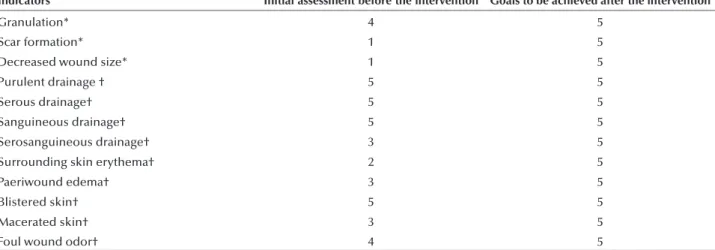
Table 1 – Initial assessment of PU and goals to be achieved through the outcome Wound Healing: secondary intention outcome from NOC – Porto Alegre, RS, Brazil, 2014.
Indicators Initial assessment before the intervention Goals to be achieved after the intervention
Granulation* 4 5
Scar formation* 1 5
Decreased wound size* 1 5
Purulent drainage † 5 5
Serous drainage† 5 5
Sanguineous drainage† 5 5
Serosanguineous drainage† 3 5
Surrounding skin erythema† 2 5
Paeriwound edema† 3 5
Blistered skin† 5 5
Macerated skin† 3 5
Foul wound odor† 4 5
In parallel with the evaluation performed with the re-sult of the NOC(12), we used the PUSH scale(11) which checks the wound area, the wound tissue type and exudate amount (Table 2).
nursing interventions – nic
To achieve the established goals, we planned and im-plemented the interventions based on the NIC: Pressure Ulcer Care and of Laser Precautions(15). hus, the lesion was treated with a daily dressing and adjuvant therapy with LLLT in order to promote accelerated wound healing and reduce the risk of clinical complications.
evaluationofHealingresults pu (noc and pusH)
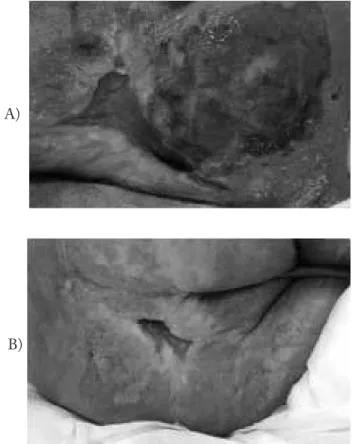
he evaluation of the results after the intervention was carried out reusing the result of NOC, called Wound Healing: secondary intention with 12 selected indica-secondary intention with 12 selected indica- 12 selected indica-tors(12), and the PUSH scale(11). his evaluation took place three times a week, over ive weeks (as described in the method section), always before the patient would receive LLLT and dressing interventions. he evolution of the lesion was also photographed over this period (Figure 1).
After the sixth day of LLLT use, there was a reduc-tion in the size of the lesion, which had, at the time, 4cm of length, width of 2.8cm and with an area of 11.2cm2. here was also increased granulation and epithelial tis-sue (but with the presence of devitalized tistis-sue in a part of the lesion), decreased serosanguineous secretion but still present in moderate amounts, no foul odor, increased maceration and perilesional erythema. his clinical chart demanded stronger orientation to the nursing staf and family members about the need to control moisture in diapers, opting for the use of urine collector. Patient de-cubitus changes were intensiied, though with some dif-iculty to mobilize when sitting on the chair.
On the ninth day of evaluation, the patient was anx-ious due to his transference from the ICU to a clinic in-patient unit of the hospital. However, the lesion contin-ued showing progress in the healing process. On that day, he presented a PU with 4cm length and 1.8cm width,
Table 2 – Initial assessment of the PU using PUSH scale – Porto Alegre, RS, Brazil, 2014.
PUSH Initial assessment before
intervention
Length x width* 10
Exudate amount† 2
Tissue type§ 2
* 0=0cm2; 1=<0.3cm2; 2=0.3 – 0,6 cm2; 3=0.7 – 1.0cm2; 4=1.1cm2 – 2.0cm2; 5=2.1
– 3.0cm2; 6=3.1 – 4.0cm2;7=4.1 – 8.0cm2; 8=8,1 – 12,0cm2; 9=12,1 – 24cm2;
10=>24cm2.
† 0=None; 1=Light; 2=Moderate; 3= Heavy.
§ 0= Closed/Resurfaced; 1=Epithelial tissue; 2=Granulation tissue; 3= Slough; 4=Necrotic tissue.
with an area of 7,2cm2, covered by granulation tissue, deined borders, serosanguineous secretion drainage in moderate amount, without the presence of foul odor, perilesional region with reduction of the erythema and maceration. he patient continued to use an urine col-lector and the nurse of the unit was instructed to request an airlow mattress.
On the 12th day of PU evaluation, he presented a further reduction in size, with 1.5cm length and 1.5cm width, with 3cm2 area. he borders of the lesion were via-ble and delimited with epithelialization tissue, and in the center of the lesion with granulation tissue. he lesion had a small amount of exudate serosanguinous without foul odor, skin adjacent to the lesion without erythema and without maceration. Diuresis in diapers because the “uripen” was injuring the penis and was removed.
On the 15th day of evaluation and inal LLLT use, the PU had 1.5cm length and 1.1cm width, with 1,65cm2 area. We highlight the reduction of 7cm length injury to 1.5cm and 6cm to 1.1cm width, comparing the irst and the 15th day of assessment. Epithelial tissue remained in the ascendancy, with signiicant reduction of the amount of serosanguineous secretion without foul odor. Never-theless, erythema and perilesional maceration had mild worsening, probably due to the diuresis in diapers, which increased perineal moisture. To reduce this problem the team and the family members were told about the need to increase the frequency of diaper changes.
Figure 1 – Initial assessment of the PU (a) and evaluation on day 15 of follow-up (b) – Porto Alegre, Brazil, 2014.
A)
Laser therapy in pressure ulcers: evaluation by the Pressure Ulcer Scale for Healing and Nursing Outcomes Classification
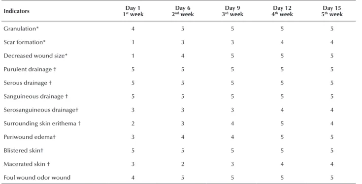
Table 3 – PU evaluation by the Wound Healing: Secondary Intention with 12 indicators from NOC. Porto Alegre, RS, Brazil, 2014.
Indicators Day 1
1st week 2Day 6 nd week 3Day 9 rd week 4Day 12th week 5Day 15 th week
Granulation* 4 5 5 5 5
Scar formation* 1 3 3 4 4
Decreased wound size* 1 4 5 5 5
Purulent drainage † 5 5 5 5 5
Serous drainage † 5 5 5 5 5
Sanguineous drainage † 5 5 5 5 5
Serosanguineous drainage† 3 3 3 4 4
Surrounding skin erithema † 2 3 4 5 4
Periwound edema† 3 4 4 5 5
Blistered skin† 5 5 5 5 5
Macerated skin † 3 2 3 4 4
Foul wound odor wound 4 5 5 5 5
*1=None; 2=Limited; 3=Moderate; 4=Substantial; 5=Extensive. †1=Extensive; 2=Substantial; 3=Moderate; 4=Limited; 5=None.
Table 3 summarizes the indicating scores of the NOC results(12) in ive distinct moments of the evaluation of PU treated with LLLT added to dressing.
he evolution of the scores on the PUSH scale(11) were also analyzed in ive distinct moments of the healing pro-cess of PU and are summarized in Table 4.
DISCUSSION
This study showed a significant decrease in the size of the PU, as its size decreased from 7cm to 1.5cm length and 6cm to 1.1cm width in a five-week period. Additionally, there was an increase in tissue granula-tion and epithelializagranula-tion, decreased secregranula-tion serosan-guineous and odor from the wound. The maceration and erythema around the wound also showed progres-sive improvement after the third day of LLLT use. This improvement can be explained by the fact that LLLT leads to release of histamine, serotonin and bradykinin, resulting in stimulation of ATP production and micro-circulation, an increase of epidermal regeneration rates,
Table 4 – PU evaluation using the Pressure Ulcer Scale for Healing – PUSH – Porto Alegre, RS, Brazil, 2014.
PUSH Day 1
1st week
Day 6 2nd week
Day 9 3rd week
Day 12 4th week
Day 15 5th week
Length x width* 10 8 7 5 4
Exudate amount† 2 2 2 1 1
Tissue type§ 2 2 2 1 1
* 0=0cm2; 1=<0.3cm2; 2=0.3 – 0,6 cm2; 3=0.7 – 1.0cm2; 4=1.1cm2 – 2.0cm2; 5=2.1 – 3.0cm2; 6=3.1 – 4.0cm2;7=4.1 – 8.0cm2; 8=8,1 – 12,0cm2; 9=12,1 – 24cm2; 10=>24cm2.
† 0=None; 1=Light; 2=Moderate; 3= Heavy.
§0= Closed/Resurfaced; 1=Epithelial tissue; 2=Granulation tissue; 3= Slough; 4=Necrotic tissue.
analgesic, anti-inflammatory, antiedema and wound healing actions(21).
The scar evolution of PU observed is conigured as an inspiration and motivation for the use of LLLT in treating patients with this type of multifactorial lesion etiology, such as metabolic disorders, extremes of age, nutritional dysfunction, urinary or fecal incontinence, hydration, mobility conditions and sensory perception(1). Critically
which constitute important elements for the development of PU(23-24). Thus, it is essential that preventive care is
introduced earlier(23-24), although in some cases
preven-tion was not able to ensure the development of the lesion, which requires effective treatment when it appears.
In the case of this patient, considering his history of paraplegia, he also presented restriction for the changes of position in bed for a long period due to tetanus complica-tions. his restricted the possibilities for prevention and treatment of PU, which performed slow healing process, despite the daily dressings, being observed an acceleration of the healing process after adjuvant use of LLLT.
he evaluation of the healing process of the PU were shown using the Wound Healing: Secondary Intention with 12 indicators from NOC(12), which were deined op-erationally, considering the magnitude of the ive points Likert scale. hus, it obtained more detailed and reliable measurement results after the LLLT intervention compared with the PUSH scale use(11).
It was found that in the PUSH scale(11), the item tissue type score equates to a diferent tissue, there is no possibil-ity of scoring the presence of two distinct tissues and even the progress of each. In the NOC(12), each indicator con-sists of a diferent tissue, thus, it is possible to mark more than one tissue type and rate the progress in each lesion.
In the item Quantity of exudate, the PUSH(11) deines the scale scores to none, light, moderate and heavy, this is a sub-jective evaluation, since it does not point parameters to deine what each of them are. Similarly, the PUSH(11) does not iden-tify the diferent types of secretion. When using the NOC indicators(12), these questions could be speciied, because ad-dressed the professional operational deinitions to assess ob-jectively each of these elements in a standardized manner(25-26). hat is, it was possible to quantify the exudate and characterize its aspect along with the PU healing process.
We highlight that this study was the irst to use NOC(12) associated with PUSH(11) and that despite these classiication
RESUMO
Objetivo: Descrever o processo de cicatrização de úlcera por pressão em paciente crítico tratado com terapêutica convencional de curativo acrescida de laserterapia de baixa intensidade avaliada pela Pressure Ulcer Scale for Healing (PUSH) e pelo resultado Cicatrização de Feridas: segunda intenção, da Nursing Outcomes Classiication (NOC). Método: Estudo de caso norteado pelo processo de enfermagem realizado com paciente de unidade de terapia intensiva. Os dados foram coletados com instrumento contendo a PUSH e o resultado da NOC. Na análise utilizou-se a estatística descritiva, considerando-se as pontuações obtidas no instrumento. Resultados: Observou-se redução nas dimensões da lesão de 7cm para 1,5cm de comprimento e de 6cm para 1,1cm de largura, além do aumento do tecido epitelial e de granulação, diminuição da secreção e odor. Conclusão: Houve melhora no processo de cicatrização da lesão tratada com terapia adjuvante e o uso da NOC permitiu uma avaliação mais detalhada e precisa do que da PUSH.
DESCRITORES
Úlcera por Pressão; Terapia a Laser; Cicatrização; Resultado do Tratamento; Processos de Enfermagem; Unidades de Terapia Intensiva.
RESUMEN
Objetivo: Describir el proceso de cicatrización de úlcera por presión en paciente crítico tratado con terapéutica convencional de curativo agregada a la laserterapia de baja intensidad evaluada por la Pressure Ulcer Scale for Healing (PUSH) y el resultado Cicatrización de Heridas por segunda intención, de la Nursing Outcomes Classiication (NOC). Método: Estudio de caso orientado por el proceso de enfermería llevado a cabo con paciente de unidad de terapia intensiva. Los datos fueron recogidos con instrumento conteniendo la PUSH y el resultado de la NOC. En el análisis se utilizó la estadística descriptiva, considerándose las puntuaciones obtenidas en el instrumento. Resultados: Se observó reducción en las dimensiones de la lesión de 7cm a 1,5cm de largo y de 6cm a 1,1cm de ancho, además del aumento del tejido epitelial y de granulación, y la reducción de la secreción y el dolor. Conclusión: Hubo mejora en el proceso de cicatrización de la lesión tratada con terapia adyuvante, y el uso de la NOC permitió una evaluación más detallada y precisa que la PUSH.
systems help the adoption of standardized terms and confer visibility to the work of nurses, it had not been explored in this way yet, which gives importance to our indings. he results showed that the NOC(12) can be used safely in patients' as-sessments for PU treatment, as their indicators favoured the evaluation and description of the healing process of the lesion treated with LLLT use, which proved promising and efective as an adjunctive therapy.
CONCLUSION
We conclude that there was signiicant improvement in the healing process of the PU treated with adjuvant LLLT, which was shown by the Healing Wounds: sec-ondary intention according to NOC, and PUSH scale, in addition to evidence of photographic record. he scores related to NOC indicators called Scar formation, De-creased wound size, Serosanguineous drainage, Surround-ing skin erythema and Periwound edema, Macerated skin and Foul wound odor after LLLT use, pointed to a lesion improvement. On the other hand, the PUSH scores de-creased, indicating a reduction in size and wound exudate, and increased tissue epithelialization.
hus we speculate the possibility of using LLLT on PU treatment protocols, because this intervention accelerated tissue proliferation and increased local vascularization, with granulation tissue formation by promoting rapid healing of the lesion. he case report study proved an important method to support the design of future clinical studies with a larger and randomized sample capable of producing greater levels of evidence of the beneits of this therapy, as this research has the limitation of using a single patient.
Laser therapy in pressure ulcers: evaluation by the Pressure Ulcer Scale for Healing and Nursing Outcomes Classification
REFERENCES
1. National Pressure Ulcer Advisory Panel (NPUAP); European Pressure Ulcer Advisory Panel (EPUAP); Pan Paciic Pressure Injury Alliance
(PPPIA). Prevention and treatment of pressure ulcers: quick reference guide. Perth, Australia: Cambridge Media; 2014.
2. Rogenski NM, Kurcgant P. The incidence of pressure ulcers after the implementation of a prevention protocol. Rev Latino Am Enfermagem. 2012;20(2):333-9.
3. Hyun S, Li X, Vermillion B, Newton C, Fall M, Kaewprag P, et al. Body mass index and pressure ulcers: improved predictability of pressure ulcers in intensive care patients. Am J Crit Care. 2014;23(6):494-500.
4. Manzano F, Navarro MJ, Roldán D, Moral MA, Leyva I, Guerrero C, et al. Pressure ulcer incidence and risk factors in ventilated intensive care patients. J Crit Care. 2010;25(3):469-76.
5. Alves P, Mota F, Ramos P, Vales L. Epidemiology of pressure ulcers: interpreting data epidemiological as an indicator of quality. Servir. 2013;58(1-2):10-8.
6. NANDA International. Nursing diagnoses: deinitions & classiication, 2015-2017. Oxford: Wiley Blackwell; 2014.
7. Costa IG, Caliri MHL. Predictive validity of the Braden Scale for patients in intensive care. Acta Paul Enferm. 2011;24(6):772-7.
8. Olyaie M, Rad FS, Elahifar MA, Garkaz A, Mahsa G. High-frequency and noncontact low-frequency ultrasound therapy for venous leg ulcer treatment: a randomized, controlled study. Ostomy Wound Manage. 2013;59(8):14-20.
9. Zhang J, Guan M, Xie C, Luo X, Zhang Q, Xue Y. Increased growth factors play a role in wound healing promoted by noninvasive oxygen-ozone therapy in diabetic patients with foot ulcers. Oxid Med Cell Longev. 2014;2014:273475.
10. Dissemond J. Physical treatment modalities for chronic leg ulcers. Hautarzt. 2010;61(5):387-96.
11. Santos VL, Azevedo MA, Silva TS, Carvalho VM, Carvalho VF. Crosscultural adaptation of the pressure ulcer scale for healing to the
portuguese language. Rev Latino Am Enfermagem. 2005;13(3):305-13.
12. Moorhead S, Johnson M, Maas ML, Swanson E. Nursing Outcomes Classiication (NOC): measurement of health outcomes. 5th ed. Philadelphia: Elsevier; 2013.
13. Lucena AF, Santos CT, Pereira AGS, Almeida MA, Dias VLM, Friedrich MA. Clinical proile and nursing diagnosis of patients at risk of pressure ulcers. Rev Latino Am Enfermagem. 2011;19(3):523-30.
14. Azzolin K, Mussi CM, Ruschel KB, Souza EN, Lucena AF, Rabelo-Silva ER. Effectiveness of nursing interventions in heart failure patients in home care using NANDA-I, NIC, and NOC. Appl Nurs Res. 2013;26(4):239-44.
15. Bulechek GM, Butcher HK, Dochterman JM, Wagner C. Nursing Interventions Classiication (NIC).6th ed. Philadelphia: Elsevier; 2013. 16. Hopkins JT, McLoda TA, Seegmiller JG, David Baxter G. Low-level laser therapy facilitates supericial wound healing in humans: a
triple-blind, shamcontrolled study. J Athl Train. 2004;39(3):223-9.
17. Pinto NC, Pereira HC, Stolf NAG, Chavantes MC. Low level laser therapy in acute dehiscence saphenectomy: therapeutic proposal. Rev Bras Cir Cardiovasc. 2009;24(1):88-91.
18. Andrade FSSD, Clark RMO, Ferreira ML. Effects of low-level laser therapy on wound healing. Rev Col Bras Cir. 2014;41(2):129-33. 19. Araújo TM, Araújo MF, Caetano JA. Using the braden scale and photographs to assess pressure ulcer risk. Rev Esc Enferm USP.
2012;46(4):858-64.
20. Yin RK. Case study research: design and methods. 4th ed. Los Angeles: Sage; 2009.
21. Franek A, Krol P, Kucharzewski M. Does low output laser stimulation enhance the healing of crural ulceration? Some critical remarks. Med Eng Phys. 2002;24(9):607-15.
22. Menegon DB, Bercini RR, Brambila MI, Scola ML, Jansen MM, Tanaka RY. Implantação do protocolo assistencial de prevenção e tratamento de úlcera de pressão do Hospital de Clínicas de Porto Alegre. Rev Hosp Clin Porto Alegre. 2007;27(2):61-4.
23. Simao CMF, Caliri MHL, Santos CB. Agreement between nurses regarding patients' risk for developing pressure ulcer. Acta Paul Enferm. 2013;26(1):30-5.
24. Tayyib N, Coyer F, Lewis PA. A two- arm cluster randomized control trial to determine the effectiveness of a pressure ulcer prevention bundle for critically ill patients. J Nurs Scholarsh. 2015;47(3):237-47.
25. Chianca TC, Salgado PO, Albuquerque JP, Campos CC, Tannure MC, Ercole FF. Mapping nursing goals of an Intensive Care Unit to the Nursing Outcomes Classiication. Rev Latino Am Enfermagem. 2012;20(5):854-62.
26. Almeida MA, Seganfredo DH, Barreto LNM, Lucena AF. Validation of indicators of the nursing outcomes classiication for hospitalized adults at risk of infection. Texto Contexto Enferm. 2014;23(2):309-17.
DESCRIPTORES