Association between serum selenium level and conversion of
bacteriological tests during antituberculosis treatment*
,**
Associações entre níveis de selênio sérico e conversão de testes bacteriológicos durante o tratamento antituberculose
Milena Lima de Moraes, Daniela Maria de Paula Ramalho, Karina Neves Delogo, Pryscila Fernandes Campino Miranda, Eliene Denites Duarte Mesquita,
Hedi Marinho de Melo Guedes de Oliveira, Antônio Ruffino-Netto, Paulo César de Almeida, Rachel Ann Hauser-Davis, Reinaldo Calixto Campos,
Afrânio Lineu Kritski, Martha Maria de Oliveira
Abstract
Objective: To determine whether serum selenium levels are associated with the conversion of bacteriological tests in patients diagnosed with active pulmonary tuberculosis after eight weeks of standard treatment. Methods: We evaluated 35 healthy male controls and 35 male patients with pulmonary tuberculosis, the latter being evaluated at baseline, as well as at 30 and 60 days of antituberculosis treatment. For all participants, we measured anthropometric indices, as well as determining serum levels of albumin, C-reactive protein (CRP) and selenium. Because there are no reference values for the Brazilian population, we used the median of the serum selenium level of the controls as the cut-off point. At 30 and 60 days of antituberculosis treatment, we repeated the biochemical tests, as well as collecting sputum for smear microscopy and culture from the patients. Results: The mean age of the patients was 38.4 ± 11.4 years. Of the 35 patients, 25 (71%) described themselves as alcoholic; 20 (57.0%) were smokers; and 21 (60.0%) and 32 (91.4%) presented with muscle mass depletion as determined by measuring the triceps skinfold thickness and arm muscle area, respectively. Of 24 patients, 12 (39.2%) were classified as moderately or severely emaciated, and 15 (62.5%) had lost > 10% of their body weight by six months before diagnosis. At baseline, the tuberculosis group had lower serum selenium levels than did the control group. The conversion of bacteriological tests was associated with the CRP/albumin ratio and serum selenium levels 60 days after treatment initiation. Conclusions: Higher serum selenium levels after 60 days of treatment were associated with the conversion of bacteriological tests in pulmonary tuberculosis patients. Keywords: Selenium; Nutritional status; Tuberculosis; Immunity.
Resumo
Objetivo: Determinar se os níveis séricos de selênio estão associados à conversão dos testes bacteriológicos em pacientes diagnosticados com tuberculose pulmonar ativa após oito semanas de tratamento-padrão. Métodos: No início do estudo, avaliamos 35 controles saudáveis, do sexo masculino, e 35 pacientes do sexo masculino com tuberculose pulmonar. Estes foram também avaliados após 30 e 60 dias de tratamento antituberculose. Todos os participantes submeteram-se a medições antropométricas e quantificação dos níveis séricos de albumina, proteína C reativa (PCR) e selênio. Como não há valores de referência para a população brasileira, usamos a mediana dos resultados de selênio sérico dos controles como ponto de corte. Aos 30 e 60 dias do tratamento antituberculose, todos os testes bioquímicos foram repetidos, e foram coletadas amostras de escarro para baciloscopia e cultura. Resultados: A média de idade dos pacientes foi de 38,4 ± 11.4 anos. Dos 35 pacientes, 25 (71,0%) referiram alcoolismo, 20 (57,0%) eram fumantes, e 21 (60,0%) e 32 (91,4%) apresentavam depleção muscular pela medição da dobra cutânea tricipital e da área muscular do braço, respectivamente. De 24 pacientes, 12 (39,2%) foram classificados em moderadamente ou gravemente magros, e 15 (62,5%) apresentaram perda de peso > 10% em até seis meses antes do diagnóstico. No início do estudo, o grupo com tuberculose apresentou menores níveis de selênio sérico que os controles. A conversão dos testes bacteriológicos associou-se à relação PCR/albumina e aos níveis de selênio sérico 60 dias após o início do tratamento. Conclusões: Níveis maiores de selênio sérico após 60 dias de tratamento associaram-se à conversão bacteriológica em pacientes com tuberculose pulmonar. Descritores: Selênio; Estado nutricional; Tuberculose; Imunidade.
*Study carried out in the Department of Clinical Medicine, Federal University of Rio de Janeiro School of Medicine, Rio de Janeiro, Brazil.
Correspondence to: Milena Lima de Moraes. Centro de Pesquisa em Tuberculose, Rua Professor Rodolpho Paulo Rocco, 255, 4º andar, Ilha do Fundão, CEP 21941-913, Rio de Janeiro, RJ, Brasil.
Tel. 55 21 2562-2426. Fax: 55 21 2550-0693. E-mail: [email protected]
Financial support: This study received financial support from the Brazilian Conselho Nacional de Desenvolvimento Científico e Tecnológico (CNPq, National Council for Scientific and Technological Development; grant MCT/CNPq/INCT 573548/2008-0 and MCT/CNPq/INCT 478033/2009-5), and from the Fundação de Amparo à Pesquisa do Estado do Rio de Janeiro (FAPERJ, Foundation for the Support of Research in the State of Rio de Janeiro; grant E 26/110974/2011).
Submitted: 5 August 2013. Accepted, after review: 30 April 2014.
admitted to either of the two referral hospitals for tuberculosis in the city of Rio de Janeiro, Brazil, namely the Hospital Estadual Santa Maria and the Instituto Estadual de Doenças do Tórax Ary Parreiras. We decided to include only male patients in the study because the great majority of the patients treated in these hospitals are males, and the inclusion of very few female patients could become a confounding factor in the data analysis. The patients enrolled in the present study had been hospitalized for clinical reasons; however, in most cases, the duration of hospital stay was prolonged for at least 60 days due to social reasons. The inclusion criteria were as follows: being 19-60 years of age; having a positive culture for M. tuberculosis or positive smear microscopy in spontaneous sputum in association with chest X-rays and symptoms indicative of tuberculosis; receiving treatment with first-line antituberculosis drugs; not having diabetes mellitus or renal disease (undergoing peritoneal dialysis or hemodialysis); having tested negative for HIV; and reporting no comorbidities. Because there are no established reference values for selenium levels in serum for the Brazilian population, we determined the serum selenium levels of 35 HIV-negative healthy subjects residing in the city of Rio de Janeiro (using similar inclusion criteria) in order to define a cut-off point. All subjects gave written informed consent. The study was approved by the Research Ethics Committee of the Federal University of Rio de Janeiro (Protocol no. 004/05, of April 28, 2005). The patients enrolled in the pilot study were not included in the present study.
Data collection
A pilot study was conducted in order to determine the adequacy of the questionnaire applied to the study subjects. The interviewers were trained regarding data collection. Anthropometric measurements taken by different interviewers showed a high level of inter-rater agreement (> 95%).
The pulmonary tuberculosis patients completed a questionnaire regarding demographic data, socioeconomic data, and tobacco use, as well as the criteria used in the Cut down, Annoyed, Guilty, and Eye-opener (CAGE) questionnaire.
(8) Anthropometric measurements were collected
at baseline, as well as at 30 and 60 days after antituberculosis treatment initiation. Blood and
Introduction
The World Health Organization considers tuberculosis a serious public health problem. In 2010, 9.4 million new tuberculosis cases occurred, with 1.7 million associated deaths, among which 500,000 were HIV-positive patients. In Brazil, tuberculosis is the leading cause of mortality among patients with HIV/AIDS, a result arising
from late diagnosis.(1) Since 2006, the Global
Plan to Stop TB has been prioritizing the critical points in the field of tuberculosis, especially the development of new diagnostic tests, vaccines, drugs, and biomarkers of therapeutic response,
of healing, and of disease recurrence.(2)
Among the risk factors associated with the occurrence of tuberculosis are precarious working conditions and changes in host defense against the infection by Mycobacterium tuberculosis, such as malnutrition, smoking, diabetes mellitus,
and alcohol abuse.(3)
The degree of malnutrition is associated with the severity of pulmonary tuberculosis in adults. Tuberculosis patients usually present malnutrition and a decrease in micronutrient levels, regardless
of their HIV status.(4)
Recently, one group of authors(5) reported
that a two-month intervention with vitamin E and selenium supplements reduced oxidative stress and increased total antioxidant capacity in patients with pulmonary tuberculosis undergoing standard treatment. A similar improvement in the immune status of patients with tuberculosis who received selenium supplementation was also
reported in another study.(6)
The objective of the present study was to determine whether serum selenium levels are associated with the conversion of bacteriological tests in patients diagnosed with active pulmonary tuberculosis after eight weeks of standard treatment. The conversion (to negative) of cultures of sputum collected eight weeks after treatment initiation has been used as a useful marker of the sterilizing activity of tuberculosis treatment,(7) and a substantial
improvement in serum selenium levels in these patients would indicate that selenium can be a biomarker of therapeutic response.
Methods
Study subjects
tubes. The samples were centrifuged at 3,000 g for 15 min for further quantification of albumin, C-reactive protein (CRP), and selenium. All quantifications were performed immediately after sample collection, except for the determination of selenium levels. In this case, a portion of the
serum obtained was stored at −70°C for later
quantification.
Albumin quantification was determined
colorimetrically (Advia®; Siemens Healthcare
Diagnostics, Eschborn, Germany). According to the manufacturer, normal albumin values should range from 3.4 to 4.8 g/dL. CRP was measured by nephelometry using a CardioPhase hsCRP assay (Dade Behring Holding GmbH, Liederbach, Germany) and a BNII nephelometer (Siemens Healthcare, Indianapolis, IN, USA). According to the manufacturer, normal values lay below 0.3 mg/dL.
In the present study, we evaluated the CRP/ albumin ratio as a substitute for the prognostic inflammatory nutritional index because it maintains the same diagnostic sensitivity regarding the
levels of complication risks.(13) According to one
study, the levels of complication risks are as follows: no risk, if the ratio is < 0.4; low risk, from 0.4 to 1.1; medium risk, from 1.2 to 2.0; and high risk, > 2.0.(13)
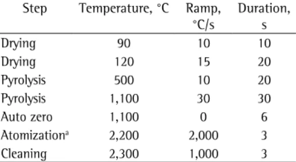
The determination of selenium levels was performed by graphite furnace atomic absorption spectrometry, using a ZEEnit 60 spectrometer (Analytik Jena, Jena, Germany) equipped with a selenium hollow cathode lamp operating at a wavelength of 196.0 nm. After the thawing and homogenizing of the serum samples, 200 mL aliquots were transferred to polyethylene tubes, free of trace elements, and 1 mL of a 0.1% v/v Triton ×100 solution was added. This solution (10 mL) was used for the instrumental analysis, together with a mixture (10 mL) containing palladium (0.15% m/v) and magnesium (0.10% m/v) as matrix modifier. External calibration was performed with calibration solutions prepared in the stock solution, and the temperature protocol is shown in Table 1. All measurements were conducted at least in triplicate.
Sputum samples of the subjects included in the study were collected in disposable vials. Smear microscopy and cultures for mycobacteria were performed in accordance with the recommendations
by the Brazilian National Ministry of Health.(14)
sputum samples were also collected at the same time points. At 30- and 60-day sample collection time points, some of the patients no longer presented sputum production, and therefore no sputum smear microscopy/culture were performed for those patients. The healthy subjects also completed the questionnaire, underwent anthropometric assessment, and had their blood samples collected.
The anthropometric evaluation consisted of two body weight measurements using a calibrated platform scale with a stadiometer (Filizola, São Paulo, Brazil) with a sensitivity of 100 g and maximum weight of 150 kg. The subjects were weighed barefoot and wearing light clothing. Height was measured twice (stadiometer with a sensitivity of 0.5 cm and maximum height of 191 cm).
The body mass index (BMI) was calculated
by the formula weight/height2 and classified
according to the World Health Organization
recommendations: underweight, < 18.5 kg/m2;
normal weight, 18.5-24.9 kg/m2; and overweight,
≥ 25.0 kg/m2.(9) All measurements were collected
in accordance with the techniques recommended
by Gibson(10) in order to avoid possible bias.
The patients with pulmonary tuberculosis also reported their usual weight (in the last 6 months) so that their weight loss until the beginning of the study (baseline) could be estimated.
The triceps skinfold thickness (TST) was measured three times with an adipometer (Lange Beta Technology Inc., Cambridge, MD, USA) with a sensitivity of 0.5 mm. Measurements were taken at the midpoint of the back of the non-dominant arm, between the acromion and olecranon, with the subjects standing with their arms relaxed and extended alongside the body.
The measurement of arm circumference (AC) was performed twice, with a flexible and inelastic millimeter tape at the same height as the midpoint used for the TST measurement. After that, the arm muscle area (AMA) was calculated using the
following equation(11):
AMA (cm²) = [(AC(cm) − π × TST(mm) ÷ 10)²
− 10]/4π.
Mean TST and AMA results were calculated, and
the cut-off values used were those by Frisancho.(12)
Statistical analysis
The Kolmogorov-Smirnov test was used in order to verify the normality of the variables, and the Levene test was used in order to determine the equality of variances. A logarithmic transformation was used for the variables that showed non-normal distribution. We used Tukey’s test and Games-Howell test to compare pairs of groups with equal and different variances, respectively. When appropriate, ANOVA and Student’s t-test were used in order to estimate differences between quantitative variables. To evaluate the association between categorical variables, we used the chi-square test with continuity correction when indicated. A p-value < 0.05 was considered significant. The Statistical Package for the Social Sciences, version 16.0 (SPSS Inc., Chicago, IL, USA), was used for data analysis.
Results
We included 35 pulmonary tuberculosis patients in the study group at baseline. Among these, 6 were recurrent tuberculosis patients. After 30 days of treatment, only 29 patients presented spontaneous sputum production, and, after 60 days of treatment, 34 patients showed spontaneous sputum production (Figure 1).
The general characteristics of the pulmonary tuberculosis patients are presented in Table 2. The
mean age of the patients was 38.4 ± 11.4 years.
Among the 35 male study subjects included in the study, 25 (71%) reported alcoholism according to the CAGE questionnaire, and 20 (57%) were smokers. We determined the BMI of 24 of the patients, and 12 (39%) were classified as being severely or moderately emaciated. Of the 35 patients, 21 (60%) and 32 (91%) were found to have with muscle mass depletion on the basis of their TST and AMA, respectively. Of the 24 patients who provided information regarding their weight by 6 months prior to their inclusion in the study, 15 (63%) had lost > 10% of their body weight. Statistically significant differences were found between the pulmonary tuberculosis patients and the healthy controls at baseline.
When we compared the three study time points (baseline, 30 days, and 60 days), we found that the conversion to a negative-culture status was associated with the CRP levels and the CRP/ albumin ratio results at 30 and 60 days, as well Cultures contaminated by other microorganisms
were designated as contaminated and considered negative in the data analysis. The strains were identified as M. tuberculosis on the basis of the characteristics of the colonies (rough, opaque, and creamy) and biochemical testing (ability to produce niacin, nitrate reduction, and thermal
inactivation of catalase).(14) In the present study,
the individuals were diagnosed with pulmonary tuberculosis at baseline when cultures were positive for M. tuberculosis or when there were positive results in sputum smear microscopy associated with X-ray findings and symptoms indicative of tuberculosis. Patients who presented with X-ray findings and symptoms indicative of tuberculosis but negative cultures or smear results at baseline were not included in the study.
Susceptibility testing was performed on the clinical specimens from 28 patients who had positive cultures using the method of proportions, which is considered the gold standard. In addition, we used the indirect proportion method (one strain per patient) in order to determine the susceptibility of the M. tuberculosis strains to isoniazid, rifampin, streptomycin, and ethambutol. All of the tested strains were susceptible to the drugs tested.
New sputum samples were collected 30 and 60 days after treatment initiation, and new smear microscopy testing and cultures for mycobacteria were performed. Depending on the results of the tests, the patients could be reallocated to either of the two groups: tuberculosis-positive (TB+) group, when smears or cultures were positive
for M. tuberculosis; and tuberculosis-negative
(TB−) group, when smears and cultures were
negative for M. tuberculosis. The individuals who were unable to produce sputum spontaneously at the moments of collection were not included in either group.
Table 1 - Temperature program used in order to determine selenium levels in serum.
Step Temperature, °C Ramp,
°C/s
Duration, s
Drying 90 10 10
Drying 120 15 20
Pyrolysis 500 10 20
Pyrolysis 1,100 30 30
Auto zero 1,100 0 6
Atomizationa 2,200 2,000 3
Cleaning 2,300 1,000 3
as with albumin and selenium levels at 60 days (Table 3). No differences were observed between
the TB+ and TB− groups for any of these variables
at 30 days.
Table 4 presents the distribution of patients in
the TB+ and TB− groups in relation to the results
of the biochemical tests and serum selenium levels at the three study time points in order to determine the existence of any associations. In order to evaluate the association between the results of bacteriological tests (culture and smear microscopy) and serum selenium levels, we used the cut-off point based on the median of the results obtained in the healthy control group.
Discussion
In the present study, the clinical characteristics of the patients are similar to those described in other studies carried out in referral hospitals for the treatment of tuberculosis in developing nations,
with high rates of alcoholism and tobacco use.(15)
TB+ at baseline (study group)
(n = 35)
TB+ at baseline (study group)
(n = 35)
TB+ at baseline (study group)
(n = 35)
Baseline
30 day
s
60 day
s
Healthy adults (control group)
n = 35
TB-n = 15
TB+ n = 14
No sputum n = 6
No sputum n = 1
TB-n = 23
TB+ n = 11
Figure 1 - Study and control groups at baseline, at 30 days after antituberculosis treatment initiation, and at 60 days after antituberculosis treatment initiation. TB+: positive sputum culture or positive sputum smear
microscopy results at that study time point; and TB−:
negative sputum culture and negative sputum smear microscopy results at that study time point.
Table 2 - General characteristics of the patients with pulmonary tuberculosis (N = 35).a
Characteristic Result
Age, yearsb 38.43 ± 11.42
Alcoholism 25 (71)
Smoking status
Smokers 20 (57)
Former smokers 7 (20)
Never smokers 8 (23)
Weight loss, kgb,c 11.03 ± 9.69
Weight loss, %c
> 10 15 (63)
5-10 4 (17)
< 5% 2 (8)
No loss 3 (12)
Classification according to BMId
Severe thinness 6 (19.4)
Moderate thinness 6(19.4)
Mild thinness 6 (19.4)
Normal weight 12 (38.7)
Overweight or obese 1 (3.2) Classification according to TST
Depletion 21 (60.0)
Normal 14 (40.0)
Classification according to AMA
Depletion 32 (91.4)
Normal 3 (8.6)
BMI: body mass index; TST: triceps skinfold thickness;
and AMA: arm muscle area. aValues expressed as n (%),
except where otherwise indicated. bValues expressed as
mean ± SD. cn = 24. dn = 31
The relationship between tuberculosis and malnutrition has been revisited, since malnutrition may predispose to the development of active tuberculosis, and tuberculosis can contribute to
malnutrition.(16) The mean weight loss in the study
group prior to antituberculosis treatment initiation
was 11.03 ± 9.69 kg. This can be considered
even more significant when categorized by the percentage of body weight loss, because 63%
of the patients presented with a weight loss ≥
10%, which is considered a predisposing factor for tuberculosis.(17)
In the present study, the assessment of the nutritional status based on anthropometric parameters (BMI, TST, and AMA) confirmed the depleted nutritional status in the study
group, as described in the literature.(18) For any
Table 3 - Anthropometric variables, biochemical test results, and serum selenium levels in the groups studied at the three study time points.a
Variable Baseline 30 days after treatment initiation 60 days after treatment initiation Control TB+ group TB−
group TB+ group TB+ group at baseline TB− group TB+ group TB+ group at baseline (n = 35) (n = 35) (n = 15) (n = 14) (n = 35) (n = 23) (n = 11) (n = 35) BMI, kg/m2 25.27 ±
3.59
18.21 ± 2.53*
19.60 ± 2.18*
19.40 ± 2.46*
19.49 ± 2.86*
20.64 ± 3.25*
20.41 ± 3.10*
20.53 ± 3.11* TST, mm 12.71 ±
4.99
5.11 ± 2.51*
6.28 ± 2.48*
5.87 ± 1.82*
6.13 ± 2.57*
7.42 ± 4.32*
7.15 ± 2.83*
7.35 ± 3.80* AMA, cm2 55.15 ±
12.11
26.10 ± 7.92*
28.54 ± 9.86*
29.07 ± 9.24*
28.74 ± 9.69*
32.29 ± 12.37*
30.52 ± 12.09*
31.77 ± 11.94* Alb, g/dL 4.86 ±
0.19
3.64 ± 0.62*
3.99 ± 0.38*
4.02 ± 0.60*
4.02 ± 0.47*
4.27 ± 0.50*†
3.95 ± 0.37*
4.16 ± 0.48*†
CRP, mg/dL 0.16 ± 0.16
6.35 ± 4.12*
2.31 ± 1.88*†
4.33 ± 3.36*
3.66 ± 3.55*
1.95 ± 1.70*†
4.43 ± 3.69*‡
2.68 ± 2.71*†
CRP/alb ratio 0.03 ± 0.03
1.93 ± 1.58*
0.60 ± 0.52*†
1.22 ± 1.20*
0.99 ± 1.11*†
0.48 ± 0.44*†
1.20 ± 1.06*‡
0.70 ± 0.76 Se, μg/L 100.12 ±
12.11
80.13 ± 46.92*
93.55 ± 56.40*
77.31 ± 40.64*
88.26 ± 54.56*
104.53 ± 55.35
70.89 ± 38.66*‡
97.60 ± 54.59††
TB+: positive sputum culture or positive sputum smear microscopy results at that study time point; TB−: negative sputum
culture and negative smear sputum microscopy results at that study time point; BMI: body mass index; TST: triceps
skinfold thickness; AMA: arm muscle area; Alb: albumin; CRP: C-reactive protein; Se: selenium. aValues expressed as
mean ± SD. *p < 0.05 vs. control. †p < 0.05 vs. TB+ group at baseline. ‡p < 0.05 TB+ group vs. TB− group. ††p < 0.05
TB+ group vs. TB+ group at baseline. Tukey test (equal variances), Games-Howell test (different variances).
Table 4 - Distribution of the patients in the TB+ and TB− groups in relation to the results of the biochemical
tests and serum selenium levels at the three study time points.a
Variable Baseline 30 days after treatment initiation 60 days after treatment initiation TB+ group TB− group TB+ group p* TB− group TB+ group p*
(n = 35) (n = 15) (n = 14) (n = 23) (n = 11)
Albumin, g/dL 0.792 0.338
< 3.4 11 (100) 1 (33.3) 2 (66.7) 2 (100) 0 (0.0) 3.4-4.8b 24 (100) 13 (54.2) 11 (45.8) 19 (63.3) 11 (36.7)
>4.8 0 (0.0) 1 (50.0) 1 (50.0) 2 (100) 0 (0.0)
CRP, mg/dL 0.617 0.683
< 0.3b 1 (100) 2 (100) 0 (0.0) 2 (100) 0 (0.0)
≥ 0.3 34 (100) 13 (50.0) 13 (50.0) 21 (65.6) 11 (34.4)
CRP/albumin ratioc 0.206 0.041
< 0.4 2 (100) 3 (30.0) 7 (70.0) 12 (75.0) 4 (25.0) 0.4-1.1 11 (100) 5 (50.0) 5 (50.0) 9 (81.8) 2 (18.2) 1.2-2.0 8 (100) 2 (50.0) 2 (50.0) 2 (50.0) 2 (50.0)
> 2.0 13 (100) 3 (100) 0 (0.0) 0 (0.0) 3 (100)
Selenium
< cut-off pointd 24 (100) 9 (47.4) 10 (52.6) 0.518 12 (54.5) 10 (45.5) 0.027 ≥ cut-off pointd 11 (100) 6 (60.0) 4 (40.0) 11 (91.7) 1 (8.3)
TB−: negative sputum culture and negative sputum smear microscopy results at that study time point; TB+: positive
sputum culture or positive sputum smear microscopy results at that study time point; and CRP: C-reactive protein.
aValues expressed as n (%). bNormal values. cUsed in order to determine the level of complication risks.(13) dBased on
the median of the results obtained in the healthy control group. *Chi-square test.
tissue loss. In tuberculosis patients, reduced appetite, malabsorption of macronutrients and micronutrients, and altered metabolism lead to
cachexia.(16) However, no association between
confirming the association between bacteriological conversion and decreased in CRP levels.
One group of authors evaluated CRP levels in patients with pulmonary tuberculosis during 6 months of treatment; at 3 and 6 months after treatment initiation, there was a significant
reduction in CRP levels.(22) CRP has been identified
as an important indicator in the diagnosis of individuals with suspected tuberculosis and positive
smear microscopy.(24) In our study, a statistically
significant association between lower CRP/albumin ratio values and negative cultures for mycobacteria was also found. The CRP/albumin ratio has been described to be increased in patients with other
APR-related diseases.(14)
The tuberculosis infection is a condition known to induce oxidative stress in the infected organism, such as the production of reactive oxygen species (ROS) derived from free radicals. These ROS are associated with dysfunction in pulmonary tuberculosis. A way of suppressing these ROS is by means of antioxidant enzymes, which scavenge free radicals and protect cells from oxidative damage. Various of these enzymes, such as glutathione peroxidase, have selenium
as an essential element.(25) Thus, a reduction in
micronutrient intake (such as vitamins, zinc, and selenium) leads to impaired immune responses.
Studies show that patients with active tuberculosis have lower concentrations of various micronutrients, including selenium, in blood. (26) In the present study, the healthy subjects
showed higher selenium levels when compared with the study group at baseline. Among the pulmonary tuberculosis patients, we found an association between positive culture results and low selenium levels even after 60 days of treatment. Micronutrient deficiency is a frequent cause of secondary immunodeficiency and morbidity due to related infections, including tuberculosis. This trace element has an important role in the maintenance of immune processes and, therefore, may have a fundamental role in the defense against the mycobacteria. Low selenium levels have been considered a significant risk factor for the development of mycobacterial disease
in HIV-positive patients.(27) In one study with
22 pulmonary tuberculosis patients who were
newly diagnosed with positive sputum,(28) the
authors found a significant difference between selenium levels between the control and study groups at baseline, as we found in the present patients (even in those whose results converted
to negative) after 60 days of treatment. The use of BMI as an indicator of nutrition in the relationship between nutritional status and
tuberculosis has been reported.(19) The evaluation
of TST and AMA in patients with tuberculosis, however, is less often described in the literature.
Nevertheless, one group of authors(20) described
differences in lean body mass and fat mass gain in tuberculosis patients after 6 months of treatment. This fact points to the importance of not only evaluating the overall weight gain, but also differentiating it between lean and fat body mass.
Regarding the biochemical tests studied, we found that albumin levels improved during antituberculosis treatment. Patients with newly diagnosed tuberculosis have been described to present with lower albumin levels when compared
with healthy control groups,(18) which corroborates
the results in the present study. In a study in Tanzania, the albumin levels of patients with tuberculosis also increased significantly after 60 days of antituberculosis treatment, equaling to the levels found in the control group, which is
at odds with our findings.(21) In another study
conducted in Brazil, tuberculosis patients were followed for 6 months, and no improvement in albumin levels throughout the study was observed.(22)
Higher levels of albumin have been considered as a predictor of a better outcome in patients with pulmonary tuberculosis. Albumin has also been identified as an indicator of protein status when
tuberculosis is diagnosed.(23) However, cytokines
present during the acute phase response (APR) to the infection can suppress the synthesis of albumin, thereby reducing its circulating levels. Therefore, it is difficult to interpret low albumin levels in patients with active tuberculosis without other parameters to assess APR and malnutrition, since low albumin levels may reflect both APR to infection and protein deficiency. Thus, the discrepancy across studies might be due to variations in nutritional status, the intensity of APR in the studied populations, or the small number of patients included.
Because CRP synthesis is increased in the host systemic response to infection, statistically significant differences were observed between
the TB+ and TB− groups at baseline, at 30
despite the limitations of the present study (small sample of tuberculosis patients and inclusion of male patients only), our results suggest that selenium levels and CRP/albumin ratio can be used as biomarkers of therapeutic response in pulmonary tuberculosis. Further studies are necessary in order to confirm or refute our results. In addition, studies on the interaction between M. tuberculosis and serum selenium levels are needed in order to help us understand whether (and how) tuberculosis modulates selenium levels.
References
1. World Health Organization [homepage on the Internet]. Geneva: World Health Organization [cited 2013 May 8].
Global tuberculosis control: WHO report 2011. [Adobe
Acrobat document, 258p.]. Available from: http://www.
who.int/tb/publications/global_report/2011/gtbr11_full.pdf 2. Wallis RS, Kim P, Cole S, Hanna D, Andrade BB, Maeurer M, et al. Tuberculosis biomarkers discovery: developments, needs, and challenges. Lancet Infect Dis. 2013;13(4):362-72. http://dx.doi.org/10.1016/S1473-3099(13)70034-3 3. Lönnroth K, Jaramillo E, Williams BG, Dye C, Raviglione M.
Drivers of tuberculosis epidemics: the role of risk factors and social determinants. Soc Sci Med. 2009;68(12):2240-6. http://dx.doi.org/10.1016/j.socscimed.2009.03.041 4. van Lettow M, Harries AD, Kumwenda JJ, Zijlstra EE,
Clark TD, Taha TE, et al. Micronutrient malnutrition and wasting in adults with pulmonary tuberculosis with and without HIV co-infection in Malawi. BMC Infect Dis. 2004;4(1):61. http://dx.doi.org/10.1186/1471-2334-4-61 5. Seyedrezazadeh E, Ostadrahimi A, Mahboob S,
Assadi Y, Ghaemmagami J, Pourmogaddam M. Effect of vitamin E and selenium supplementation on oxidative stress status in pulmonary tuberculosis patients. Respirology. 2008;13(2):294-8. http://dx.doi. org/10.1111/j.1440-1843.2007.01200.x
6. Villamor E, Mugusi F, Urassa W, Bosch RJ, Saathoff E, Matsumoto K, et al. A trial of the effect of micronutrient supplementation on treatment outcome, T cell counts, morbidity, and mortality in adults with pulmonary tuberculosis. J Infect Dis. 2008;197(11):1499-505. http://dx.doi.org/10.1086/587846
7. Wilson D, Nachega J, Morroni C, Chaisson R, Maartens G. Diagnosing smear-negative tuberculosis using case definitions and treatment response in HIV-infected adults. Int J Tuberc Lung Dis. 2006;10(1):31-8. 8. Ewing JA. Detecting alcoholism: the CAGE questionnaire.
JAMA. 1984;252(14):1905-7. http://dx.doi.org/10.1001/ jama.1984.03350140051025
9. World Health Organization. Physical status: the use and interpretation of anthropometry. Report of a WHO Expert Committee. WHO Technical Report Series 854. Geneva: World Health Organization; 1995.
10. Gibson RS. Principles of nutritional assessment. 2nd ed. New York: Oxford University Press; 2005. p. 245-93. 11. Heymsfield SB, McManus C, Smith J, Stevens V, Nixon
DW. Anthropometric measurement of muscle mass: revised equations for calculating bone-free arm muscle area. Am J Clin Nutr. 1982;36(4):680-90. PMID: 7124671
study. However, in that study, no bacteriological tests were performed 60 days later. In the present study, it is noteworthy that the selenium levels remained low in the TB+ group individuals. One group of authors in India evaluated the circulating concentrations of antioxidant enzymes that have selenium as an essential component and are markers of oxidative stress in patients with
pulmonary tuberculosis.(29) The results showed
lower antioxidant potential as determined by low levels of superoxide dismutase, catalase, and glutathione, as well as increased lipid peroxidation (malonaldehyde), in the patients with tuberculosis. However, the antioxidant potential and selenoenzymes levels increased with the treatment, as observed in the present study.
In another study, conducted in Malawi(30)
and involving 500 newly diagnosed pulmonary tuberculosis patients (including 370 coinfected with HIV), it was observed that micronutrient deficiencies were common in all patients, and 88% of the sample was deficient in selenium. These decreased selenium concentrations were also associated with the severity of anemia, which is common in active tuberculosis patients. It is thus suggested that selenium deficiency might contribute to anemia via increased oxidative stress in tuberculosis patients. According to one
group of authors,(5) a two-month intervention
with vitamin E and selenium supplementation reduced oxidative stress and increased the total antioxidant capacity in patients with treated
pulmonary tuberculosis. However, in that study,(5)
the association between selenium supplementation and negative smear microscopy results or cultures at the end of 2 months of treatment was not reported.
In summary, in our study, we found poor nutritional status (based on BMI, TST, and AMA) in patients with pulmonary tuberculosis, but these parameters were not associated with sputum culture conversion at 60 days of antituberculosis treatment. The relationship between CRP and albumin levels might be a useful tool for assessing the bacteriological conversion in patients with tuberculosis. In addition, low serum selenium levels after 60 days of treatment were associated with positive sputum culture and positive sputum smear microscopy. Our results corroborate the findings in other studies that showed improvement of the immune status of tuberculosis patients who
and asymptomatic HIV-infected individuals, Dar es Salaam, Tanzania. Int J Tuberc Lung Dis. 2003;7(8):804-7. 22. Peresi E, Silva SM, Calvi SA, Marcondes-Machado J.
Cytokines and acute phase serum proteins as markers of inflammatory regression during the treatment of pulmonary tuberculosis. J Bras Pneumol. 2008;34(11):942-9. http:// dx.doi.org/10.1590/S1806-37132008001100009 23. Mehta JB, Fields CL, Byrd RP Jr, Roy TM. Nutritional
status and mortality in respiratory failure caused by tuberculosis. Tenn Med. 1996;89(10):369-71. 24. Wilson D, Badri M, Maartens G. Performance of serum
C-reactive protein as a screening test for smear-negative tuberculosis in an ambulatory high HIV prevalence population. PLoS One. 2011;6(1):e15248. http://dx.doi. org/10.1371/journal.pone.0015248
25. Kassu A, Yabutani T, Mahmud ZH, Mohammad A, Nguyen N, Huong BT, et al. Alterations in serum levels of trace elements in tuberculosis and HIV infections. Eur J Clin Nutr. 2006;60(5):580-6. http://dx.doi.org/10.1038/ sj.ejcn.1602352
26. Papathakis PC, Piwoz E. Nutrition and tuberculosis: a review of the literature and consideration for TB control programs. Washington (DC): U.S. Agency for International Development; 2008. p. 46.
27. Shor-Posner G, Miguez MJ, Pineda LM, Rodriguez A, Ruiz P, Castillo G, et al. Impact of selenium status on the pathogenesis of mycobacterial disease in HIV-1-infected drug users during the era of highly active antiretroviral therapy. J Acquir Immune Defic Syndr. 2002;29(2):169-73. http://dx.doi.org/10.1097/00042560-200202010-00010 28. Ciftci TU, Ciftci B, Yis O, Guney Y, Bilgihan A, Ogretensoy
M. Changes in serum selenium, copper, zinc levels and cu/zn ratio in patients with pulmonary tuberculosis during therapy. Biol Trace Elem Res. 2003;95(1):65-71. http://dx.doi.org/10.1385/BTER:95:1:65
29. Reddy YN, Murthy SV, Krishna DR, Prabhakar MC. Role of free radicals and antioxidants in tuberculosis patients. Indian J Tuberc. 2004;51:213-8.
30. van Lettow M, West CE, van der Meer JW, Wieringa FT, Semba RD. Low plasma selenium concentrations, high plasma human immunodeficiency virus load and high interleukin-6 concentrations are risk factors associated with anemia in adults presenting with pulmonary tuberculosis in Zomba district, Malawi. Eur J Clin Nutr. 2005;59(4):526-32. http://dx.doi.org/10.1038/sj.ejcn.1602116 12. Frisancho AR. New norms of upper limb fat and muscle
areas for assessment of nutritional status. Am J Clin Nutr. 1981,34(11):2540-5. PMID: 6975564
13. Corrêa CR, Angeleli AY, Camargo NR, Barbosa L, Burini RC. Comparação entre a relação PCR/albumina e o índice prognóstico inflamatório nutricional (IPIN). J Bras Patol Med Lab. 2002;38(3):183-90. http://dx.doi.org/10.1590/ S1676-24442002000300004
14. Brasil. Ministério da Saúde. Secretaria de Vigilância em Saúde. Departamento de Vigilância Epidemiológica. Guia de vigilância epidemiológica. Brasília: Ministério da Saúde; 2005.
15. Rehm J, Samokhvalov AV, Neuman MG, Room R, Parry C, Lönnroth K, et al. The association between alcohol use, alcohol use disorders and tuberculosis (TB). A systematic review. BMC Public Health. 2009;9:450 http://dx.doi. org/10.1186/1471-2458-9-450
16. Macallan DC, Mcnurlan MA, Kurpad AV, de Souza G, Shetty PS, Calder AG, et al. Whole body protein metabolism in human pulmonary tuberculosis and undernutrition: evidence for anabolic block in tuberculosis. Clin Sci (Lond). 1998;94(3):321-31. PMID: 9616267
17. American Thoracic Society; Centers for Disease Control and Prevention; Infectious Diseases Society of America. American Thoracic Society/Centers for Disease Control and Prevention/Infectious Diseases Society of America: controlling tuberculosis in the United States. Am J Respir Crit Care Med. 2005;172(9):1169-227. http://dx.doi. org/10.1164/rccm.2508001
18. Wiid I, Seaman T, Hoal EG, Benade AJ, Van Helden PD. Total antioxidant levels are low during active TB and rise with anti-tuberculosis therapy. IUBMB Life. 2004;56(2):101-6. http://dx.doi.org/10.1080/152165 40410001671259
19. Lönnroth K, Williams BG, Cegielski P, Dye C. A consistent log-linear relationship between tuberculosis incidence and body mass index. Int J Epidemiol. 2010;39(1):149-55. http://dx.doi.org/10.1093/ije/dyp308
20. Schwenk A, Hodgson L, Wright A, Ward LC, Rayner CF, Grubnic S, et al. Nutrient partitioning during treatment of tuberculosis: gain in body fat mass but not in protein mass. Am J Clin Nutr. 2004;79(6):1006-12.
21. Mugusi FM, Rusizoka O, Habib N, Fawzi W. Vitamin A status of patients presenting with pulmonary tuberculosis
About the authors
Milena Lima de Moraes
Postdoctoral Student. McGill University, Montreal, QC, Canada.
Daniela Maria de Paula Ramalho
Doctoral Student. Graduate Program in Clinical Medicine, Federal University of Rio de Janeiro School of Medicine, Rio de Janeiro, Brazil.
Karina Neves Delogo
Master’s Student in Science. Graduate Program in Clinical Medicine, Federal University of Rio de Janeiro School of Medicine, Rio de Janeiro, Brazil.
Pryscila Fernandes Campino Miranda
Doctoral Student in Science. Graduate Program in Clinical Medicine, Federal University of Rio de Janeiro School of Medicine, Rio de Janeiro, Brazil.
Eliene Denites Duarte Mesquita
Hedi Marinho de Melo Guedes de Oliveira
Physician. Tuberculosis Research Center, Academic Program in Tuberculosis, Federal University of Rio de Janeiro School of Medicine, Rio de Janeiro, Brazil.
Antônio Ruffino-Netto
Full Professor. University of São Paulo at Ribeirão Preto School of Medicine, Ribeirão Preto, Brazil.
Paulo César de Almeida
Adjunct Professor. Graduate Course in Nutrition, Ceará State University, Fortaleza, Brazil.
Rachel Ann Hauser-Davis
Biologist. Pontifical Catholic University of Rio de Janeiro, Rio de Janeiro, Brazil.
Reinaldo Calixto Campos
Associate Professor. Department of Chemistry, Pontifical Catholic University of Rio de Janeiro, Rio de Janeiro, Brazil.
Afrânio Lineu Kritski
Full Professor. Academic Program in Tuberculosis, Federal University of Rio de Janeiro School of Medicine, Rio de Janeiro, Brazil.
Martha Maria de Oliveira