Up at 37°C on the Cellular Stress Response
Thibaut Neutelings
*, Charles A. Lambert, Betty V. Nusgens, Alain C. Colige
Laboratory of Connective Tissue Biology, Interdisciplinary Grouping of Applied Genoproteomic - Research, University of Liège, Liège, Belgium
Abstract
Temperature variations in cells, tissues and organs may occur in a number of circumstances. We report here that reducing temperature of cells in culture to β5°C for 5 days followed by a rewarming to γ7°C affects cell biology and induces a cellular stress response. Cell proliferation was almost arrested during mild hypothermia and not restored upon returning to γ7°C. The expression of cold shock genes, CIRBP and RBMγ, was increased at β5°C and returned to basal level upon rewarming while that of heat shock protein HSP70 was inversely regulated. An activation of pro-apoptotic pathways was evidenced by FACS analysis and increased Bax/Bclβ and BclXS/L ratios. Concomitant
increased expression of the autophagosome-associated protein LCγII and AKT phosphorylation suggested a simultaneous activation of autophagy and pro-survival pathways. However, a large proportion of cells were dying β4 hours after rewarming. The occurrence of DNA damage was evidenced by the increased phosphorylation of p5γ and HβAX, a hallmark of DNA breaks. The latter process, as well as apoptosis, was strongly reduced by the radical oxygen species (ROS) scavenger, N-acetylcysteine, indicating a causal relationship between ROS, DNA damage and cell death during mild cold shock and rewarming. These data bring new insights into the potential deleterious effects of mild hypothermia and rewarming used in various research and therapeutical fields.
Citation: Neutelings T, Lambert CA, Nusgens BV, Colige AC (β01γ) Effects of Mild Cold Shock (β5°C) Followed by Warming Up at γ7°C on the Cellular Stress Response. PLoS ONE 8(7): e69687. doi:10.1γ71/journal.pone.0069687
Editor: Georg Stoecklin, German Cancer Research Center, Germany
Received September 14, β01β; Accepted June 17, β01γ; Published July βγ, β01γ
Copyright: © β01γ Neutelings et al. This is an open-access article distributed under the terms of the Creative Commons Attribution License, which permits unrestricted use, distribution, and reproduction in any medium, provided the original author and source are credited.
Funding: Prodex/BelSPo grant n° PEA 400010β800 Prodex 9 Belgian Fonds de la Recherche Scientifique médicale (FRNS) n° γ.4587.05. The funders had no role in study design, data collection and analysis, decision to publish, or preparation of the manuscript.
Competing interests: The authors have declared that no competing interests exist. * E-mail: [email protected]
Introduction
While heat shock has been intensively investigated, cold shock has retained relatively less attention. Cooling at various temperatures and subsequent rewarming however happen even for homeothermic animals or cultured cells, tissues and organs in a number of physiological or accidental situations. Hypothermia is largely used during cardiac surgery or treatment of brain damage. Preservation and transportation of organs and cells usually take place at low temperature, and the production of recombinant proteins is improved by lowering temperature [1–4]). The return to normothermia after cooling induces at least some of the phenotypical changes observed upon hyperthermia, suggesting that cells somehow acclimatize to mild temperature and sense a relative, rather than an absolute, hypothermia [5].
Mild cold stress (β5-γ5°C) and heat shock induce somehow similar phenotypical modifications. A general decrease of transcription and translation rate has been reported, although the expression of a subset of temperature-sensitive proteins is not modified or even increased [6,7]. Regulations affecting mRNA stability, alternative transcription start site and splicing
decisions have also been documented [8,9]. Reduced metabolism [10,11], cell cycle arrest [1β], activation of apoptotic program, disassembly of the cytoskeleton and altered composition or fluidity of lipidic membranes have been reported [4,11,1γ]. Contrasting to these features common to both temperature shifts, heat shock can also induce autophagy, a process protecting cells from death [14–16]. Hypothermia has been reported to reduce the level of intracellular reactive oxygen species (ROS) while hyperthermia would stimulate their production [17].
the effect of weightlessness on cells phenotype during space flights in autonomous capsules or on the ISS (for detailed descriptions of the experiments, see Erasmus Experiment Archive database from ESA at http://eea.spaceflight.esa.int). The usual experimental time schedule implies a delay of several days between the preparation of the cell cultures on Earth, their transportation to the site of take off (Cape Canaveral, Baikonour...), integration in the space vessel, launch, orbiting and transfer to the ISS. During this period, cells are usually kept at ββ-β7°C, considered to provide a “sleeping mode” avoiding disturbances due to vibrations and short periods of hypergravity during launch [βγ]. Thereafter, cultures are transferred to an incubator at γ7°C for experiment in microgravity. During preparatory experiments, we observed that morphology was affected by rewarming cells at γ7°C after several days at β5°C. These observations suggested the induction of a cellular stress that might represent a confounding bias in the interpretation of the microgravity data. We therefore thought to investigate the phenotype of cells during storage at β5°C followed by rewarming at γ7°C. These data bring new cellular and molecular mechanisms that might benefit to research and therapeutical fields using hypothermia. They will further allow to set up a more appropriate experimental design for future space experiments.
Materials and Methods
Cell culture and proliferation assays
WIβ6 cells (SV40 transformed human lung fibroblasts, ATCC: CCL-95.1), MG-6γ (human osteosarcoma cell line, ATCC: CRL-14β7), HeLa (epithelial containing HPV cell line, CRM-CCL- β), HMEC (human dermal microvascular endothelial cells, ATCC: CRL-106γ6) and HBME-1 (SV40 immortalized human bone marrow endothelial cells [β4]) were routinely grown at γ7°C in Dulbecco’s Modified Eagle’s Medium (DMEM, Lonza, Verviers, Belgium) buffered with sodium bicarbonate and supplemented with 10% Foetal Bovine Serum (Lonza), 100 U/ml penicillin/ streptomycin (Lonza) under 5% COβ. For cold shock and rewarming experiments, cells were
trypsinized, seeded at 10,000 cells/cmβ and cultured for β4h at
γ7°C. Samples were then collected and frozen at -80°C prior to protein or RNA extraction and constituted the control group (T0). For mild hypothermia experiments, culture medium was replaced with the same medium supplemented by β5 mM
After the indicated times of culture at β5°C and γ7°C, 10% TCA-insoluble radioactivity was measured by liquid scintillation radioluminescence using Aqualuma plus (Lumac LSC BV, Groningen, the Netherlands) and a -counter (TriCarb 1900 TR, Packard BioScience, Vic, Australia).
Western-Blot analysis
Antibodies against JNK (#9β5β), phospho-JNK (#9β515), AKT (#9β7β), phospho-AKT (#9β71), phospho-p5γ (#9β84), HβAX (#9718) and phospho-histone HβAX (#9718) were purchased from Cell Signaling Technology, Inc. (Beverly, MA, USA), anti-p5γ (#sc-6β4γ) from Santa Cruz (Santa Cruz, CA, USA), anti-LCγ (#PM0γ4) from MBL (Naka-ku Nagoya, Japan), anti-MAP kinase (p4β/44) (# M-5670) and anti-phospho-MAP kinase (#M8159) from Sigma-Aldrich (St-Louis, MO, USA), GAPDH (#MABγ74) from Millipore (Temecula, CA, USA). Secondary horseradish peroxydase-conjugated antibodies (rabbit anti-mouse IgG P0β60 and swine anti-rabbit P0β17) were from DAKO (Glostrup, Denmark). Cells were lysed in Laemmli buffer supplemented with 50 mM dithiothreitol (DTT). Proteins were run on a 15% acrylamide gel and transferred to PVDF transfer membrane (NEN Life Science Products, Boston, MA, USA) at β0V overnight. Membranes were blocked for 1 hour with γ% dry milk in PBS-Tween (0.05% Tween β0 in PBS), incubated for β hours or overnight with the primary antibody, washed three times with PBS-Tween and incubated for one hour in horseradish peroxydase-conjugated secondary antibody. After washing with PBS-Tween, immunoreactivity was revealed by chemoluminescence using an ECL kit (Amersham Biosciences, Buckinghamshire, UK) and X-ray film exposure. To control proteins loading on the gels, the membranes were further probed with extracellular signal-regulated kinase p4β/44 (MAPK) (Erk1/β) or GAPDH antibodies.
Analysis of mRNA expression
primers (Eurogentec, Seraing, Belgium) in an automated thermal cycler (GeneAmp PCR System β400 or 9600, PerkinElmer, Norwalk, CT, USA). The RT step was at 70°C for 15 minutes. Denaturation of RNA/DNA heteroduplexes for β minutes incubation at 95°C was followed by PCR amplification for adequate number of cycles (β5 to γ5) and a final elongation step of β minutes at 7β°C. The PCR conditions for amplification of the various genes were 15s of denaturation at 94°C; β0s of primer annealing at 66°C and 10s of polymerization at 7β°C, except for HSP70: 15s at 95°; γ0s at 60° and γ0s at 7β°. The primers sequence, number of PCR cycles and size of the expected RT-PCR products are described in Table S1. Primer pairs allow the RT-PCR amplification of all known splice variants of Bcl-X, BAX and VEGF-A mRNA and their discrimination by electrophoresis on the basis of the size of their amplification products.
The PCR products were analyzed by electrophoresis on 10% polyacrylamide. After staining with GelStar dye (FCM Bioproducts, Rockland, ME, USA), the signals were quantified using Fluor-S Multimager and the software Quantity One 4.6 (BioRad, Hercules, CA, USA). Each RT-PCR experiment included a no template control which showed no signal. The results were expressed in arbitrary units per unit of β8S rRNA used as calibrator. For the measurement of β8S rRNA a synthetic RNA (β8S Ctrl) reverse transcribed and amplified with the same primers as the cellular RNA was added in each reaction tube to monitor reaction efficiencies [β6,β7].
Cell survival and apoptosis
Cell survival was quantified by trypan blue exclusion assay. Adherent cells were detached with β.5% trypsin-EDTA and added to the culture supernatant to take account of the floating cells. Cells were pelleted and suspended in 0.5% trypan blue (BDH Chemicals Ltd, Poole, England) in PBS. Dead and viable cells were counted on a Thoma cell counting chamber (Marienfeld, Germany).
Apoptosis was evaluated by fluorescence-activated cell sorting after annexin V–FITC and propidium iodide staining. Adherent cells were detached with β.5% trypsin-EDTA and added to the culture supernatant. Cells were pelleted, suspended in Annexin binding buffer (Annexin V-FITC Apoptosis Detection kit, Sigma) and incubated for 10 min with Annexin V-FITC (β70 ng/ml) and propidium iodide (1.1 µg/ml). Flow cytometry was performed on a FACSCanto II double LASER flow cytometer (UV, 488 nm, 6γγ nm) (BD Biosciences) and data were analyzed using FACSDiva Software (BD Biosciences).
ROS Measurements
The accumulation of intracellular ROS was determined by measuring fluorescence after dichlorofluorescein (DCF) loading [β8]. Cells (10,000/well of 96 wells multidish) were incubated in PBS containing β5µM of β′, 7′-dichlorodihydrofluorescein diacetate (HβDCF) (Sigma-Aldrich, St. Louis, Mo, USA) for βh
at γ7°C. The fluorescence emitted by the oxidized dye was measured in a spectrometer SpectraMax Gemini XS apparatus at 485 nm excitation and 5γ0 nm emission wavelengths. When
indicated, 15mM of N-Acetylcysteine (Sigma-Aldrich, St. Louis, Mo, USA), was added to cultures.
Immunofluorescence staining
Cells fixed with γ% paraformaldehyde in PBS were permeabilized with 0.1% Triton X-100 in PBS for γ minutes and incubated in normal goat serum 1/40 in PBS for β0 minutes. They were probed with anti-LCγ or anti HβAX (Ser1γ9) antibodies (1/500 in PBS) for 1 hour, washed with PBS and revealed with Alexa Fluor 488-conjugated goat anti-rabbit IgG (Molecular Probes, Eugene, Oregon, USA) (1/1000 in PBS) for 1 hour. Fibrillar actin and nuclei were labeled with 1/β00 TRITC-conjugated phalloidin (Sigma-Aldrich, St-Louis, MO, USA) and 1/1000 bis-benzimide (Hoechst GmbH, Francfort, Germany), respectively, for β0 minutes. After extensive washing in PBS the coverslips were mounted on histoslides (Labonord, France) and observed by inverted fluorescence microscopy (Zeiss Axiovert β5, Carl Zeiss Co., Oberkochen, Germany) with single-channel excitation and photographed using a CCD camera.
Statistical analysis
The results are expressed as the mean values ± standard deviation. The statistical analysis was performed using the t-test of Student. The experiments were made in triplicate except otherwise indicated. Significant modulations are indicated by *p<0.05; **p<0.01; ***p<0.001, versus T0.
Results
1: Mild hypothermia and rewarming affect cell morphology, proliferation, survival and expression of temperature-dependent genes
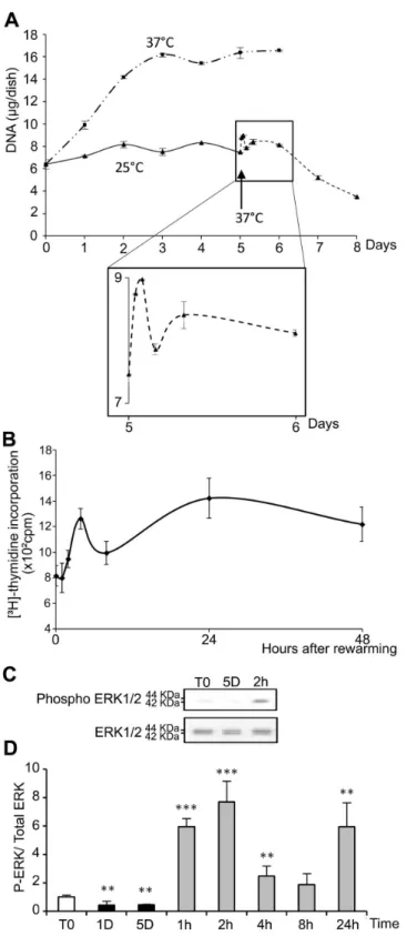
As compared to cells kept at γ7°C (Figure 1A), morphological alterations were observed in WIβ6 cells after 5 days of storage at β5°C (Figure 1B). After warming-up at γ7°C, cells did not recover a normal morphology after β4h (Figure 1C). Instead, some were rounded, refringent and detached, suggesting induction of apoptosis. At β5°C, the number of cells as measured as the DNA content remained constant during the 5 days with no observed significant loss (Figure βA). Upon rewarming after 5 days at β5°C, DNA content in the cultures slightly and transiently increased to drop down thereafter (insert in Figure βA). A similar wave-shaped curve was observed for [γH]-thymidine incorporation, although with a slight
To support the induction of apoptosis suggested by the morphological alterations illustrated in Figure 1, the expression of several pro- and anti-apoptotic factors was investigated at the mRNA level and the phosphorylation of Akt was evaluated by western blot. The expression of both the anti-apoptotic factor Bcl-β and the pro-apoptotic Bcl-β-associated X protein (BAX) was decreased at β5°C and remained low for several hours after warming-up before returning to control level (Figure S1, A, B and C). However, the BAX to Bcl-β ratio, considered as indicative of apoptosis, was increased during cold shock and early rewarming (Figure S1, D). Bcl-X exists under β alternatively spliced isoforms: a short pro-apoptotic form (Bcl-XS) and a long anti-apoptotic transcript (Bcl-XL). Total Bcl-X
mRNA level was largely increased by cold shock and rewarming (Figure Sβ, B). A shift towards the short pro-apoptotic isoform, illustrated by the Bcl-XS/L ratio, was observed
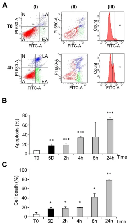
at β5°C, and up to 8 hours after transition to γ7°C (Figure Sβ, C). Phosphorylation of Akt, a kinase involved in survival pathways by inhibiting apoptotic processes, was first decreased after 1 day at β5°C, largely increased above control levels after 5 days at β5°C and remained elevated after warming-up (Figure Sγ, A and B). Together these data indicate that both pro- and anti-apoptotic processes are triggered during hypothermia and warming-up. Cells were however ultimately committed to apoptosis as shown by FACS analysis (Figure 4A and B) and trypan blue exclusion (Figure 4C). FACS data showed that a significant apoptosis was induced after five days at β5°C and more dramatically upon rewarming (Figure 4B, grey bars). Similar observations were made by measuring cell death using trypan blue exclusion test (Figure 4C). A similar induction of apoptosis was observed in osteoblastic MG-6γ cells (Figure S4) suggesting that apoptosis is a general cellular response to hypothermia and rewarming.
2: Mild hypothermia and rewarming induce autophagy
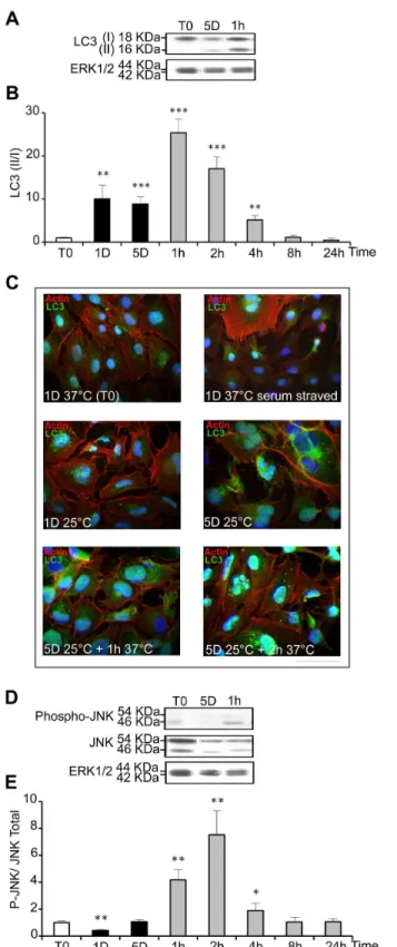
The microtubule-associated protein 1 light chain γ (LCγ) is present in cells as two isoforms: LCγ I (18kDa) located in the cytoplasm, and LCγ II (16kDa) associated with the inner membrane of autophagosomes [β9], the LCγ II/I ratio being
correlated to the level of autophagy. It was measured by western-blot in WIβ6 cells experiencing mild cold shock and warming-up (Figure 5A). LCγ II/I ratio was low in control samples, increased up to 10 fold after 1 and 5 days at β5°C and further transiently increased, up to β5 fold, during rewarming (Figure 5B). Intracellular localization of LCγ was visualized by immunostaining. Serum-starved cells as positive and cells kept at γ7°C in the presence of serum were used as positive and negative controls, respectively. A clear punctuated labeling typical of autophagosomes vesicles was seen in serum-starved cells and in cells cultured for 1 and 5 days at β5°C, and was even more evident after warming-up to γ7°C for 1 and β hours (Figure 5C). Together, these data indicate that autophagy is triggered by both hypothermia and relative hyperthermia.
Jun N-terminal kinase (JNK) activation, known to be induced by cellular stress and to mediate autophagy [γ0], was transiently increased after warming-up to γ7°C for 1, β and 4 hours (Figure 5D and E). However, it was not increased after 1 day and 5 days at β5°C, suggesting that it might participe in the autophagy induced by warming up but not by cold shock.
3: Mild cold shock and rewarming induce ROS production and DNA damage
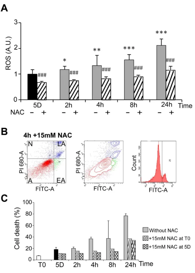
Heat shock has been shown to induce an oxidative stress in HEKβ9γ cells [γ1]. Oxidative stress was analysed here during cold shock and rewarming. The level of ROS in cells kept at β5°C for 5 days was similar to that of control cells cultured at γ7°C (not illustrated) and progressively increased after rewarming (plain grey bars in Figure 6A). It was significantly reduced by the ROS scavenger N-acetylcysteine (NAC) as expected (dashed bars in Figure 6A). As oxidative stress may lead to apoptosis, the effect of NAC on induction of apoptosis by cold shock and rewarming was investigated by FACS analysis. NAC, whether added at the beginning or at the end of the period in mild hypothermia, reduced cell death (compare Figure 6B to Figure 4A) at all time points (Figure 6C). Similar induction of ROS and reduction of cell apoptosis by NAC were Figure 1. Phase contrast micrographs of WI26 cells. Cells were continuously cultured at γ7°C (A), at β5°C for 5 days (B) or at β5°C for 5 days followed by a rewarming to γ7°C for β4h (C). Arrows point to refringent, apoptotic-like cells. Bar: 100µm.
Figure 2. Temperature dependence of cell proliferation. (A) WIβ6 cells maintained at γ7°C for 1 to 6 days (— - - —) or at β5°C for 1 to 5 days (——) and then warmed at γ7°C for 1 to 7β hours (- - - -), and DNA content was measured. The black arrow indicates the transition from β5°C to γ7°C. Insert provides an enlarged view of the first β4 hours of rewarming at γ7°C. (B) [γH]-thymidine
incorporation by WIβ6 cells maintained for 5 days at β5°C and warmed at γ7°C for 1 to 48 h. (C) Representative western blot probed with antibodies specific for phospho ERK 1/β or total ERK 1/β. (D) Quantification of the western blots. Data are expressed as the mean ratio of P-ERK/ total ERK normalized to ratio in control cells (T0), taken as 1. T0: 1 day at γ7°C; 1D and 5D: 1 or 5 days at β5°C; 1h, βh, 4h, 8h and β4h: 5 days at β5°C followed by 1 to β4h at γ7°C.
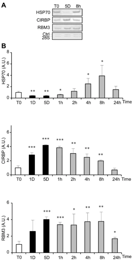
Figure 3. Cold-shock and rewarming affect heat and cold shock genes expression. The expression of heat shock (HSP70) and cold shock genes (CIRBP and RBMγ) was quantified at the mRNA level by RT-PCR in WIβ6 cells cultured at β5°C for 1 and 5 days before warming up at γ7°C for 1h to β4h. (A) Representative gels showing the RT-PCR amplification products. (B) HSP70, CIRBP and RBMγ mRNA levels are expressed as mean ± SD (n=γ) after normalization to the β8S rRNA content used as calibrator. Values at T0 were arbitrary taken as 1. Legend for culture schedule is the same as in Figure β.
Figure 4. Cold shock and rewarming affect cell viability. WIβ6 cells were cultured at β5°C for 5 days and then warmed-up at γ7°C for β to β4h. Cells kept at γ7°C were used as control (T0). FACS analysis was performed after labeling with FITC-annexin V (FITC-A, X-axes) and propidium iodide (PI, Y-axes). 1β.000 to 18.000 events were collected for each experiment. (A) Example of dots graphs (I), contour graphs (II) and annexin V curves (III) of control cells and cells maintained 5 days at β5°C and then warmed-up at γ7°C for 4h (4h). Alive cells [A] (double negative staining), cells in early apoptosis [EA] (annexin V positive, PI negative), in late apoptosis [LA] (double positive) and necrotic [N] (annexin V negative, PI positive) are indicated on the graphs. Annexin V curves (III) were used to define the gating allowing to discriminate the populations. (B) Percentage of apoptotic cells as measured by FACS analysis. Cells were cultured at γ7°C (T0) or β5°C for 5 days and subsequent warming-up at γ7°C for β to β4h. (C) Percentage of dead cells as measured by trypan blue exclusion assay. Cells were cultured in duplicate for 5 days at β5°C and then rewarmed for β to β4h at γ7°C.
Figure 5. Cold shock and rewarming induce autophagy and a cellular stress response. The level of LCγ I and II (A), phospho-JNK (P-JNK) and total JNK (D) was analyzed by western blot in cells cultured at β5°C for 1 and 5 days and then warmed-up at γ7°C for 1 to β4h. The levels of ERK1/β were taken as calibrator and used to monitor protein loading. Results are expressed as the mean ratio of LCγ II/I (B) and of P-JNK/ total JNK (D) taking T0 as 1. (C) Immunostaining of LCγ was performed in WIβ6 cells maintained at γ7°C in the presence or in the absence of serum or at various time points during the cold shock and rewarming. Actin stress fibers appear in red, LCγ in green and nucleus in blue. Bar: 100µm.
observed in another cell line, namely MG6γ cells (Figure S5, A, B and C).
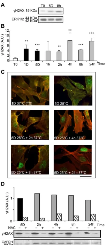
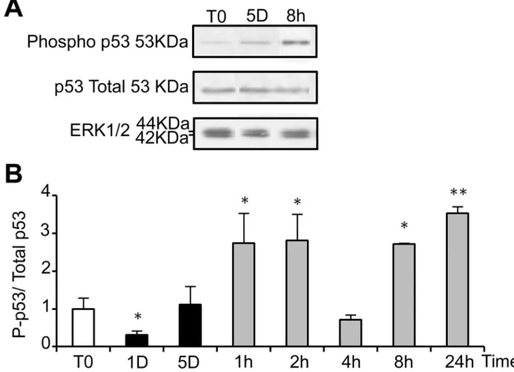
ROS are known inducers of oxidative DNA damage. To see if cold shock and rewarming may have the unexpected property to induce DNA damage, we analyzed the C-terminal phosphorylation of HβA histone family member X (HβAX), a double strand breaks tracer which participates in the DNA damage response and mediates DNA repair [γβ]. Mild hypothermia and rewarming induced a significant increase of HβAX phosphorylation ( HβAX) as shown by western-blot (Figure 7A and B) and immunostaining that showed an increased nuclear labeling (Figure 7C). A similar response was observed in osteoblastic cell lines (MG-6γ) and endothelial cells (HBME-1 and HMEC), showing that temperature-induced DNA damage is not restricted to WIβ6 cells (Figure S6). As illustrated in Figure 7D, HβAX was strongly reduced upon NAC treatment, indicating a causal relationship between oxidative stress and DNA damage. We further investigated the phosphorylation of p5γ and the expression of the alternative spliced VEGF111 variant [γγ], two processes known to be induced in response to DNA damage. Phospho-p5γ signal was indeed significantly increased upon rewarming after 5 days of hypothermia (Figure 8). The expression of VEGF111, barely detectable after 5 days at β5°C, was strongly increased upon rewarming in WIβ6 cells (Figure S7, A and B). A similar induction was observed in other cell types of different lineage such as Hela cells, HMEC and MG6γ (Figure S7, C), again underscoring the generalized cellular response.
Discussion
Our work highlights deleterious effects on cell phenotype during mild hypothermia and return to normothermia. In agreement with others, we observed that hypothermia induces a re-programming of gene expression, RNA processing and intracellular signaling, and affects cell shape, growth and survival. Additionally, far from reducing the effects of cold shock, the return to physiological temperature (γ7°C) rather sustains or exacerbates some of them, in accordance with previous reports [1,γ,11]. Rewarming also induces a burst of ROS, an effect likely induced by the sudden increase of mitochondrial electron transport [5].
General transcription and translation are reduced in hypothermia but remain able to proceed at a sufficient rate to ensure expression of at least some specific genes [β,γ4]. Hypothermia can also modulate the level and stabilization of specific transcripts as described for CIRBP [γ5]. In agreement with previous finding [5,11,γ6], the return to normothermia after a cold shock induced a heat shock-like response, as observed by the increased expression of HSP70 largely exceeding that of normothermic conditions during the first 8 hours of rewarming. This suggests that the sensing mechanism of heat shock detects a relative, instead of an absolute, hyperthermia.
It was reported that the proliferation rate of mammary cells significantly decreases with temperature [γ7] and human normal fibroblasts undergo a cell cycle arrest around β8°C. Our data only partly agree those of Matijasevic et al. The authors postulated that the cold-induced inhibition of cycle progression
was linked to the activation of p5γ [γ7]. Accordingly, we found that hypothermia resulted in a general inhibition of DNA synthesis of WI-β6 cells. However, in our model, increased phosphorylation of p5γ was not observed at β5°C. Furthermore, p5γ deficient MG-6γ cells, showed the same growth arrest at β5°C as cells proficient in p5γ (data not shown). During rewarming, we systematically observed an early and transient increase of DNA content in WIβ6 and several other cell types (not shown). A similar “wave-patterned” [γH]-thymidine incorporation also occurred. In accordance with
these data, we found that cells maintained at β5°C were arrested in G1 and Gβ phase (data not shown) while only cells in Gβ were observed within two hours of rewarming confirming a burst of DNA synthesis early after temperature transition. This observation is in agreement with previous reports [γ8,γ9] showing that cold-induced growth arrest might not only occur at a single check point. It is further supported by the pattern of ERK1/β phosphorylation, known as a main regulator of the G1 to S transition [40]. However, this burst of DNA synthesis is transient and rapidly followed by a progressive loss of cells.
Divergent data have been described concerning the effects of mild hypothermia on apoptosis. Some studies showed induction of apoptosis upon cold shock while others did not or even suggested it had a protective effect against subsequent heat-induced cell death [41–4γ]. These discrepancies may be due to different experimental conditions in terms of treatment duration and/or range of temperatures. In our model, a low level of apoptosis and cell death was induced during hypothermia but sharply increased during rewarming, likely explaining the significant cell loss observed after two and three days of culture at γ7°C.
Figure 6. Rewarming-induced ROS production and ROS-dependant apoptosis. (A) ROS were measured in WIβ6 cells cultured at β5°C for 5 days and then warmed-up at γ7°C for β to β4h in absence (-) and in presence (+) of 15mM N-Acetylcysteine (NAC). Data are expressed in arbitrary units taking T0 as 1. Significant inhibition by NAC versus condition without NAC is indicated # # # (p<0.001). (B) Cell viability was measured by FACS analysis after labeling with FITC-annexin V (FITC-A, X-axes) and propidium iodide (PI, Y-axes) of cells maintained 5 days at β5°C and then warmed-up at γ7°C for 4h (4h) in the presence or in the absence of 15mM of NAC added at T0. (C) Percentage of apoptotic cells in the indicated culture conditions.
Figure 7. Cold shock and rewarming induce H2AX phosphorylation through ROS production. The levels of phosphorylated HβAX ( HβAX) were analyzed in WIβ6 cells cultured at β5°C for 1 and 5 days and then warmed-up at γ7°C for 1 to β4h. (A) Representative western blots. (B) HβAX was quantified by western blotting. Data are expressed in arbitrary units after normalization by ERK1/β, taking T0 as 1. (C) HβAX in WIβ6 cells in normal conditions (1Dγ7°C) and during cold shock for 5 days and rewarming for β to β4h was evidenced by immunostaining. Actin stress fibers are stained in red and HβAX in green. Bar: 100µm. (D) HβAX was measured by western blot in WIβ6 cells cultured at β5°C for 5 days and then warmed-up at γ7°C for β to β4h in absence (-) and in presence (+) of NAC 15mM added at T0. After normalization by GAPDH, results are expressed in arbitrary units taking 5D as 1.
rewarming of the cells induced an oxidative burst and we hypothesize that it may induce DNA damage, trigger the DDR and provoke apoptosis. According to this hypothesis, scavenging ROS by N-acetylcysteine significantly reduced apoptosis and cell death as well as HβAX levels. These data strongly suggest a causative relationship between ROS production, induction of DDR, apoptosis and ultimately cell death in our model of mild hypothermia and rewarming. The mechanisms leading to DNA damage during the cold-shock phase remain however elusive.
The observation that hypothermia and, more importantly, rewarming induce an oxidative burst, apoptosis and a DDR might have practical consequences in biology and medicine and should be taken in consideration for cells, clinical samples or organs conservation and transportation prior to transplantation. They could also bring new insights for investigating adaptation to low temperatures and rewarming during therapeutic or accidental hypothermia.
Supporting Information
Table S1. Sequences of the primers used for RT-PCR analyses. The number of PCR cycles and the size of the RT-PCR products are also indicated. β8S: ribosomal RNA β8S subunit (cellular: endogenous RNA; synth: synthetic control co-amplified with cellular β8S to monitor the efficiency of the reaction); BAX: BCLβ-associated X protein; BCL-β: B-cell CLL/ lymphoma β; BCL-X: bcl-β-related gene (long - short: long and short isoforms of the gene); CIRBP: cold inducible RNA binding protein; HSP-70: heat shock protein 70; RBMγ: RNA biding motif protein γ; VEGF: vascular endothelial growth factor. (TIF)
Figure S1. Cold shock and rewarming affect markers of apoptosis. The expression of the anti-apoptotic Bcl-β and pro-apoptotic BAX genes was quantified by RT-PCR in WIβ6 cells routinely maintained at γ7°C or cultured at β5°C for 1 and 5 Figure 8. Return to normal temperature after a cold shock affects the phosphorylation of p53. Total and phosphorylated p5γ (P-p5γ) were analyzed in WIβ6 cells cultured in normal conditions or at β5°C for 1 and 5 days and then warmed-up at γ7°C for 1 to β4h. (A) Representative western blot. (B) Quantifications of the western-blots are expressed as the mean ratio P-p5γ/total p5γ, taking T0 as 1. Total protein loading was monitored by ERK1/β.
days before warming up at γ7°C for 1h to β4h (abbreviations as in Figure β). (A) Representative western blot showing the RT-PCR products. (B) Levels (mean ± SD) of Bcl-β, BAX mRNA and BAX to Bcl-β ratio.
(TIF)
Figure S2. Cold shock and rewarming affect the splicing of Bcl-X. The expression of the Bcl-X gene was quantified by RT-PCR. (A) Representative example in WIβ6 cells routinely maintained at γ7°C or cultured at β5°C for 1 to 5 days before warming up at γ7°C for 1h to β4h. (B) Mean total Bcl-X mRNA, obtained by summing the signal of the two isoforms, is expressed in arbitrary units after normalization for the β8S mRNA, taking T0 as 1. (C) Mean Bcl-XS/L ratio, taking T0 as 1.
(TIF)
Figure S3. Cold shock and rewarming affect Akt
activation. Akt, phospho-Akt and ERK1/β were quantified by western blot in WIβ6 cells cultured at β5°C for 1 and 5 days before warming up at γ7°C for 1h to β4h. ERK1/β was used to monitor equal protein loading. (A) Representative western Blot. (B) Results are expressed as the ratio (mean ± SD) of phospho-Akt (P-Akt)/ total Akt. T0 sample was arbitrary set at 1.
(TIF)
Figure S4. Cold shock and rewarming affect MG63 cells viability. Cells were cultured at β5°C for 5 days and then warmed-up at γ7°C for β to β4h. Cells kept at γ7°C were used as control (T0). Analysis was performed after cell labeling with FITC-annexin V (FITC-A, X-axes) and propidium iodide (PI, Y-axes) on 11.600 to γ0.000 events collected for each experiment. (A) Examples of dots graphs (I) and annexin V curves (II) of control cells (T0) and cells maintained 5 days at β5°C and then warmed-up at γ7°C for 4h (4h). Alive cells [A] (double negative staining), in early apoptosis [EA] (annexin V positive, PI negative), in late apoptosis [LA] (double positive) and necrotic [N] (annexin V negative, PI positive) are indicated on the graphs by using (II) Annexin V curves were used to define the gating allowing to discriminate the populations. (B) Quantification of cell death in MG6γ cultured at γ7°C (T0) or β5°C for 5 days and subsequent warming-up at γ7°C for β to β4h.
(TIF)
Figure S5. Rewarming-induced ROS production and
ROS-dependant apoptosis in MG63 cells. (A) ROS were
measured in MG6γ cells cultured at β5°C for 5 days and then warmed-up at γ7°C for β to β4h in absence (-) and in presence
(+) of 15mM of NAC. Cells kept at γ7°C were used as control (T0). Data are expressed in arbitrary units taking control cells as 1. Significant inhibition by NAC is indicated (# # p<0.01; # # # p<0.001). (B) Cell viability was measured by FACS analysis after cell labeling with FITC-annexin V (FITC-A, X-axes) and propidium iodide (PI, Y-axes) of cells maintained 5 days at β5°C and then warmed-up at γ7°C for 4h (4h) in the presence or in absence of 15mM of NAC added at T0 (compare with Fig. 4SA, 4 hours). (C) Percentage of dead cells in the indicated culture conditions.
(TIF)
Figure S6. Cold shock and rewarming induce H2AX
phosphorylation in different cell types. HβAX
phosphorylation was measured by western blot in HBME-1, HMEC and MG6γ cells after 5 days at β5°C and warming up at γ7°C for 1h and βh. Cells kept at γ7°C were used as control (T0). Data were normalized using GAPDH as calibrator. Results are expressed in arbitrary units taking control cells (T0) as 1 (n=1).
(TIF)
Figure S7. Cold shock and rewarming affect the splicing of VEGF pre-mRNA. The levels of VEGF variants and of the β8S rRNA were measured by RT-PCR in WIβ6, Hela, HMEC and MG6γ cells cultured in the indicated conditions. (A) Representative gel showing the various VEGF splice variants in WIβ6. Arrow indicated the VEGF111. The β8S rRNA was used as calibrator. (B) Levels of the VEGF111 variant (mean ± SD) in WIβ6 cells cultured in the indicated conditions. VEGF111 level was expressed in % of total VEGF, taking T0 as 1. (C) Expression of VEGF111 in epithelial (Hela), endothelial (HMEC) and osteoblastic (MG6γ) cells after 5 days at β5°C and warming up at γ7°C for 1h and βh taking T0 as 1 (n=1).
(TIF)
Acknowledgements
We acknowledge the Imaging platform of the GIGA-Research for its support.
Author Contributions
Conceived and designed the experiments: TN CAL BVN ACC. Performed the experiments: TN CAL. Analyzed the data: TN CAL BVN ACC. Contributed reagents/materials/analysis tools: TN CAL BVN ACC. Wrote the manuscript: TN CAL BVN ACC.
References
1. Al-Fageeh MB, Marchant RJ, Carden MJ, Smales CM (β006) The cold-shock response in cultured mammalian cells: harnessing the response for the improvement of recombinant protein production. Biotechnol Bioeng 9γ: 8β9-8γ5. doi:10.100β/bit.β0789. PubMed: 16γβ914β. β. Al-Fageeh MB, Smales CM (β006) Control and regulation of the cellular
responses to cold shock: the responses in yeast and mammalian systems. Biochem J γ97: β47-β59. doi:10.104β/BJβ0060166. PubMed: 1679β5β7.
γ. Roobol A, Carden MJ, Newsam RJ, Smales CM (β009) Biochemical insights into the mechanisms central to the response of mammalian cells to cold stress and subsequent rewarming. FEBS J β76: β86-γ0β. doi:10.1111/j.174β-4658.β008.06781.x. PubMed: 19054067.
10. Plesofsky N, Brambl R (1999) Glucose metabolism in Neurospora is altered by heat shock and by disruption of HSPγ0. Biochim Biophys Acta 1449: 7γ-8β. doi:10.1016/S0167-4889(98)0017β-4. PubMed: 1007605β.
11. Sonna LA, Fujita J, Gaffin SL, Lilly CM (β00β) Invited review: Effects of heat and cold stress on mammalian gene expression. J Appl Physiol 9β: 17β5-174β. PubMed: 1189604γ.
1β. Shapiro IM, Lubennikova EI (1968) Population kinetics of cells in tissue culture incubated at low temperature. Exp Cell Res 49: γ05-γ16. doi: 10.1016/0014-48β7(68)9018β-1. PubMed: 5761γ87.
1γ. Creagh EM, Sheehan D, Cotter TG (β000) Heat shock proteins--modulators of apoptosis in tumour cells. Leukemia 14: 1161-117γ. doi: 10.10γ8/sj.leu.β401841. PubMed: 109145γ8.
14. Yang Y, Xing D, Zhou F, Chen Q (β010) Mitochondrial autophagy protects against heat shock-induced apoptosis through reducing cytosolic cytochrome c release and downstream caspase-γ activation. Biochem Biophys Res Commun γ95: 190-195. doi:10.1016/j.bbrc. β010.0γ.155. PubMed: β0γ619γ1.
15. Swanlund JM, Kregel KC, Oberley TD (β008) Autophagy following heat stress: the role of aging and protein nitration. Autophagy 4: 9γ6-9γ9. PubMed: 18758βγ5.
16. Nivon M, Richet E, Codogno P, Arrigo AP, Kretz-Remy C (β009) Autophagy activation by NFkappaB is essential for cell survival after heat shock. Autophagy 5: 766-78γ. PubMed: 1950β777.
17. Hsu YL, Yu HS, Lin HC, Wu KY, Yang RC et al. (β011) Heat shock induces apoptosis through reactive oxygen species involving mitochondrial and death receptor pathways in corneal cells. Exp Eye Res 9γ: 405-41β. doi:10.1016/j.exer.β011.06.005. PubMed: β171β0γ1. 18. Ladomery M (1997) Multifunctional proteins suggest connections
between transcriptional and post-transcriptional processes. Bioessays 19: 90γ-909. doi:10.100β/bies.950191010. PubMed: 9γ6γ684. 19. Nishiyama H, Higashitsuji H, Yokoi H, Itoh K, Danno S et al. (1997)
Cloning and characterization of human CIRP (cold-inducible RNA-binding protein) cDNA and chromosomal assignment of the gene. Gene β04: 115-1β0. doi:10.1016/S0γ78-1119(97)005γ0-1. PubMed: 94γ417β.
β0. Artero-Castro A, Callejas FB, Castellvi J, Kondoh H, Carnero A et al. (β009) Cold-inducible RNA-binding protein bypasses replicative senescence in primary cells through extracellular signal-regulated kinase 1 and β activation. Mol Cell Biol β9: 1855-1868. doi:10.11β8/ MCB.01γ86-08. PubMed: 19158β77.
β1. Valko M, Leibfritz D, Moncol J, Cronin MT, Mazur M et al. (β007) Free radicals and antioxidants in normal physiological functions and human disease. Int J Biochem Cell Biol γ9: 44-84. doi:10.1016/j.biocel. β006.07.001. PubMed: 16978905.
ββ. Jian Z, Li K, Liu L, Zhang Y, Zhou Z et al. (β011) Heme oxygenase-1 protects human melanocytes from HβOβ-induced oxidative stress via the Nrfβ-ARE pathway. J Invest Dermatol 1γ1: 14β0-14β7. doi:10.10γ8/ jid.β011.56. PubMed: β141ββ59.
βγ. Hunt L, Hacker DL, Grosjean F, De Jesus M, Uebersax L et al. (β005) Low-temperature pausing of cultivated mammalian cells. Biotechnol Bioeng 89: 157-16γ. doi:10.100β/bit.β0γβ0. PubMed: 155840β5. β4. Lehr JE, Pienta KJ (1998) Preferential adhesion of prostate cancer
cells to a human bone marrow endothelial cell line. J Natl Cancer Inst 90: 118-1βγ. doi:10.109γ/jnci/90.β.118. PubMed: 9450571.
β5. Sa Russell (β001) Molecular Cloning: A Laboratory Manual (γrd ed.). Cold Spring Harbor Laboratory Press.
β6. Lambert CA, Colige AC, Munaut C, Lapière CM, Nusgens BV (β001) Distinct pathways in the over-expression of matrix metalloproteinases in human fibroblasts by relaxation of mechanical tension. Matrix Biol
γ1. Kim HJ, Kang BS, Park JW (β005) Cellular defense against heat shock-induced oxidative damage by mitochondrial NADP+ -dependent
isocitrate dehydrogenase. Free Radic Res γ9: 441-448. doi: 10.1080/10715760500066β65. PubMed: 160β8γ69.
γβ. Mahrhofer H, Bürger S, Oppitz U, Flentje M, Djuzenova CS (β006) Radiation induced DNA damage and damage repair in human tumor and fibroblast cell lines assessed by histone HβAX phosphorylation. Int J Radiat Oncol Biol Phys 64: 57γ-580. doi:10.1016/j.ijrobp. β005.09.0γ7. PubMed: 16414γ7β.
γγ. Mineur P, Colige AC, Deroanne CF, Dubail J, Kesteloot F et al. (β007) Newly identified biologically active and proteolysis-resistant VEGF-A isoform VEGF111 is induced by genotoxic agents. J Cell Biol 179: 1β61-1β7γ. doi:10.108γ/jcb.β0070γ05β. PubMed: 180869β1. γ4. Burdon RH (1987) Temperature and animal cell protein synthesis.
Symp Soc Exp Biol 41: 11γ-1γγ. PubMed: γγγβ481.
γ5. Al-Fageeh MB, Smales CM (β009) Cold-inducible RNA binding protein (CIRP) expression is modulated by alternative mRNAs. RNA 15: 1164-1176. doi:10.1β61/rna.1179109. PubMed: 19γ98494.
γ6. Laios E, Rebeyka IM, Prody CA (1997) Characterization of cold-induced heat shock protein expression in neonatal rat cardiomyocytes. Mol Cell Biochem 17γ: 15γ-159. doi:10.10βγ/A:1006844114γ48. PubMed: 9β78β66.
γ7. Matijasevic Z, Snyder JE, Ludlum DB (1998) Hypothermia causes a reversible, p5γ-mediated cell cycle arrest in cultured fibroblasts. Oncol Res 10: 605-610. PubMed: 10γ6794β.
γ8. Kaufmann H, Mazur X, Fussenegger M, Bailey JE (1999) Influence of low temperature on productivity, proteome and protein phosphorylation of CHO cells. Biotechnol Bioeng 6γ: 57γ-58β. doi:10.100β/ (SICI)1097-0β90(19990605)6γ:5. PubMed: 10γ9781γ.
γ9. Rieder CL, Cole RW (β00β) Cold-shock and the Mammalian cell cycle. Cell Cycle 1: 169-175. PubMed: 1β4β99β7.
40. Meloche S, Pouysségur J (β007) The ERK1/β mitogen-activated protein kinase pathway as a master regulator of the G1- to S-phase transition. Oncogene β6: γββ7-γβγ9. doi:10.10γ8/sj.onc.1β10414. PubMed: 17496918.
41. Sakurai T, Itoh K, Higashitsuji H, Nonoguchi K, Liu Y et al. (β006) Cirp protects against tumor necrosis factor-alpha-induced apoptosis via activation of extracellular signal-regulated kinase. Biochim Biophys Acta 176γ: β90-β95. doi:10.1016/j.bbamcr.β006.0β.007. PubMed: 1656945β.
4β. Guedez L, Stetler-Stevenson WG, Wolff L, Wang J, Fukushima P et al. (1998) In vitro suppression of programmed cell death of B cells by tissue inhibitor of metalloproteinases-1. J Clin Invest 10β: β00β-β010. doi:10.117β/JCIβ881. PubMed: 98γ56β6.
4γ. Gregory CD, Milner AE (1994) Regulation of cell survival in Burkitt lymphoma: implications from studies of apoptosis following cold-shock treatment. Int J Cancer 57: 419-4β6. doi:10.100β/ijc.β910570γβ1. PubMed: 8169005.
44. Maiuri MC, Zalckvar E, Kimchi A, Kroemer G (β007) Self-eating and self-killing: crosstalk between autophagy and apoptosis. Nat Rev Mol Cell Biol 8: 741-75β. doi:10.10γ8/nrmββγ9. PubMed: 17717517. 45. Høyer-Hansen M, Jäättelä M (β007) AMP-activated protein kinase: a
universal regulator of autophagy? Autophagy γ: γ81-γ8γ. PubMed: 174570γ6.
46. Yorimitsu T, Nair U, Yang Z, Klionsky DJ (β006) Endoplasmic reticulum stress triggers autophagy. J Biol Chem β81: γ0β99-γ0γ04. doi:10.1074/ jbc.M607007β00. PubMed: 16901900.
lipopolysaccharide-induced peritonitis in rats. Am J Pathol 179: β8ββ-β8γ4. doi:10.1016/ j.ajpath.β011.08.01γ. PubMed: ββ001γ49.
48. Lu Z, Dono K, Gotoh K, Shibata M, Koike M et al. (β005) Participation of autophagy in the degeneration process of rat hepatocytes after transplantation following prolonged cold preservation. Arch Histol Cytol 68: 71-80. doi:10.1679/aohc.68.71. PubMed: 158β7γ80.
49. Kroemer G, Mariño G, Levine B (β010) Autophagy and the integrated stress response. Mol Cell 40: β80-β9γ. doi:10.1016/j.molcel. β010.09.0βγ. PubMed: β09654ββ.
50. Shkreta L, Froehlich U, Paquet ER, Toutant J, Elela SA et al. (β008) Anticancer drugs affect the alternative splicing of Bcl-x and other
human apoptotic genes. Mol Cancer Ther 7: 1γ98-1409. doi: 10.1158/15γ5-716γ.MCT-08-019β. PubMed: 18566β1β.
51. Nicholls CD, Shields MA, Lee PW, Robbins SM, Beattie TL (β004) UV-dependent alternative splicing uncouples p5γ activity and PIGγ gene function through rapid proteolytic degradation. J Biol Chem β79: β4171-β4178. doi:10.1074/jbc.M401049β00. PubMed: 15067011. 5β. Rauen U, Polzar B, Stephan H, Mannherz HG, de Groot H (1999)
Cold-induced apoptosis in cultured hepatocytes and liver endothelial cells: mediation by reactive oxygen species. FASEB J 1γ: 155-168. PubMed: 987β940.