The Brazilian Journal of
INFECTIOUS DISEASES
w w w . e l s e v i e r . c o m / l o c a t e / b j i d
Original article
Methicillin-resistant
Staphylococcus aureus
nasal carriage in
neonates and children attending a pediatric outpatient
clinics in Brazil
Maria Aparecida Vieira
a, Ruth Minamisava
b,∗, Vicente Pessoa-Júnior
c,
Juliana Lamaro-Cardoso
d, Yves Mauro Ternes
c, Maria Cláudia Porfirio Andre
d,
Sabrina Sgambatti
e, André Kipnis
d, Ana Lúcia Andrade
c aDepartment of Nursing, Pontifícia Universidade Católica de Goiás, Goiânia, GO, Brazil bFaculty of Nursing, Universidade Federal de Goiás, Goiânia, GO, BrazilcDepartment of Community Health, Institute of Tropical Pathology and Public Health, Universidade Federal de Goiás, Goiânia, GO, Brazil dDepartment of Microbiology, Institute of Tropical Pathology and Public Health, Universidade Federal de Goiás, Goiânia, GO, Brazil eDepartment of Medicine, Pontifícia Universidade Católica de Goiás, Goiânia, GO, Brazil
a r t i c l e
i n f o
Article history:
Received 1 April 2013 Accepted 25 April 2013
Available online 25 September 2013
Keywords:
Staphylococcus aureuscarriage Methicillin-resistantStaphylococcus aureus
Neonates Children
a b s t r a c t
Background:In Latin America, few studies have been carried out on methicillin-resistant
Staphylococcus aureuscarriage in the pediatric population. We conducted a survey of nasal
S. aureuscarriage in neonates and in children attending the pediatric outpatient clinics in a large Brazilian city with high antimicrobial consumption.
Methods:Pernasal swabs of neonates were collected upon admission and at discharge in four neonatal intensive care units and of children less than five years of age during outpatient visits. Methicillin-resistantS. aureusisolates were characterized for antibiotic susceptibil-ity, mec gene presence, pulsed-field gel electrophoresis,spatype, SCCmec-type, multilocus sequence type, and presence of Panton-Valentine leukocidin genes.
Results: S. aureuswas carried by 9.1% and 20.1% of the 701 neonates and of 2034 children attending the outpatient clinics, respectively; methicillin-resistantS. aureuscarriage was detected in 0.6% and 0.2%, of the these populations, respectively. Healthcare-associated methicillin-resistantS. aureusstrains found in neonates from neonatal intensive care units and outpatients were genetically related to the Brazilian (SCCmec-III, ST239) and to the Pediatric (SCCmec-IV, ST5) clones. Community-associated methicillin-resistantS. aureuswas only detected in outpatients. None of the methicillin-resistantS. aureusstrains contained the Panton-Valentine leukocidin gene. Methicillin-resistantS. aureusstrains related to the Brazilian clone showed multidrug resistance pattern.
Conclusions:Despite the high antibiotic pressure in our area, and the cross transmission of the healthcare-associated methicillin-resistantS. aureusclones between neonatal intensive care units and outpatients, the prevalence of methicillin-resistantS. aureuscarriage is still low in our setting.
© 2013 Published by Elsevier Editora Ltda.
∗ Corresponding author at: Rua 227, Qd. 68, S/N, Setor Leste Universitário, Goiânia, Goiás 74605-080, Brazil. E-mail address: [email protected] (R. Minamisava).
Introduction
Staphylococcus aureusis a common cause of community and healthcare-associated illness. Carriage ofS. aureusis frequent in children and the anterior nares are the most consis-tent site forS. aureuscolonization.1 Children colonized with
methicillin-resistantS. aureus(MRSA) are potential reservoirs for the spread of MRSA in the community.2,3The relationship
between MRSA nasal carriage and invasive staphylococcal
infection has been well documented.4 A limited number of
MRSA clonal lineages has been responsible for the majority of MRSA infections in several regions.
Since the emergence and rapid spread of
community-associated MRSA (CA-MRSA) in previously healthy hosts,5
attention has been given to studies reporting changes in the epidemiology of MRSA colonization especially CA-MRSA in
hospitalized patients6and healthcare-associated MRSA
(HA-MRSA) in healthy subjects.7,8
In neonatal intensive care units (NICUs), neonates are at high risk for MRSA infections. Patients colonized by MRSA in NICUs present significant rates of bloodstream infection9and
are often transferred to other hospitals, contributing to the spread of MRSA in health facilities. Studies on MRSA carriage in neonates, however, are usually conducted under outbreak situations in NICUs.10,11
Despite the severity and rapid global expansion of MRSA strains, the epidemiology of MRSA nasal carriage remains unclear. In Latin America, few molecular studies are available on MRSA carriage in the pediatric population.12,13 To
deter-mine the extent the nasal carriage of MRSA is widespread in the pediatric population, we carried out a survey in neonates and in outpatient children in a large Brazilian city. We inves-tigated molecular characteristics of MRSA isolates.
Materials and methods
Study area
The study was conducted in the municipality of Goiânia, cap-ital of Goiás state, located in Central Brazil, a region with high rates of antibiotics consumption (Andrade 2012). In the 2010 census, the population of Goiânia was estimated at 1,302,001 inhabitants, with 20,014 live births, infant mortality at 14.6 deaths per 1000 live births, and 84,465 children under five years.14Public health care is provided free of charge by
the Brazilian Unified Health System, and an estimated 75%
of the population use the public health system.15 In
Goia-nia, children are first seen at the public healthcare centers (pediatric outpatient clinics) and referred to hospitalization whenever necessary. For this study, two pediatric populations were screened for MRSA, as described below.
Neonates admitted to NICUs
From June 2007 through November 2008 were screened for MRSA carriage all neonates admitted to four NICUs, which attend children from public and private health insurance. Tak-ing together these NICUs comprise the majority of beds (n= 69)
for neonates in the city. Data were collected on demograph-ics (gender, date of birth, and age at NICU admission), each neonate’s clinical history (length of hospital stay, congenital malformation, birth weight, preterm, timing of swab collec-tion, nasogastric intubacollec-tion, and diagnoses upon admission and at discharge), and variables related to their mothers (type of delivery, schooling, number of prenatal visits, and placental abruption).
Outpatient children
The carriage survey was conducted from July 2007 through July 2008 in children less than five years of age attending the major outpatient clinic of the city, served by the Brazilian pub-lic health insurance (Brazilian Unified Health System). Data were collected on child (gender, age, congenital malforma-tion, skin and soft-tissue infections, hospitalization in the last six months, antibiotic use in the last three months, day care attendance in the last six months, acute otitis media in the last 12 months, pneumonia in the last 12 months, and tonsil-litis in the last 12 months) and the family (mother’s schooling, smoking at home, number of family members, and health worker in the family). We calculated that a sample size of 2000 children would be necessary to ascertain MRSA
preva-lence assuming a 1.2% prevapreva-lence of MRSA nasal carriers16
with a 95% confidence interval (95% CI), a precision of 0.7%, design effect of 1.8, and a refusal rate of 10%. The number of children who were recruited at the outpatient clinics per day took into account the laboratory capability for processing nasal specimens (approximately 10 swabs/day).
Collection of nasal specimens
Pernasal flocked swabs were collected by rotating the swab twice in the vestibule of the anterior nostrils of each child. In the outpatients, a single sample was collected from each child. At the NICUs, two samples were collected per child; the first collected upon admission and the second at discharge or death. After collection, the swabs were placed in a liquid medium (ESwab, Copan Diagnostics, Inc., Murrieta, CA, USA) and sent to the Microbiology Laboratory of the Federal Univer-sity of Goiás for immediate processing.
Study ethics
The Institutional Review Board approval was given by the Ethics Committee of the Federal University of Goiás (# 008/2007). All parents or legal guardians signed copies of the informed consent form before recruitment.
Microbiological techniques
The specimens were plated onto mannitol salt agar (Difco®
Laboratories Inc., Detroit, MI, USA) forS. aureusisolation. Tryp-ticase soy agar plates, which were supplemented with 4% NaCl
and oxacillin at 6g/mL, were used to screen MRSA isolates.
After 24–48 h of incubation at 37◦C, the colonies that were
catalase, coagulase, and PCR amplification of thefemB gene (Jonas 1999).
Antimicrobial susceptibility testing
Susceptibility tests were determined by disk diffusion (Oxoid Ltd., Basingstoke, Hampshire, England) for oxacillin, cefox-itin, clindamycin, ciprofloxacin, erythromycin, rifampicin, tetracycline, trimethoprim–sulfamethoxazole, linezolid, and quinupristin/dalfopristin according to the Clinical Laboratory Standards Institute guideline.17Isolates resistant to cefoxitin
and oxacillin were submitted to the Etest®(AB Biodisk, Slona,
Sweden) to determine the sensitivity to vancomycin.S. aureus strains ATCC 25923 and ATCC 29213 were used as quality con-trols.
MRSA definition
Isolates showing inhibition zones of ≤10 mm and≤21 mm
around 1 mg oxacillin (Oxoid, Cambridge, UK) and 30g cefox-itin (Oxoid, Cambridge, UK) disks, respectively, and that were
positive formecA gene by PCR, were characterized as MRSA.
Molecular typing
Detection ofmecA gene and SCCmectypes were investigated
by multiplex PCR18using COL (SCCmectype I), N315 (SCCmec
type II), ANS46 (SCCmectype III), and MW2 (SCCmectype IV) as positive controls. Multilocus sequence typing was carried out
as previously reported19 and submitted to the international
database (http://www.mlst.net) to assign the sequence type (ST). Sequencing of the polymorphic X region of the protein A gene (spa) and the Panton-Valentine leukocidin (PVL) gene
were performed as described elsewhere.20,21 The sequences
were analyzed using the Ridom Staph Type software (Ridom,
Germany) to determine thespa types. The following
inter-national MRSA clones were included as controls: ST5-II (New-York/Japan, USA100), ST239-III (Brazilian/Hungarian), ST5-VI (Pediatric, USA800), and ST22-IV (EMRSA-15). The clonal relatedness was investigated by pulsed-field gel
elec-trophoresis (PFGE) with SmaI macrorestriction according to
standard procedures.22 The PFGE patterns were interpreted
according to the criteria proposed by Tenover et al.,23and the
resulting dendrogram was constructed using Dice coefficients with BioNumerics software (version 5.0). Isolates were defined as epidemiologically related if they shared≥80% similarity on
the dendrogram.
Data analysis
Prevalence of MRSA and the corresponding 95% confidence interval (95% CI) were calculated. Multidrug resistance (MDR) was defined for isolates with resistance to three classes of
antimicrobials and to-lactams. Moving-average was used to
calculate the percentage of colonization byS. aureus; the value attributed to a given month was the average colonization in that month, and the ones of the months prior and after the given month. Differences in lengths of stay in NICUs between
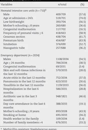
Table 1 – Characteristics of children screened for
Staphylococcus aureusand MRSAanasal colonization.
Goiânia, Brazil, 2007–2008.
Variables n/total (%)
Neonatal intensive care units (n = 710)b
Male 409/709 (57.6)
Age at admission < 24 h 519/701 (74.0)
Low birthweight 393/704 (56.5)
Mother’s schooling≤8 years 260/680 (36.6)
Congenital malformation 117/702 (16.5)
Frequency of prenatal visits≤6 418/663 (58.9)
Cesarean section 438/704 (61.7)
Premature birth 454/687 (63.9)
Intubation 374/690 (52.7)
Nasogastric tube 257/688 (36.2)
Emergency department (n = 2034)
Males 1108/2034 (54.5)
Age≥24 months 784/2034 (38.5)
Congenital malformation 69/2031 (3.4)
Skin and soft-tissue infections in the last 12 months
570/2033 (28.0)
Acute otitis in the last 12 months 752/2034 (37.0) Pneumonia in the last 12 months 623/2033 (30.6) Tonsillitis in the last 12 months 1139/2033 (56.0) Hospitalization in the last 6
months
586/2031 (28.8)
Antibiotic use in the last 3 months
948/1821 (46.6)
Day care attendance in the last 6 months
388/2033 (19.1)
Mother’s schooling≤8 years 893/2028 (43.9)
Smoking at home 695/2033 (34.2)
Health worker in the family 109/2034 (5.4) Number of family members > 4 676/2031 (33.2)
a Methicillin-resistantStaphylococcus aureus.
b The total does not always equals to 710.
neonates colonized and non-colonized by MRSA strains were evaluated by the Mann–Whitney test.
Results
Neonates
During the study period, a total of 710 neonates were admit-ted to the four NICUs, and nasal swabs were obtained from 701 (98.7%) of them. The characteristics of the neonates and their mothers are described in Table 1.The overall preva-lence of S. aureusand MRSA was 9.1% (95% CI 7.2–11.4) and 0.6% (95% CI 0.2–1.4), respectively, and varied among the NICUs (Table 2). Four neonates carried MRSA strains; three of them acquired MRSA during the hospitalization period and had higher lengths of stay (median = 20 days) compared to neonates who were non-colonized by MRSA (median = 10 days) (p= 0.072).
Children attending the outpatients
Table 2 – Prevalence ofStaphylococcus aureusand methicillin-resistantS. aureuscarriage according to the neonatal intensive care unit.
Neonatal intensive care units S. aureus MRSA
n % (95% CI) n % (95% CI)
#1 (n= 93) 5 5.4 (2.0–11.5) 0 0.0 –
#2 (n= 80) 2 2.5 (0.4–8.0) 0 0.0 –
#3 (n= 426) 41 9.6 (7.1–12.7) 2 0.5 (0.1–1.5)
#4 (n= 102) 16 15.7 (9.6–23.7) 2 2.0 (0.3–6.3)
Total (n= 701) 64 9.1 (7.2–11.4) 4 0.6 (0.3–1.6)
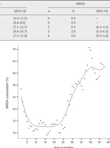
of age. The main clinical features of the participant children are shown in Table 1. Four hundred and eight children (20.1%; 95% CI: 18.4–21.9%) were colonized byS. aureus.Colonization byS. aureus showed an age-related distribution, with peak prevalence at 47 months of age (Fig. 1). Four children (0.2%; 95% CI: 0.1–0.5%) carried MRSA strains.
Antimicrobial resistance of MRSA isolates
We identified 16 MRSA isolates from eight children. Six children had more than one isolate. MDR MRSA isolates were found in three children. Eleven MRSA isolates did not meet criteria for MDR, but they were resistant to differ-ent classes of antibiotics. MRSA isolates showed resistance to erythromycin (43.8%), clindamycin (37.5%), ciprofloxacin (25.0%) and trimethoprim–sulfamethoxazole (18.8%). All MRSA strains were susceptible to vancomycin.
MRSA molecular findings
The majority of the MRSA strains clustered into two genomic groups; within each group we identified children from both, NICUs and outpatient clinics carrying MRSA strains genetically related to classical pandemic HA-MRSA Brazil-ian (SCCmec-III, ST239) and Pediatric (SCCmec-IV, ST5) clones (Fig. 2). CA-MRSA strains were only detected in outpatient children and were clonally related to Western Australian 1 clone/WA-1 (SCCmec-IV, ST128). Only the HA-MRSA Brazilian clone showed MDR pattern. None of the MRSA strains con-tained the PVL gene.
45 40 35 30 25 20 15 10
6 12 18 24 30
Age in months
36 42 48 54 60
MRSA colonization (%)
Fig. 1 – Age-related prevalence ofStaphylococcus aureus
colonization by age. Each dot represents a child colonized byS. aureus. Vertical arrows indicate the age of child colonized by the corresponding HA-MRSA and CA-MRSA clones.
BR936
ID CenterDate of swabs Previous MIC MDR SCCmec
Outpatient
NICU - 1 05.14.08 06.05.08 08.03.07 06.15.08 07.22.08 02.14.08 11.21.08 08.29.07 Outpatient
NICU - 2
Outpatient Outpatient
NICU - 2
NICU - 1 BR902 BR415 HU25 BR1419 BR2044 BR682 BR811 HDE288 BR457 NA NA NA
NA NA NA
NA NA NA NA NA NA No No No No No No Yes Yes Yes Yes Yes Yes >256 >256 >256 >256 24 24 16 16 8 III III III III IV IV IV IV IV IV (–) (–) (–) (–) (–) (–) (–) (–) 2223 t037 t037 t037 t037 t128 t128 t045 t045 t002 t002 585 770 239 641 5 5 1 1 5
SLV - HU25
SLV - HU25
SLV - HU25
Brazilian
Western Australian (WA - 1)
Western Australian (WA - 1)
Pediatric (USA800)
Pediatric (USA800)
Pediatric (USA800)
Pediatric (USA800) 94.4
% of similarity
91.7 87.6 76.9 83.9 80.3 100.0 100.0 80 85 90 95 100
75 PVL MLST Clone
Spa type
hospitalization collection
Discussion
Despite evidence that the MRSA incidence is increasing world-wide, studies on MRSA carriage in Latin America are still scarce. This study provides information onS. aureusand MRSA nasal carriage in outpatient children and in neonates admit-ted to NICUs in an urban area of Brazil with high antimicrobial pressure and infection control lapses.
We found that the frequency ofS. aureusby age in
chil-dren attending outpatients resembled a “J” curve, with higher prevalence in infants less than six months of age and in children older than 30 months of age. This result is consis-tent with studies by Bogaert et al.,24which reported similar
shape of age distribution ofS. aureuscarriage in healthy chil-dren.
The rates of MRSA carriage in NICUs herein were lower compared to studies from the US, Israel, and Taiwan.9,11,25One
possible explanation for the low MRSA carriage rate is that the present investigation was performed out of an outbreak, while the majority of studies in NICUs are usually conducted as a result of an outbreak of MRSA infection.
We also found lower prevalence of MRSA nasal coloniza-tion in outpatients when comparing to reports in children
attending well-child health care services in the US3 and in
Taiwan.26 Our findings, however, were comparable to those
obtained in Southern Israel, where the prevalence of MRSA carriage in healthy infants aged 2–12 months ranged from 0.0% to 0.7%.27Previous investigation in Brazil conducted in
outpatient children16detected a higher prevalence of MRSA
nasopharyngeal carriage (1.0%) than that found in the cur-rent study (0.2%), possibly because in the previous study the nasopharyngeal specimens were obtained during the win-ter period, when bacwin-terial colonization of respiratory tract is expected to be higher compared to colonization rates obtained from non-seasonal periods. Nevertheless, comparison of our results on MRSA carriage with other studies is not straight-forward due to differences in study designs, inclusion criteria, epidemiological scenarios, and age groups.
Despite the low colonization rates by MRSA found herein, this might turn out to be a serious concern. We identified three pandemic clones of MRSA (Brazilian, Pediatric, and WA1). The CA-MRSA WA-1 clone, detected in outpatients, has been identified in patients with bloodstream infections from South and Southeast regions of Brazil,28 indicating the
dissemina-tion of this clone across the country. The HA-MRSA Brazilian clone was detected in an outpatient child. This clone has been disseminated through hospitals in many countries of Latin America.29Previously, we described the presence of the
Brazilian clone in healthy children.16In all these cases, the
children had been admitted to hospitalization in the previous six months.
One interesting finding of this study is the presence of the HA-MRSA Pediatric clone recovered from outpatient children. This clone has been identified in hospitals of the Southeast and Northeast Regions of Brazil30,31but this is the first
detec-tion of the Pediatric clone in Central Brazil, suggesting that this clone is settling in Brazilian hospitals. This observation confirms that the Pediatric clone is spread in the community, increasing the chances of expanding its reservoir.
Some limitations of our study should be mentioned. First, as we collected nasal swabs of neonates only at admittance and discharge, we were not able to evaluate children who were intermittently colonized by MRSA in NICUs. Second, the small number of MRSA isolates resulted in an insufficient precision of the prevalence rates of MRSA carriage. Third, in outpatients, we did not enroll children aged higher than five years of age, as the peak of MRSA carriage is expected to be at 11 years of age.24
In conclusion, we found low rates of MRSA carriage among children in an area with high antimicrobial pressure. However, we detected CA-MRSA, and HA-MRSA clones in the commu-nity, which could contribute for an invisible MRSA reservoir, requiring a close monitoring of the MRSA epidemiology in both community and healthcare environment.
Conflicts of interest
The authors declare no conflicts of interest.
Acknowledgments
This work was supported by the Brazilian National Council for Scientific and Technological Development/CNPq. We are grateful to all pediatricians at the participant hospitals. We would also like to thank Copan Diagnostics Inc. for swab dona-tions. A.L.A. (Grants 482646/2007-1; 306096/2010-2) and A.K. (Grant 301199/2009-8) are research fellows of CNPq.
r e f e r e n c e s
1. Wertheim HF, Melles DC, Vos MC, et al. The role of nasal carriage inStaphylococcus aureusinfections. Lancet Infect Dis. 2005;5:751–62.
2. Chen CJ, Hsu KH, Lin TY, et al. Factors associated with nasal colonization of methicillin-resistantStaphylococcus aureus among healthy children in Taiwan. J Clin Microbiol. 2011;49:131–7.
3. Creech 2nd CB, Kernodle DS, Alsentzer A, Wilson C, Edwards KM. Increasing rates of nasal carriage of methicillin-resistant Staphylococcus aureusin healthy children. Pediatr Infect Dis J. 2005;24:617–21.
4. Lo WT, Lin WJ, Tseng MH, et al. Methicillin-resistant Staphylococcus aureusin children, Taiwan. Emerg Infect Dis. 2006;12:1267–70.
5. David MZ, Daum RS. Community-associated
methicillin-resistantStaphylococcus aureus: epidemiology and clinical consequences of an emerging epidemic. Clin Microbiol Rev. 2010;23:616–87.
6. Carey AJ, Della-Latta P, Huard R, et al. Changes in the molecular epidemiological characteristics of
methicillin-resistantStaphylococcus aureusin a neonatal intensive care unit. Infect Control Hosp Epidemiol. 2010;31:613–9.
7. Ho PL, Chiu SS, Chan MY, et al. Molecular epidemiology and nasal carriage ofStaphylococcus aureusand
methicillin-resistantS. aureusamong young children attending day care centers and kindergartens in Hong Kong. J Infect. 2012;64:500–6.
children attending day care centers in Singapore. Pediatr Infect Dis J. 2012;31:213–4.
9. Huang YC, Chou YH, Su LH, Lien RI, Lin TY.
Methicillin-resistantStaphylococcus aureuscolonization and its association with infection among infants hospitalized in neonatal intensive care units. Pediatrics. 2006;118:469–74. 10. Sax H, Posfay-Barbe K, Harbarth S, et al. Control of a cluster of
community-associated, methicillin-resistantStaphylococcus aureusin neonatology. J Hosp Infect. 2006;63:93–100. 11. Regev-Yochay G, Rubinstein E, Barzilai A, et al.
Methicillin-resistantStaphylococcus aureusin neonatal intensive care unit. Emerg Infect Dis. 2005;11:453–6.
12. Bartoloni A, Pallecchi L, Fernandez C, et al. Low prevalence of methicillin-resistantStaphylococcus aureusnasal carriage in urban and rural community settings in Bolivia and Peru. Int J Infect Dis. 2012.
13. Gardella N, Murzicato S, Di Gregorio S, et al. Prevalence and characterization of methicillin-resistantStaphylococcus aureus among healthy children in a city of Argentina. Infect Genet Evol. 2011;11:1066–71.
14. Brazilian Ministry of Health. Informac¸ões de Saúde. Brasília, DF: DATASUS; 2010. Available at: http://www2.datasus.gov.br/ DATASUS/index.php?area=02 [accessed 01.07.12].
15. Paim J, Travassos C, Almeida C, Bahia L, Macinko J. The Brazilian health system: history, advances, and challenges. Lancet. 2011;377:1778–97.
16. Lamaro-Cardoso J, Castanheira M, de Oliveira RM, et al. Carriage of methicillin-resistantStaphylococcus aureusin children in Brazil. Diagn Microbiol Infect Dis. 2007;57:467–70. 17. Clinical and Laboratory Standards Institute. Performance
standards for antimicrobial susceptibility testing. Supplement M100-S19. 10th ed. Wayne, PA: CLSI; 2009. 18. Milheiric¸o C, Oliveira DC, de Lencastre H. Multiplex PCR
strategy for subtyping the staphylococcal cassette chromosomemectype IV in methicillin-resistant Staphylococcus aureus: ‘SCCmecIV multiplex’. J Antimicrob Chemother. 2007;60:42–8.
19. Enright MC, Day NP, Davies CE, Peacock SJ, Spratt BG. Multilocus sequence typing for characterization of methicillin-resistant and methicillin-susceptible clones of Staphylococcus aureus. J Clin Microbiol. 2000;38:1008–15. 20. Koreen L, Ramaswamy SV, Graviss EA, et al.spatyping method
for discriminating amongStaphylococcus aureusisolates: implications for use of a single marker to detect genetic micro- and macrovariation. J Clin Microbiol. 2004;42:792–9.
21. Lina G, Piemont Y, Godail-Gamot F, et al. Involvement of Panton-Valentine leukocidin-producingStaphylococcus aureus in primary skin infections and pneumonia. Clin Infect Dis. 1999;29:1128–32.
22. Chung M, de Lencastre H, Matthews P, et al. Molecular typing of methicillin-resistantStaphylococcus aureusby pulsed-field gel electrophoresis: comparison of results obtained in a multilaboratory effort using identical protocols and MRSA strains. Microb Drug Resist. 2000;6:189–98.
23. Tenover FC, Arbeit RD, Goering RV, et al. Interpreting chromosomal DNA restriction patterns produced by pulsed-field gel electrophoresis: criteria for bacterial strain typing. J Clin Microbiol. 1995;33:2233–9.
24. Bogaert D, van Belkum A, Sluijter M, et al. Colonisation by Streptococcus pneumoniaeandStaphylococcus aureusin healthy children. Lancet. 2004;363:1871–2.
25. Sarda V, Molloy A, Kadkol S, et al. Active surveillance for methicillin-resistantStaphylococcus aureusin the neonatal intensive care unit. Infect Control Hosp Epidemiol. 2009;30:854–60.
26. Huang YC, Hwang KP, Chen PY, Chen CJ, Lin TY. Prevalence of methicillin-resistantStaphylococcus aureusnasal colonization among Taiwanese children in 2005 and 2006. J Clin Microbiol. 2007;45:3992–5.
27. Adler A, Givon-Lavi N, Moses AE, Block C, Dagan R. Carriage of community-associated methicillin-resistantStaphylococcus aureusin a cohort of infants in southern Israel: risk factors and molecular features. J Clin Microbiol. 2010;48:531–8. 28. Ribeiro A, Coronado AZ, Silva-Carvalho MC, et al. Detection
and characterization of international community-acquired infections by methicillin-resistantStaphylococcus aureus clones in Rio de Janeiro and Porto Alegre cities causing both community- and hospital-associated diseases. Diagn Microbiol Infect Dis. 2007;59:339–45.
29. Rodriguez-Noriega E, Seas C, Guzman-Blanco M, et al. Evolution of methicillin-resistantStaphylococcus aureusclones in Latin America. Int J Infect Dis. 2010;14:e560–6.
30. Sousa-Junior FC, Silva-Carvalho MC, Fernandes MJ, et al. Genotyping of methicillin-resistantStaphylococcus aureus isolates obtained in the Northeast region of Brazil. Braz J Med Biol Res. 2009;42:877–81.